Meticorten
Ed Gane, M.D., F.R.A.C.P.
- Associate Professor
- Faculty of Medicine
- University of Auckland
- Consultant Hepatologist
- Liver Unit
- Auckland City Hospital
- Auckland, New Zealand
Meticorten dosages: 40 mg, 20 mg, 10 mg, 5 mg
Meticorten packs: 30 pills, 60 pills, 90 pills, 120 pills, 180 pills, 270 pills, 360 pills
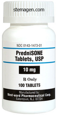
Buy 10mg meticorten free shipping
Surface anatomy of the spleen and pancreas relative to the diaphragm and posterior belly viscera allergy symptoms only at night generic 5 mg meticorten visa. Concavities on the visceral floor are impressions formed by the buildings involved with the spleen allergy x for dogs safe 10 mg meticorten. It follows a tortuous course posterior to the omental bursa allergy treatment in toddlers cheap meticorten 40 mg without a prescription, anterior to the left kidney, and alongside the superior border of the pancreas. Between the layers of the splenorenal ligament, the splenic artery divides into 5 or more branches that enter the hilum. The entry of the bile duct and pancreatic duct into the duodenum via the hepatopancreatic ampulla. The inside of the descending a part of the duodenum reveals the most important and minor duodenal papillae. The pancreaticosplenic nodes relate to the posterior surface and superior border of the pancreas. The pancreas produces: � an exocrine secretion (pancreatic juice from the acinar cells) that enters the duodenum through the primary and accessory pancreatic ducts. Because of the shut relationship of the pancreas and duodenum, their blood vessels are the identical in whole or in part. Except for the inferior part of the pancreatic head (including uncinate process), the spleen and pancreas obtain blood from the celiac artery. It firmly attaches to the medial side of the descending and horizontal parts of the duodenum. The physique of the pancreas continues from the neck and lies to the left of the superior mesenteric vessels, passing over the aorta and L2 vertebra, continuing just above the transpyloric airplane posterior to the omental bursa. The nerves of the pancreas are autonomic nerves from the celiac and superior mesenteric plexuses. A dense network of nerve fibers passes from the celiac plexus along the splenic artery to the spleen. Most are postsynaptic sympathetic fibers to smooth muscle of the splenic capsule, trabeculae, and intrasplenic vessels. The sphincter of the pancreatic duct (around the terminal a part of the pancreatic duct), the sphincter of the bile duct (around the termination of the bile duct), and the hepatopancreatic sphincter (of Oddi)-around the hepatopancreatic ampulla-are easy muscle sphincters that control the circulate of bile and pancreatic juice into the ampulla and stop reflux of duodenal content material into the ampulla. As many as 10 branches could move from the splenic artery to the physique and tail of the pancreas. Most vessels end within the pancreaticosplenic lymph nodes, which lie alongside the splenic artery. The parasympathetic and sympathetic fibers attain the pancreas by passing along the arteries from the celiac plexus and superior mesenteric plexus (see additionally "Summary of Innervation of Abdominal Viscera," p. Liver the liver is the largest gland in the physique and, after the skin, the largest single organ. Except for fat, all vitamins absorbed from the gastrointestinal tract are initially conveyed to the liver by the portal venous system. In addition to its many metabolic activities, the liver shops glycogen and secretes bile, a yellow-brown or green fluid that aids within the emulsification of fats. The liver produces bile constantly; nonetheless, between meals it accumulates and is saved in the gallbladder, which additionally concentrates the bile by absorbing water and salts. The regular liver lies deep to ribs 7�11 on the right aspect and crosses the midline toward the left nipple. The liver moves with the excursions of the diaphragm and is positioned more inferiorly when one is erect due to gravity. The liver has a convex diaphragmatic floor (anterior, superior, and some posterior) and a comparatively flat and even concave visceral floor (postero-inferior), which are separated anteriorly by its sharp inferior border that follows the best costal margin. Sagittal magnetic resonance imaging part demonstrating the relationships featured in A in a living particular person. Subphrenic recesses-superior extensions of the peritoneal cavity (greater sac)-exist between diaphragm and the anterior and superior elements of the diaphragmatic floor of the liver. The subphrenic recesses are separated into proper and left recesses by the falciform ligament, which extends between the liver and the anterior belly wall. The portion of the supracolic compartment of the perito- neal cavity immediately inferior to the liver is the subhepatic house. The hepatorenal recess (hepatorenal pouch; Morison pouch) is the posterosuperior extension of the subhepatic area, lying between the proper a part of the visceral surface of the liver and the proper kidney and suprarenal gland. The peritoneal reflections (ligaments) and cavity associated to the liver are proven diagrammatically. In the anatomical place, the visceral floor of the liver is directed inferiorly, posteriorly, and to the left. In embalmed specimens, impressions remain the place this floor is contacted by adjacent structures. Of the two gravity-dependent recesses of the abdominopelvic cavity in the supine position, the hepatorenal recess is the upper one, receiving drainage from the omental bursa and higher stomach (supracolic) portions of the higher sac. The anterior layer of the coronary ligament is steady on the left with the best layer of the falciform ligament, and the posterior layer is steady with the proper layer of the lesser omentum. In distinction to the smooth diaphragmatic floor, the visceral surface bears multiple fissures and impressions from contact with different organs. The umbilical (left sagittal) fissure is the continual groove fashioned anteriorly by the fissure for the spherical ligament and posteriorly by the fissure for the ligamentum venosum. The round ligament and small para-umbilical veins course in the free edge of the falciform ligament. The four anatomical lobes of the liver are defined by external options (peritoneal reflections and fissures). The anterior sagittal reduce is made in the aircraft of the fossa for the gallbladder, and the posterior sagittal minimize is in the airplane of the fissure for the ligamentum venosum. These cuts have been joined by a slim coronal cut within the plane of the porta hepatis. The portal triad passes between the layers of the hepatoduodenal ligament to enter the liver at the porta hepatis. The common hepatic artery passes between the layers of the hepatogastric ligament. The clinical significance of hepatic segments is explained in the blue box "Hepatic Lobectomies and Segmentectomy" on p. The caudate lobe (segment I, bringing the whole variety of segments to eight) is provided by branches of each divisions and is drained by its own minor hepatic veins. The hepatic portal vein carries nearly all the nutrients absorbed by the alimentary tract to the sinusoids of the liver. The exception is lipids, that are absorbed into and bypass the liver by way of the lymphatic system. Arterial blood from the hepatic artery, accounting for less than 20�25% of blood received by the liver, is distributed initially to nonparenchymal constructions, particularly the intrahepatic bile ducts. The hepatic portal vein, a brief, broad vein, is formed by the superior mesenteric and splenic veins posterior to the neck of the pancreas. Between one quarter and one half of the lymph getting into the thoracic duct comes from the liver.
Order meticorten 20 mg without prescription
The planes of those transverse research allergy forecast hong kong cheap 40 mg meticorten with visa, oriented in the same path as part C allergy symptoms on one side of face meticorten 40mg otc, pass superior (D) and inferior (E) to the rima glottidis allergy attack purchase meticorten 40 mg with visa. The shape of the rima glottidis, the aperture between the vocal folds, varies based on the place of the vocal folds. During normal respiration, the laryngeal muscle tissue are relaxed and the rima glottidis assumes a slender, slit-like position. During phonation, the arytenoid muscle tissue adduct the arytenoid cartilages at the identical time that the lateral crico-arytenoid muscle tissue moderately adduct. The decrease vary of pitch of the voice of postpubertal males results from the larger size of the vocal folds. The lateral recesses between the vocal and the vestibular folds are the laryngeal ventricles. The infrahyoid muscles are depressors of the hyoid and larynx, whereas the suprahyoid muscular tissues (and the stylopharyngeus, a pharyngeal muscle discussed later on this chapter) are elevators of the hyoid and larynx. The principal adductors are the lateral crico-arytenoid muscles, which pull the muscular processes anteriorly, rotating the arytenoid cartilages in order that their vocal processes swing medially. These fibers represent the thyro-epiglottic muscle, which widens the laryngeal inlet. This is the position of whispering when the breath is modified into voice within the absence of tone. The sole abductors are the posterior crico-arytenoid muscular tissues, which pull the muscular processes posteriorly, rotating the vocal processes laterally and thus widening the rima glottidis. This action occurs reflexively in response to the presence of liquid or particles approaching or inside the laryngeal vestibule. This will increase the distance between the thyroid prominence and the arytenoid cartilages. Because the anterior ends of the vocal ligaments attach to the posterior facet of the prominence, the vocal ligaments elongate and tighten, elevating the pitch of the voice. The vocalis muscular tissues lie medial to the thyro-arytenoid muscular tissues and lateral to the vocal ligaments within the vocal folds. The vocalis muscles produce minute changes of the vocal ligaments, selectively tensing and relaxing the anterior and posterior elements, respectively, of the vocal folds throughout animated speech and singing. The posterior branch provides the posterior crico-arytenoid and transverse and indirect arytenoid muscle tissue. Because it supplies all of the intrinsic muscular tissues except the cricothyroid, the inferior laryngeal nerve is the primary motor nerve of the larynx. The inferior laryngeal vein joins the inferior thyroid vein or the venous plexus of veins on the anterior aspect of the trachea, which empties into the left brachiocephalic vein. The laryngeal lymphatic vessels superior to the vocal folds accompany the superior laryngeal artery through the thyrohyoid membrane and drain into the superior deep cervical lymph nodes. The nerves of the larynx are the interior and exterior branches of the superior laryngeal nerve and the inferior laryngeal nerve from the recurrent laryngeal nerve. The trachea, extending from the larynx into the thorax, terminates inferiorly as it divides into right and left primary bronchi. The pharynx is widest (approximately 5 cm) reverse the hyoid and narrowest (approximately 1. Alimentary Layer of Cervical Viscera In the alimentary layer, cervical viscera take part in the digestive capabilities of the body. In this dissection, the posterior wall has been incised alongside the midline and spread apart. The posterior border of the taste bud types the anterior margin of the pharyngeal isthmus through which the 2 spaces communicate posteriorly. Deglutition (swallowing) is the advanced process that transfers a meals bolus from the mouth through the pharynx and esophagus into the stomach. The lymphoid tissue is aggregated in sure areas to kind lots called tonsils. It covers the salpingopharyngeus muscle, which opens the pharyngeal orifice of the pharyngotympanic tube during swallowing. The oral cavity and palatine tonsils in a young youngster, with the mouth broad open and the tongue protruding as far as potential. The tongue is pulled anteriorly, and the inferior (lingual) attachment of the superior pharyngeal constrictor muscle is minimize away. The nasopharynx is sealed off and the larynx is elevated, enlarging the pharynx to obtain food. Of the three pharyngeal constrictor muscular tissues, the inferior muscle overlaps the center one and the center one overlaps the superior one. The narrowest and least distensible a part of the alimentary tract is the pharyngo-esophageal junction, the place the laryngopharynx turns into the esophagus. This mucosa-lined fossa is separated from the laryngeal inlet by the ary-epiglottic fold. Most of the alimentary tract is composed of easy muscle, with a layer of longitudinal muscle external to a circular layer. The inside longitudinal muscle tissue consists of the palatopharyngeus, stylopharyngeus, and salpingopharyngeus. These muscle tissue elevate the larynx and shorten the pharynx throughout swallowing and speaking. The tonsillar nerves are derived from the tonsillar plexus of nerves formed by branches of the glossopharyngeal and vagus nerves. Superior to the superior pharyngeal constrictor, the levator veli palatini, pharyngotympanic tube, and ascending palatine artery cross through a gap between the superior pharyngeal constrictor and the cranium. A hole inferior to the inferior pharyngeal constrictor allows the recurrent laryngeal nerve and inferior laryngeal artery to cross superiorly into the larynx. The tonsil also receives arterial twigs from the ascending palatine, lingual, descending palatine, and ascending pharyngeal arteries. The giant exterior palatine vein (paratonsillar vein) descends from the taste bud and passes near the lateral surface of the tonsil before it enters the pharyngeal venous plexus. The inferior pharyngeal constrictor also receives some motor fibers from the external and recurrent laryngeal branches of the vagus. Sensory fibers in the esophagus is a muscular tube that connects the pharynx to the stomach. It begins immediately posterior to , and on the degree of, the inferior border of the cricoid cartilage in the median plane. Externally, the pharyngo-esophageal junction appears as a constriction produced by the cricopharyngeal a half of the inferior pharyngeal constrictor muscle (the superior esophageal sphincter) and is the narrowest part of the esophagus. When a meals bolus descends in it, the lumen expands, eliciting reflex peristalsis in the inferior two thirds of the esophagus.
Order meticorten 10mg with visa
For instance allergy kit discount meticorten 10 mg without prescription, silencing mutations responsible for S-s-U- phenotypes are common in African black ethnic teams allergy symptoms swollen glands discount meticorten 5 mg on line, and markers for these should be included when typing these populations allergy medicine for cats generic meticorten 40 mg amex. Silencing mutations related to loss of Kidd antigen expression occur more often in Asians, while nucleotide modifications encoding amino acid modifications that weaken Kidd expression are seen in blacks. In transfusion-dependent sufferers who produce alloantibodies, an extended antigen profile is necessary to decide further blood group antigens to which the patient can turn into sensitized. Determination of D Status: Altered expression of D antigen happens in 2% of Caucasians, less than 1% of Asians, and approximately 4% of black and Hispanic teams. The samples may have a novel amino acid change in the protein carrying the blood group antigen. These lead to new epitopes and altered (or partial) expression of the standard antigen. More than a third of sufferers with this hybrid gene encoding a C+ phenotype make anti-C or -Ce. Transfusion with e- blood will expose them to the E antigen, and most are E- and at risk of anti-E. Alternatively fetal antigen status may be carried out by non-invasive fetal testing from the maternal plasma. Non-invasive fetal testing from the maternal plasma has been reported and may become extra available sooner or later. The syndrome may be under-diagnosed and the physical characteristics, which regularly develop solely after the fourth decade of life, embrace muscular and neurological problems. Antibodies to these antigens are clinically vital but affected person serum antibodies are sometimes weak, have poor avidity, disappear over time, and are nearly all the time current with different blood group specificities that interfere with screening donor models. Integrating molecular technologies for pink blood cell typing and compatibility testing into blood centers and transfusion providers. The H antigen defines the O blood group and is the precursor for A and B antigens. Having synthesized H in secretions, they subsequently convert the H antigen to A and B in the presence of the suitable transferases. In distinction, 20% of people have a defective fucosyltransferase gene indicated as sese (non-secretor phenotype). A large number of completely different subgroup and O alleles have been reported, now numbering over a hundred. In addition, the same genotype may give rise to completely different phenotypes even inside families, which additional adds to the complexity. Mutations in the transferase genes that trigger reduced enzyme effectivity lead to a decreased number of antigens and altered branching construction answerable for subgroup phenotypes. Approximately 80% of group A individuals are A1, while approximately 20% are A2 which is the primary A subgroup, whereas subgroups A3, Ael, Ax and so forth. The terminal galactose residues differ solely in that the A antigen has substituted the amino-acetyl group on carbon number 2. These antibodies are produced in response to environmental stimuli, such as plant and bacterial moieties. Antibody manufacturing begins after start and is normally detectable by 4�6 months of age, reaches a peak at age 5�10 years, and then declines with increasing age. Immunodeficient patients may not produce detectable levels of anti-A and/or anti-B. Typically these are group O parts, with important quantities of plasma, similar to apheresis platelets. Anti-A and/or anti-B titers are carried out on group O platelet components by some establishments; a crucial titer cut-off is used to determine elements that are at larger danger for producing acute hemolysis. Alternatively, some establishments volume-reduce plasma from group O platelets if transfusion is deliberate to a non-group O recipient. For kidney transplantation, Group A2 or weaker subgroup donors have been shown to be equal to group O organ donors for transplantation into non-O recipients. Passenger lymphocyte syndrome happens when lymphocytes within the strong organ produce antibodies towards the recipient (such as a group O organ into a group A patient). Hemolysis can be severe and fatal, especially in group A sufferers with a group O donor, but that is minimized in patients receiving methotrexate or related medicine. Negative outcomes may be obtained when constructive results are anticipated, or optimistic outcomes may be seen when adverse results are expected. Resolving Discrepancies Due To Absence of Expected Antigens: Acquired weak A and B antigen expression may be seen in sufferers with hematologic ailments and in other circumstances. Acquired B may be associated with bacteremia secondary to intestinal obstruction, or gastric or intestinal malignancy. All normal human sera comprise anti-T, and due to this fact exposure of the T antigen results in polyagglutination. This is a transient situation which resolves upon elimination of the causal organism. There are additionally other infectious and non-infectious causes for polyagglutination beyond the scope of this chapter. Anti-A1 may be current in the plasma of A2 or A2B people or those with different subgroups. Nonfatal intravascular hemolysis in a pediatric patient after transfusion of a platelet unit with high-titer anti-A. Hematopoietic stem cell transplantation between purple cell incompatible donor-recipient pairs. The system is more advanced in some ethnic groups, specifically African blacks and Hispanics, and point mutations and genetic change between the two genes generate new epitopes on the Rh proteins liable for the massive variety of antigens. The N-glycan on the primary extracellular loop of the Rh-associated glycoprotein is indicated by the branched construction. D Antigen: It is the presence or absence of the D antigen that confers the Rh-positive or Rh-negative status commonly used in lay and scientific parlance. A very weak type of D, termed Del, is only detected by adsorption and elution of anti-D, and is more prevalent in Asians. It is important to examine if a D-negative patient given an apparent D-negative product makes anti-D. Unfortunately, in practice most are frequently typed as optimistic and are recognized solely after they form alloanti-D. C/c and E/e Antigens: C and c differ by six nucleotide substitutions causing four amino acid changes. Only the Ser103Pro polymorphism strictly correlates with C/c antigenicity, while Pro102 can additionally be critical to strong expression of the c antigen. E and e differ by one nucleotide substitution, leading to one amino acid difference, Pro226Ala.
Buy meticorten 5 mg without prescription
Approximately 30% of sufferers who sustain anticoagulant-related hemorrhage have beforehand undiagnosed predisposing lesions allergy vs side effect purchase meticorten 10 mg on line, significantly of the gastrointestinal and genitourinary tracts allergy forecast marble falls tx purchase meticorten 10 mg line. The estimated every day frequency of deadly allergy now order 10mg meticorten fast delivery, major, and all kinds of hemorrhage in patients receiving therapeutic anticoagulation is zero. The underlying mechanism most likely entails the action of heparin as a mild platelet aggregator. Recovery of the platelet rely often happens inside per week however can occasionally be prolonged. This antibody binds the heparinplatelet factor 4 complex, which is able to binding to platelets to produce two separate results. First, the immune complexes coat platelets and enhance their elimination from the circulation by the reticuloendothelial system. Second, the immune complexes activate platelets and the coagulation cascade, resulting in a hypercoagulable state. A high index of suspicion is necessary as a outcome of solely immediate withdrawal of heparin reduces mortality and morbidity. Arterial and venous thrombosis can happen both alone or together, and multiple sites are often concerned. Miscellaneous Heparin administration may cause anaphylactic reactions, osteoporosis after long-term high-dose therapy, suppression of aldosterone synthesis, delayed transient alopecia, priapism, and rebound hyperlipidemia on withdrawal. Urinary excretion of anti-Xa activity for enoxaparin, dalteparin, and nadroparin, all given at doses for prevention of venous thrombosis, is between 3% and 10% of the injected dose. These variations additionally result in major problems in offering a reference commonplace to assess potency. In patients present process prophylaxis after elective hip surgical procedure or hip fracture surgery, the clearance of fondaparinux is 25% decrease in sufferers with gentle renal impairment (creatinine clearance [CrCl] 50�80 mL/min), 40% decrease in patients with reasonable renal impairment (CrCl 30�50 mL/min), and 55% lower in patients with extreme renal impairment (CrCl < 30 mL/ min) compared with sufferers with regular renal operate. Pentasaccharide Basic Pharmacology Structure Activity and Mechanism of Action Fondaparinux is an artificial preparation of the pentasaccharide sequence found in heparin manufactured to a high diploma of purity and uniformity. However, the next incidence of hemorrhage has been noted in topics with reasonable hepatic impairment than in regular topics. Age and Frailty Metabolism and Elimination In wholesome people with regular kidney perform as much as 75 years of age, about 80% of a single subcutaneous dose is eliminated in urine as unchanged fondaparinux in seventy two hours with an elimination half-life of 17 to 21 hours. Since the overwhelming majority of the administered dose is eradicated unchanged, metabolism of fondaparinux has not been investigated. Clinical Pharmacology Pharmacokinetics Fondaparinux elimination is prolonged in sufferers older than 75 years with whole clearance 25% lower compared with sufferers younger than sixty five years. The second-generation pentasaccharides, idraparinux and idrabiotaparinux, which have a longer half-life, have had their improvement suspended mainly due to an increased threat of bleeding. Vitamin K Antagonists Historical Considerations Following an outbreak of a previously unrecognized hemorrhagic cattle illness within the northern United States and Canada in the 1920s, a Canadian veterinary pathologist determined that the cattle have been ingesting moldy candy clover that functioned as a potent anticoagulant. In 1933, Link, working on the University of Wisconsin, isolated and characterised the hemorrhagic agent as 3,3-methylene-bis(4-hydroxycoumarin), later named dicoumarol. Over the subsequent few years, numerous similar chemical compounds have been found to have the same anticoagulant properties. The first drug within the class to be widely commercialized was dicoumarol itself, patented in 1941 and later used as a pharmaceutical. Link continued working on creating more potent coumarin-based anticoagulants for use as rodent poisons, leading to warfarin in 1948. Studies on the usage of warfarin as a therapeutic anticoagulant found it to be typically superior to dicoumarol, and in 1954 it was approved for medical use in humans. The actual mechanism of action remained unknown till it was demonstrated in 1978 that Fondaparinux administered by subcutaneous injection is rapidly and utterly absorbed (absolute bioavailability is 100%). Pharmacodynamics Anti-Xa exercise is used to define the pharmacology of this agent. Anti-Xa activity will increase with growing drug focus, reaching most values in 2 to three hours. The glutamate residues of sure coagulation elements require carboxylation by gamma-glutamyl carboxylase (carboxylase) to obtain full activity. The carboxylation response proceeds only if the reduced form of vitamin K is out there as a cosubstrate for conversion to vitamin K epoxide (oxidized vitamin K). The vitamin K epoxide is in flip recycled back to reduced vitamin K by vitamin K epoxide reductase. Warfarin inhibits the epoxide reductase, thereby blocking the carboxylase reaction. Also shown are the principal cytochrome P450 enzymes within the metabolic pathways of the 2 enantiomers of warfarin. Warfarin has a protracted half-life (~35 hours) and due to this fact needs to be given only once daily. It takes several days for warfarin to produce a therapeutic effect as a outcome of it impacts only newly synthesized but not circulating coagulation factors. This additionally signifies that it remains efficient for several days after administration is stopped. Initiation of warfarin remedy can promote clot formation temporarily as a end result of anticoagulant protein C and protein S are also dependent on vitamin K activity. The precursors of those factors require carboxylation of particular glutamic acid residues to enable the coagulation elements to bind to phospholipid surfaces such as that of activated platelets. The enzyme that carries out carboxylation of glutamic acid is gamma-glutamyl carboxylase. The carboxylation response proceeds only if this enzyme is ready to convert reduced vitamin K (vitamin K hydroquinone) to vitamin K epoxide. Coagulation factors not carboxylated are incapable of binding to floor phospholipids and are thus biologically inactive. The pharmacokinetic and pharmacodynamic properties of warfarin in addition to its slim therapeutic index make it notably prone to interactions with other medicine (Table 45. Therapeutic Effects Warfarin is utilized in numerous persistent thrombotic and thromboembolic circumstances. Adverse Effects Hemorrhage Metabolism Warfarin consists of a racemic combination of two lively enantiomers: R and S forms. S-warfarin has five times the efficiency of the R-isomer with respect to vitamin K antagonism. The efficacy of warfarin is affected primarily when metabolism of S-warfarin is altered. Potential warfarin-drug interactions occur the one frequent facet effect of warfarin is hemorrhage. Any profit must outweigh this significant danger when warfarin is considered as a therapeutic measure. This may cause hemoptysis, extreme bruising, bleeding from the nostril or gums, or blood in the urine or stool. Because warfarin initially decreases protein C ranges quicker than the coagulation elements, it can paradoxically improve coagulation when remedy is initiated, resulting in thrombosis sometimes manifesting as skin necrosis and peripheral gangrene.
Trusted meticorten 5mg
A psoas abscess should all the time be thought-about when edema occurs in the proximal a part of the thigh allergy symptoms home remedies purchase 5 mg meticorten fast delivery. Such an abscess could additionally be palpated or observed within the inguinal region allergy medicine making me dizzy buy 5 mg meticorten with amex, simply inferior or superior to the inguinal ligament allergy testing gluten order meticorten 20 mg with mastercard, and could also be mistaken for an oblique inguinal hernia or a femoral hernia, an enlargement of the inguinal lymph nodes, or a saphenous varix. Chondromalacia patellae may also outcome from a blow to the patella or excessive flexion of the knee. Ossification abnormalities are almost at all times bilateral; due to this fact, diagnostic images should be examined from either side. Afferent impulses from the spindles journey in the femoral nerve to the L2�L4 segments of the spinal twine. Diminution or absence of the patellar tendon reflex could result from any lesion that interrupts the innervation of the quadriceps. Patellar ligament Abnormal Ossification of Patella the patella is cartilaginous at delivery. It ossifies during the 3rd�6th years, regularly from more than one ossification heart. Because the gracilis is a comparatively weak member of the adductor group of muscular tissues, it might be removed with out noticeable lack of its actions on the leg. The proximal attachments of those muscle tissue are within the inguinal area (groin), the junction of the thigh and trunk. Some vascular surgeons refer to this a half of the femoral artery because the widespread femoral artery and to its continuation distally because the superficial femoral artery. The femoral artery could also be cannulated just inferior to the midpoint of the inguinal ligament. In left cardial (cardiac) angiography, a long, slender catheter is inserted into the artery and passed up the exterior iliac artery, widespread iliac artery, and aorta to the left ventricle of the guts. This same strategy is used to visualize the coronary arteries in coronary arteriography. Blood may also be taken from the femoral artery for blood gas evaluation (the dedication of oxygen and carbon dioxide concentrations and pressures with the pH of the blood by laboratory tests). In some cases, an arteriovenous shunt happens because of communication between the injured vessels. Some major care physicians could not have been taught and/or might not understand that the so-called superficial femoral vein is actually a deep vein, and that acute thrombosis of this vessel is probably life threatening. Under fluoroscopic management, the catheter is passed superiorly by way of the external and customary iliac veins into the inferior vena cava and proper atrium of the heart. A saphenous varix could also be confused with different groin swellings, corresponding to a psoas abscess; nonetheless, a varix must be thought of when varicose veins are current in different elements of the lower limb. In skinny people, the femoral vein may be close to the surface and may be mistaken for the nice saphenous vein. It is essential therefore to know that the femoral vein has no tributaries at this stage, apart from the great saphenous vein that joins it approximately 3 cm inferior to the inguinal ligament. Strangulation of a femoral hernia may occur because of the sharp, inflexible boundaries of the femoral ring, notably the concave margin of the lacunar ligament. Strangulation of a femoral hernia interferes with the blood provide to the herniated gut. � Major muscular tissues of this compartment atrophy quickly with illness or disuse, requiring bodily therapy to retain or restore perform. � these muscular tissues are adductors of the thigh, innervated primarily by the obturator nerve. � the first neurovascular bundle of the thigh, like that of the arm, is positioned on the medial aspect of the limb for protection. Neurovascular structures and relationships in anteromedial thigh: In the higher third of the thigh, the neurovascular bundle is most superficial because it enters deep to the inguinal ligament. � the femoral vessels bisect the femoral triangle, the place the primary vessels of the thigh, the profunda femoris artery and vein, arise and terminate, respectively. � the vascular buildings then move via the adductor hiatus, becoming popliteal in name and location in the distal thigh/posterior knee region. The higher sciatic foramen is the passageway for buildings coming into or leaving the pelvis. These muscle tissue all have proximal attachments to the posterolateral (external) floor and margins of the ala of the ilium, and are primarily extensors, abductors, and medial rotators of the thigh. The gluteal muscles (gluteus maximus, medius, and minimus and tensor fasciae latae) form the majority of the region. The posterior sacro-iliac ligament is continuous inferiorly with the sacrotuberous ligament. The sacrotuberous and sacrospinous ligaments convert the larger and lesser sciatic notches into foramina. These muscular tissues all have distal attachments on or adjacent to the intertrochanteric crest of the femur. These muscular tissues are lateral rotators of the thigh, however in addition they stabilize the hip joint, working with the robust ligaments of the hip joint to regular the femoral head in the acetabulum. The gluteus maximus slopes inferolaterally at a 45� angle from the pelvis to the buttocks. Three bursae (trochanteric, gluteofemoral, and ischial) often separate the gluteus maximus from underlying bony prominences. The gluteus maximus functions primarily between the flexed and standing (straight) positions of the thigh, as when rising from the sitting place, straightening from the bending position, strolling uphill and up stairs, and operating. It is used only briefly throughout informal strolling and usually by no means when standing motionless. Testing the gluteus maximus is carried out when the person is susceptible with the lower limb straight. The person tightens the buttocks and extends the hip joint as the examiner observes and palpates the gluteus maximus. Bursae are membranous sacs lined by a synovial membrane containing a capillary layer of slippery fluid resembling egg white. This bursa is usually the most important of the bursae formed in relation to bony prominences and is present at delivery. The gluteofemoral bursa separates the iliotibial tract from the superior part of the proximal attachment of the vastus lateralis. The gluteus minimus and many of the gluteus medius lie deep to the gluteus maximus on the external surface of the ilium. The tensor fasciae latae and the superficial and anterior a half of the gluteus maximus share a standard distal attachment to the anterolateral tubercle of the tibia by way of the iliotibial tract, which acts as an extended aponeurosis for the muscles. Most of the gluteus maximus and medius are eliminated, and segments of the hamstrings are excised, to reveal the neurovascular buildings of the gluteal area and proximal posterior thigh. The sciatic nerve runs deep (anterior) to and is protected by the overlying gluteus maximus initially after which the biceps femoris. The lines of pull of the rotators of the hip are indicated by arrows, demonstrating the antagonistic relationship ensuing from their positions relative to the lever and the center of rotation (fulcrum).
Generic 20mg meticorten free shipping
One platelet apheresis unit contains 30 to 50 � 1010 platelets in 250 to 300 mL of plasma allergy symptoms ears order meticorten 20mg free shipping. Platelet concentrates are agitated and saved at room temperature (20�24�C) for as much as allergy medicine 180 mg purchase 10mg meticorten otc 5 days (see Table forty four allergy forecast jackson ms buy 20 mg meticorten otc. Clinical Uses Platelet transfusion is used to forestall or deal with bleeding as a end result of platelet dysfunction or thrombocytopenia. Platelet transfusion can be required even with a normal platelet rely if platelet dysfunction is clinically suspected or recognized by platelet perform testing. The lack of virus discount procedures for platelet concentrates has been a significant concern, and transmissions of Zika virus by way of platelet transfusion were lately reported. Further clinical validations are needed as to its indications and efficacies in opposition to rising pathogens. Problems due to platelet alloimmunization include refractoriness to platelet transfusion and post-transfusion purpura. This sometimes happens after group O platelet transfusion in non-group O recipients. The increment in platelet depend was greater after apheresis platelets compared to pooled concentrates, but with out impacting clinical bleeding in hemato-oncology�related thrombocytopenia. However, other plasma merchandise have been increasingly used to make up the shortfall in plasma provide in the United States. The restoration of coagulation elements after each plasma unit is about 2% to 3% in the adult but can vary with donors, scientific hemorrhage, and/ or ongoing consumption. The bottles of plasma are sealed and refrozen (< -30�C) and subsequently dehydrated under vacuum and steadily rising temperature. Leukoreduced plasma undergoes pathogen reduction steps, together with amotosalen and ultraviolet gentle (Intercept Blood System). The plasma is subsequently aliquoted in an individual flask and freeze-dried over 4 days. Each bottle of powdered plasma is reconstituted with 200 mL of sterile water earlier than transfusion. Plasma can be used as a alternative fluid (plasma exchange) in sufferers present process therapeutic plasma trade (apheresis). The danger of fluid overload owing to a big volume of plasma transfusion must be thought of in sufferers with restricted cardiovascular reserve. Hypocalcemia may result from citrate accumulation after plasma transfusion, and is handled with calcium chloride or gluconate. Risk of viral transmission has been lowered considerably for the reason that Nineteen Nineties by implementing nucleic acid testing for human immunodeficiency virus and hepatitis C virus. Use of pathogeninactivated plasma (S/D or methylene blue�treated plasma) may additional scale back viral transmission risks. The minimal level of plasma fibrinogen to reduce perioperative bleeding has not been established. More lately, larger fibrinogen ranges (150 to 200 mg/dL) have been really helpful in European tips for perioperative transfusion10,eleven based on clinical knowledge supporting fibrinogen higher than 200 mg/dL in postpartum hemorrhage,93 coronary bypass grafting surgery,forty six,94�96 and cystectomy. Each unit incorporates 150 to 250 mg of fibrinogen; 5 to 10 models are thawed and pooled earlier than infusion (Table 44. Each unit of cryoprecipitate will increase plasma fibrinogen by roughly 100 mg/dL per 5 kg body weight. The quantity of cryoprecipitate required to Side Effects Exposure to multiple donors from pooled cryoprecipitate models is a major concern since no viral inactivation process is clinically out there. Fibrinogen Concentrate Fibrinogen concentrate is a lyophilized product ready from plasma. It may be quickly reconstituted and administered intravenously as a end result of no thawing or blood type matching is required. The incidence of thromboembolic problems seems to be nearly 7-fold higher in these without congenital factor deficiency. Several small retrospective medical studies have demonstrated hemostatic results of fibrinogen substitute after complicated cardiac surgical cases,104,105 however the results of recent potential randomized trials for fibrinogen alternative in cardiac surgery are blended (Table 44. Four-factor prothrombin advanced focus versus plasma for fast vitamin K antagonist reversal in sufferers needing pressing surgical or invasive interventions: a part 3b, open-label, non-inferiority, randomised trial. Elevated systemic thrombin exercise thus will increase protein C activation as observed in thrombophilia,138 sepsis,139 and traumatic harm. Local and systemic regulation of coagulation and fibrinolysis at a site of vascular harm are shown. Lessons from the aprotinin saga: current perspective on antifibrinolytic therapy in cardiac surgery. The infectious threat of plasma-derived protein C is low owing to viral inactivation steps, together with polysorbate-80, vapor-heat, and ion change chromatography. Precautions to be used embrace bleeding, sodium overload, rare allergic reactions and heparin-induced thrombocytopenia due to trace quantities of heparin. Clinical use of aprotinin has been resumed however is proscribed to coronary bypass grafting surgery in Canada and Europe after its suspension from 2007 to 2012 owing to security issues. Antifibrinolytic remedy appears to be helpful in bleeding related to menorrhagia 149 and continual thrombocytopenia. With renal or ureteral bleeding, lysine analogs can increase the chance of ureteral obstruction due to clot formation. In gentle to moderate von Willebrand illness and hemophilia A, intravenous desmopressin (0. Desmopressin is commonly administered to patients with preexisting platelet dysfunction related to antiplatelet medicine and uremia. Disadvantages include inactivation of pure clotting enzymes, similar to thrombin, and potential for irritation and delayed wound healing. Microfibrillar collagen (Avitene, Bard) increases native platelet adhesion and activation, resulting in hemostasis inside 5 minutes. They are provided with separate vials of fibrinogen, thrombin, and calcium chloride that are mixed at the wound by a dual-syringe applicator. A patch sponge (TachoSil, Baxter) impregnated with lyophilized human fibrinogen and thrombin can be available for therapy of uncooked surface bleeding. To forestall viral transmission from the human plasma, fibrinogen and thrombin are handled with solvent-detergent, nanofiltration or vapor-heat. Gelatin varieties (Gelfoam, Pfizer; Gelfilm, Pfizer; Surgiform, Surgiform Technology, Ltd. Another gelatin-based sealant (FloSeal, Baxter) is a mix of human thrombin and bovine-derived gelatin-based matrix. Thrombin generates fibrin from blood fibrinogen and the gelatin particles broaden to tamponade bleeding. These topical agents are generally protected and helpful adjuncts for hemostasis when used for applicable indications and anatomic websites.
Cheap meticorten 10mg visa
The black dot on the dorsum of the hand indicates the place of the medial epicondyle allergy treatment pdf purchase 40 mg meticorten mastercard. The pulsations of the radial artery may be palpated throughout the forearm as it runs its superficial course from the cubital fossa to the wrist (anterior to the radial styloid process) allergy testing devices order meticorten 5mg with visa, demarcating the anterolateral boundary separating the flexor�pronator and extensor�supinator compartments of the forearm allergy itchy skin cheap 20mg meticorten free shipping. This relationship of the styloid processes is essential within the analysis of certain accidents within the wrist area. Proximal to the radial styloid process, the surfaces of the radius are palpable for a couple of centimeters. The medial cutaneous nerve of the forearm (medial antebrachial cutaneous nerve) is an impartial branch of the medial wire of the brachial plexus. Pain is felt over the lateral epicondyle and radiates down the posterior surface of the forearm. People with elbow tendinitis usually feel pain after they open a door or raise a glass. These actions avulse (tear away) the attachment of the tendon to the bottom of the distal phalanx. The reason for the cyst is unknown, but it may result from mucoid degeneration (Salter, 1999). In this case, the ulnar and radial arteries start within the superior or middle a half of the arm, and the median nerve passes between them. This variation must be saved in mind when performing venesections for withdrawing blood or making intravenous injections. If an aberrant ulnar artery is mistaken for a vein, it may be damaged and produce bleeding. If sure drugs are injected into the aberrant artery, the outcome could be deadly. Thenar muscle perform (function of the muscles on the base of the thumb) can also be lost, as in carpal tunnel syndrome (see the blue field "Carpal Tunnel Syndrome" on p. When the anterior interosseous nerve is injured, the thenar muscle tissue are unaffected, but paresis (partial paralysis) of the flexor digitorum profundus and flexor pollicis longus occurs. Pronator Syndrome Pronator syndrome, a nerve entrapment syndrome, is caused by compression of the median nerve near the elbow. This may lead to an erroneous conclusion that the median nerve has not been damaged. Injury of Ulnar Nerve at Elbow and in Forearm More than 27% of nerve lesions of the upper limb affect the ulnar nerve (Rowland, 2010). Any lesion superior to the medial epicondyle will produce paresthesia of the median part of the dorsum of the hand. Pluck your ulnar nerve on the posterior facet of your elbow together with your index finger and you may feel tingling in these fingers. Uncommonly, the ulnar nerve is compressed as it passes through the ulnar canal (see the blue box "Ulnar Canal Syndrome" on p. An harm to the nerve within the distal a half of the forearm denervates most intrinsic hand muscles. When the superficial branch of the radial nerve, a cutaneous nerve, is severed, sensory loss is normally minimal. Commonly, a coin-shaped space of anesthesia occurs distal to the bases of the first and 2nd metacarpals. The indicators and symptoms of cubital tunnel syndrome are the same as an ulnar nerve lesion in the ulnar groove on the posterior side of the medial epicondyle of the humerus. This harm is proximal to the motor branches to the long and short extensors of the wrist from the (common) radial nerve, and so wrist-drop is the primary medical manifestation of an harm at this stage (see the blue field "Injury to the Radial Nerve in Arm" on p. Muscles of posterior compartment of forearm: the extensor-supinator muscle tissue of the posterior compartment of the forearm are positioned posterolaterally in the proximal forearm, and are innervated by the radial nerve. The supinator acts on the radio-ulnar joint, while the remaining muscles extend and abduct the hand at the wrist joint and the thumb. The extensor muscle tissue turn into tendinous in the distal forearm, and cross deep to the extensor retinaculum in osseofibrous tunnels. Extension ("cocking") of the wrist is important in enabling the flexors of the fingers to grip tightly or make a fist. Neurovascular bundles of forearm: Three main (radial, median or middle, and ulnar) and two minor (anterior and posterior interosseous) neurovascular bundles happen deep to the antebrachial fascia. The middle (median nerve and variable median artery and veins) and ulnar (ulnar nerve, artery, and accompanying veins) bundles course in a fascial plane between the intermediate and the deep flexor muscle tissue. The median nerve supplies most muscular tissues in the anterior compartment, many through its anterior interosseous branch, which courses on the interosseous membrane. This, and the truth that the flexor facet of the limb is the extra protected facet, accounts for the main neurovascular buildings being situated within the anterior compartment, with only the comparatively small posterior interosseous vessels and nerve in the posterior compartment. The digits are numbered from one to 5, beginning with the thumb: digit 1 is the thumb; digit 2, the index finger; digit 3, the center finger; digit 4, the ring finger; and digit 5, the little finger. The energy grip entails the lengthy flexor muscular tissues to the digits (acting at the interphalangeal joints), the intrinsic muscular tissues within the palm (acting at the metacarpophalangeal joints), and the extensors of the wrist (acting on the radiocarpal and midcarpal joints). The "cocking" of the wrist by the extensors increases the gap over which the flexors of the fingers act, producing the identical result as a more complete muscular contraction. Casts for fractures are utilized most frequently with the hand and wrist within the place of relaxation. When gripping an unattached rod loosely (G) or firmly (H), the 2nd and third carpometacarpal joints are rigid and stable, however the 4th and fifth are saddle joints permitting flexion and extension. The skinny thenar and hypothenar fascia covers the intrinsic muscles of the thenar and hypothenar eminences, respectively. Between the thenar and the hypothenar muscle masses, the central compartment of the palm is roofed by the thick palmar aponeurosis. Similarly, a lateral fibrous septum extends deeply from the lateral border of the palmar aponeurosis to the third metacarpal. Between the hypothenar and thenar compartments is the central compartment, bounded anteriorly by the palmar aponeurosis and containing the flexor tendons and their sheaths, the lumbricals, the superficial palmar arterial arch, and the digital vessels and nerves. The deepest muscular aircraft of the palm is the adductor compartment containing the adductor pollicis. The spaces are bounded by fibrous septa passing from the perimeters of the palmar aponeurosis to the metacarpals. Between the two spaces is the especially strong lateral fibrous septum, which is attached to the 3rd metacarpal. Thenar muscle tissue within the thenar compartment: abductor pollicis brevis, flexor pollicis brevis, and opponens pollicis. The proximal finish or apex of the triangular palmar aponeurosis is continuous with the flexor retinaculum and the palmaris longus tendon. Transverse section via the center of the palm illustrating the fascial compartments of the hand. The midpalmar space underlies the central compartment of the palm and is related distally to the synovial tendon sheaths of the 3rd�5th digits and proximally to the common flexor sheath because it emerges from the carpal tunnel. This movement occurs at the carpometacarpal joint and ends in a "cupping" of the palm. Bringing the tip of the thumb into contact with the fifth finger, or any of the opposite fingers, includes significantly extra motion than may be produced by the opponens pollicis alone.
Purchase 5 mg meticorten fast delivery
When the epiphysis is positioned in its normal place during reduction allergy induced asthma generic meticorten 40 mg without a prescription, the prognosis for normal bone growth is nice allergy to sunscreen purchase meticorten 20mg without a prescription. Further allergy symptoms+swollen joints meticorten 10mg for sale, the fibrous capsule is unfastened to permit the big selection of movement that occurs at this joint. Elbow joint: Although the elbow joint seems simple due to its main operate as a hinge joint, the fact that it includes the articulation of a single bone proximally with two bones distally, certainly one of which rotates, confers surprising complexity on this compound (three-part) joint. Wrist joint: Motion on the wrist strikes the whole hand, making a dynamic contribution to a talent or movement, or allowing it to be stabilized in a specific place to maximize the effectiveness of the hand and fingers in manipulating and holding objects. The viscerocranium (facial skeleton) comprises the facial bones that mainly develop within the mesenchyme of the embryonic pharyngeal arches (Moore et al. The face includes openings and passageways, with lubricating glands and valves (seals) to shut some of them, the masticatory (chewing) units, and the orbits that house the visible equipment. Disease, malformation, or trauma of buildings in the head type the bases of many specialties, including dentistry, maxillofacial surgery, neurology, neuroradiology, neurosurgery, ophthalmology, oral surgical procedure, otology, rhinology, and psychiatry. The neurocranium is the bony case of the mind and its membranous coverings, the cranial meninges. The neurocranium has a dome-like roof, the calvaria (skullcap), and a floor or cranial base (basicranium). In some adults a frontal suture persists; this remnant is known as a metopic suture. There has additionally been confusion because some individuals have used the time period cranium for less than the neurocranium. In the anatomical position, the inferior margin of the orbit and the superior margin of the external acoustic meatus lie in the identical horizontal orbitomeatal (Frankfort horizontal) plane. The spinal twine is steady with the mind via the foramen magnum, the large opening within the basal a part of the occipital bone. The mandible is a significant part of the viscerocranium, articulating with the rest of the skull through the temporomandibular joint. The broad ramus and coronoid strategy of the mandible provide attachment for powerful muscles able to generating great drive in relationship to biting and chewing (mastication). The supra-orbital notch, the infra-orbital foramen, and the psychological foramen, giving passage to main sensory nerves of the face, are approximately in a vertical line. Just superior to the supra-orbital margin is a ridge, the superciliary arch, that extends laterally on each side from the glabella. The zygomatic bones (cheek bones, malar bones), forming the prominences of the cheeks, lie on the inferolateral sides of the orbits and relaxation on the maxillae. The maxillae kind the higher jaw; their alveolar processes include the tooth sockets (alveoli) and represent the supporting bone for the maxillary enamel. The maxillae surround a lot of the piriform aperture and kind the infra-orbital margins medially. Sutural bones occurring along the temporoparietal (B) and lambdoid (C) sutures are shown. The primary features of the neurocranial part are the temporal fossa, the external acoustic meatus opening, and the mastoid strategy of the temporal bone. The primary options of the viscerocranial half are the infratemporal fossa, zygomatic arch, and lateral elements of the maxilla and mandible. The zygomatic arch is formed by the union of the temporal means of the zygomatic bone and the zygomatic process of the temporal bone. In the anterior a part of the temporal fossa, 3�4 cm superior to the midpoint of the zygomatic arch, is a clinically necessary area of bone junctions: the pterion (G. Less commonly, the frontal and temporal bones articulate; typically all four bones meet at a degree. Occipital Aspect of Cranium the posterior or occipital side of the skull consists of the occiput (L. A craniometric point outlined by the tip of the external protuberance is the inion (G. Chapter 7 � Head 829 Vertex Parietal emissary foramina Sagittal suture Parietal eminence Lambda Lambdoid suture Squamous part of occipital bone Superior nuchal line External occipital protuberance (inion) Mastoid course of Styloid course of Inferior nuchal line Occipital condyle External occipital crest (A) Cranium Basilar part of occiptal bone (clivus) Jugular foramen Superior Temporal line Inferior Dorsum sellae Internal acoustic meatus Grooves for: Superior petrosal sinus Inferior petrosal sinus* Bones: Frontal Mandible Occipital Parietal Sphenoid Sutural Temporal Hypoglossal canal Foramen magnum Sigmoid sinus *Groove overlies petro-occipital fissure (B) Neurocranium with squamous a part of occipital bone eliminated. The squamous part of the occipital bone has been eliminated to expose the anterior a half of the posterior cranial fossa. Most irregular, extremely variable foramina that occur in the neurocranium are emissary foramina that transmit emissary veins connecting scalp veins to the venous sinuses of the dura mater (see "Scalp," p. The external surface of the cranial base features the alveolar arch of the maxillae (the free border of the alveolar processes surrounding and supporting the maxillary teeth); the palatine processes of the maxillae; and the palatine, sphenoid, vomer, temporal, and occipital bones. Superior to the posterior edge of the palate are two large openings: the choanae (posterior nasal apertures), which are separated from each other by the vomer (L. The larger and lesser wings of the sphenoid unfold laterally from the lateral features of the physique of the bone. The cranial base is formed posteriorly by the occipital bone, which articulates with the sphenoid anteriorly. On the lateral components of the occipital bone are two massive protuberances, the occipital condyles, by which the skull articulates with the vertebral column. The foramen magnum is positioned halfway between and on a stage with the mastoid processes. Parts of the thin anterior wall of the body of the sphenoid have been chipped off revealing the interior of the sphenoid sinus, which usually is inconsistently divided into separate right and left cavities. Details of the sella turcica, the midline formation that surrounds the hypophysial fossa, are proven. The higher a half of the fossa is fashioned by the orbital elements of the frontal bone, which assist the frontal lobes of the mind and type the roofs of the orbits. The center cranial fossa is postero-inferior to the anterior cranial fossa, separated from it by the sharp sphenoidal crests laterally and the sphenoidal limbus centrally. The lateral parts of the center cranial fossa support the temporal lobes of the brain. Clinoid means "bedpost," and the four processes (two anterior and two posterior) encompass the hypophysial fossa, the "mattress" of the pituitary gland, just like the posts of a four-poster bed. The tuberculum sellae (horn of saddle): a variable slight to distinguished median elevation forming the posterior boundary of the prechiasmatic sulcus and the anterior boundary of the hypophysial fossa. Only some meningeal arterial branches and small veins are transmitted vertically by way of the cartilage, completely traversing this foramen. The inside occipital crest ends in the inside occipital protuberance fashioned in relationship to the confluence of the sinuses, a merging of dural venous sinuses (discussed in a while page 867). The diplo� is cancellous bone containing pink bone marrow during life, by way of which run canals fashioned by diploic veins. However, along with housing the mind, the bones of the neurocranium (and processes from them) present proximal attachment for the sturdy muscular tissues of mastication that attach distally to the mandible; consequently, excessive traction forces happen across the nasal cavity and orbits which may be sandwiched between. The major buttresses are the frontonasal buttress, extending from the area of the canine tooth between the nasal and the orbital cavities to the central frontal bone, and the zygomatic arch�lateral orbital margin buttress from the area of the molars to the lateral frontal and temporal bones. Similarly, occipital buttresses transmit forces received lateral to the foramen magnum from the vertebral column. Perhaps to compensate for the denser bone required for these buttresses, some areas of the cranium not as mechanically confused turn out to be pneumatized (air-filled). The buttresses are thicker portions of cranial bone that transmit forces round weaker areas of the cranium.
References
- Kerr KM, Fraire AE, Pugatch B. Atypical adenomatous hyperplasia. In Travis WD, Brambilla E, Muller- Hemelink HK, eds. World Health Organization Classification of Tumours Pathology and Genetics of Tumours of the Lung, Pleura, Thymus and Heart. Lyon: IARC, 2004.
- Franklin SS, Larson MG, Khan SA, et al. Does the relation of blood pressure to coronary heart disease risk change with aging? The Framingham Heart Study. Circulation. 2001;103:1245-1249.
- Ferrante FM, VadeBancouer TP. Postoperative Pain Management. New York, NY: Churchill Livingstone, Inc; 1993:1-15.
- Camus J. The deadly quintet. Rev. Rhum. Mal. Osteoartic. 1966;33:10-14.
- Third report of the National Cholesterol Education Program Expert Panel on detection, evaluation, and treatment of high blood cholesterol in adults (Adult Treatment Panel III). NIH Publication No. 02-5215.
- Chambers JC, Somerville J, Stone S, Ross DN. Pulmonary autograft procedure for aortic valve disease: long-term results of the pioneer series. Circulation 1997;96:2206-2214.
- Siemieniuk R, Gregson D, Gill J. The persisting burden of invasive pneumococcal disease in HIV patients: An observational cohort study. BMC Infect Dis. 2011;11:314.

