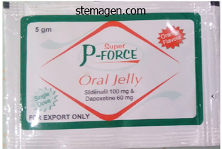
Super P-Force Oral Jelly
Yegappan Lakshmanan, MD, (FRCS Ed)
- Chief, Department of Pediatric Urology,
- Children’s Hospital of Michigan,
- Detroit, Michigan
Super P-Force Oral Jelly dosages: 160 mg
Super P-Force Oral Jelly packs: 7 sachets, 14 sachets, 21 sachets, 28 sachets, 35 sachets, 42 sachets, 49 sachets, 56 sachets, 63 sachets, 70 sachets
Cheap super p-force oral jelly 160 mg on-line
More recent advancements in plating strategies have provided many various types of plating know-how to the surgeon including nonlocking plates erectile dysfunction treatment in usa order 160 mg super p-force oral jelly free shipping, threaded locking plates erectile dysfunction caused by spinal cord injury cheap 160mg super p-force oral jelly visa, tapered locking plates erectile dysfunction drugs and hearing loss purchase super p-force oral jelly 160 mg free shipping, bicortical and monocortical fixation, rigid and semi-rigid plates. The goals of treatment should be as follows: anatomic reduction and stabilization of fractures; preservation of facial dimensions; establish and protect the occlusion; early return to function; avoidance of infection. Understanding these goals is paramount and the surgeon must weigh the dangers against the benefits of the proposed management. The objective of this chapter is to provide the clinician with an overview of the developments made in maxillofacial fixation and the forms of techniques obtainable. The patient is placed into his premorbid occlusion upon the ultimate tightening of the intra- and interarch wires. They present a flexible means of directing vectors of forces with a V�W pattern of crossbracing, to help in fracture reduction and re-establishment of the premorbid occlusion. The peak of contour position and contacts in the major dentition may prevent the appliance of circumdental wires. Many clinicians elect to use them primarily based on a decreased danger of penetrating harm for the user, ease of placement with shorter working room time, decreased trauma to the periodontium, convenience of maintaining oral hygiene, and the power to use them both intraoperatively and post-operatively, corresponding to with guiding elastics. Because they required a drilled hole for placement, there were concerns about suboptimal placement and root damage that occurred throughout placement. The second era self-drilling/selftapping screws improved tactile feedback, limiting the potential of root damage. The manufacturer recommends inserting screws above the basis apices; however, the writer has found that subapical placement led to mucosal overgrowth complicating their elimination. Instead the screws ought to be positioned in a bicortical style between the roots at the stage of the mucogingival junction. It is recommended, if possible, to get hold of a panorex film previous to placement of the screws so as to consider the foundation morphology. When attainable, no much less than one screw ought to be positioned proximal and distal to the fracture. However, a quantity of screws may be placed to direct the vectors of the interarch wires appropriately, which in flip aids in fracture reduction and stabilization. Recent reports have illustrated a quantity of inherent dangers and limitations which embrace root damage, screw loosening, screw shearing and aspiration. The self-drilling function offers a greater diploma of tactile suggestions throughout placement, permitting the operator to change insertion location before root damage happens. However, via the developments made with rigid fixation, as properly as microvascular reconstructive surgical procedure, this system has waned in its use. Once delicate tissue stabilization is achieved, the definitive restore can take place and the exterior fixation eliminated. First-generation exterior fixators utilized transcutaneous bicortical pins (placed alongside the inferior border) which were then bridged by filling an endotracheal tube with acrylic resin. A curvilinear incision relies a minimum of 2 cm inferior to the inferior of the mandible inside a skin crease. The marginal mandibular department is superior to the inferior border of the mandible as quickly as it passes to the anterior facial vessels. Incision is made through the skin and subcutaneous tissue to the level of the platysma. Dissection continues by way of the platysma to the level of the superficial layer of the deep cervical fascia. The superficial layer of the deep cervical fascia is incised, the facial vein is recognized and is then clamped, divided and ligated. The distal side of the vein is left on a long silk tie and is gently retracted superiorly with this suture. This manoeuvre retracts the nerve (which is superficial to the vein in the cervical fascia) superiorly defending it from harm. The facial artery is identified on the posterior/superior facet of the submandibular gland. In the submental area, the fascial plane overlying the anterior stomach of the digastric is adopted superiorly to its attachment at the symphysis. The periosteum and masseteric sling is then incised and a subperiosteal airplane of dissection is then performed to expose the mandible. A plating system is defined by the diameter of the screws; relying on the manufacturer, the diameters used for mandibular repair will range from 2. The nonlocking plates/screws are designed to act as separate units working at the side of one another to set up a stabilized fracture. When nonlocking screws are tightened, the heads of the screws exert pressure upon the plate which provides stabilization across the fracture. If this happens prior to the time required for primary bone therapeutic, the hardware can then fail, increasing the risk of nonunion, malunion, malocclusion and infection. This occurs when the screw head comes into contact with the plate in the course of the tightening process. This motion pulls the fracture segments to the plate, resulting in displacement of the segments and malalignment. The locking plate/screws have two sets of threads, bone threads and plate threads, which are threads in the head of the screw. This second set of threads unifies (locks) the screws to the bone resorption hardware failure 7. It creates a system which eliminates strain being translated via the plate and underlying bone, thereby lowering the likelihood of bone resorption and hardware failure. In addition, should any gaps exist between the plate and bone as a result of imprecise bending, this method is theoretically more forgiving, with less likelihood of fracture displacement, though studies indicate displacement remains to be attainable with locking plates. There are two kinds of locking designs out there, the threaded and tapered methods. Each uses the identical principle of locking the screw to the plate; nevertheless, the precise mechanics range, giving every a relative benefit and drawback. In the threaded design, the plate itself also has machined threads, so that these screws are placed perpendicular to the plate. The second set of screw head threads mesh with the plate threads creating a locking system. When utilizing locking know-how in ablative surgery, the manufacturers advocate limiting the variety of instances the same screw is reinserted to three attempts. In addition, though these screws ought to be positioned perpendicular to permit the threads to mesh appropriately. Some producers state that the screws may be angled in limited entry instances, essentially permitting the screw/plate to cross-thread. The threads in the head of these screws will both cut its own thread sample into the plate as the screw is being seated, or upon the final turn of the screw a single thread will have interaction the plate, offering a locking mechanism. It is typically recommended that one of many advantages of the tapered locking design is the freedom to place the screws at as a lot as a 10� angulation from the airplane perpendicular to the plate. This alleviates the risk of cross-threading and security of the locking mechanism. This could also be advantageous when entry is restricted, though the writer rarely finds this to be a difficulty. A theoretical advantage of the tapered locking design is its ability to compensate for thread/hole distortion.
Diseases
- Lupus anticoagulant, familial
- Erb Duchenne palsy
- Hereditary pancreatitis
- Eec syndrome
- Xanthic urolithiasis
- Progressive supranuclear palsy atypical
- Spasmodic dysphonia
Buy 160 mg super p-force oral jelly with amex
Although it might be potential to run the flap on one set of vessels impotence from blood pressure medication order super p-force oral jelly 160 mg overnight delivery, ideally all these vessels erectile dysfunction psychological treatment techniques buy super p-force oral jelly 160 mg mastercard, which are normally less than 2 mm in diameter erectile dysfunction injections youtube buy super p-force oral jelly 160mg without a prescription, ought to be anastomosed. The pedicle ought to now be traced backwards in the path of the ilium, separating the pedicle from the underlying psoas muscle. If the anterior crest is to be harvested after detaching the inguinal ligament, the gluteus medius muscle should be stripped from the outer aspect of the ilium to match the template. If the anterior crest is to not be harvested, the periosteum should be stripped from the inside facet of the ilium to where the medial bone cut is to be made. It is necessary to depart the gluteus medius hooked up to the outer aspect, otherwise the anterior crest can turn into avascular and may dehisce via the wound! The bone is minimize with a saw and the medial and lateral beginning and end cuts must be made with a small copper retractor or periosteal elevator protecting the underlying periosteum and pedicle. The horizontal cut can now be made with a retractor located medially to shield the peritoneum. Bone harvesting the point at which bone harvest ought to start along the size of the ilium is dependent on three elements: 1 the amount of bone required: this flap will attain the contralateral ramus with total mandibular reconstruction, but the complete size of the ilium that may be harvested with the muscle and pedicle shall be required. Note that the 2 incomplete osteotomy cuts that shall be greenstick fractured later. Operation 239 Once it has been ascertained that the flap together with the bone is bleeding, attention ought to be drawn to the donor web site, most of which may be closed earlier than detaching the pedicle. To assist this, a series of holes are made roughly 1 cm aside along the inner and outer cortex of the bony defect in the ilium. It is beneficial to use 1 or 1/0 nylon sutures and quickly hold them with artery clips until all the sutures have been positioned earlier than tying the knots. A piece of nonresorbable mesh ought to be trimmed to the dimensions of the inner indirect defect. A suction drain with a minimal intraluminal diameter of 3 mm must be placed over this layer and an epidural catheter teased by way of one of the sutured muscle layers. Both of these gadgets must be secured to the skin with a suture instantly after introduction. Closure of the muscle layers medial to the ilium has to be delayed till pedicle division. If detached, the inguinal ligament is sutured to the ilium and the exterior indirect muscle is closed to itself medially and to the gluteus medius muscle laterally. Even if a large pores and skin flap has been harvested, direct pores and skin and subcutaneous tissue closure is assured. In contrast to the fibula, the iliac bone is contoured using opening osteotomies with easy splitting of the bone prior to spreading the bone across the cut. The muscle and pores and skin ought to just be tacked in place to orientate the pedicle, and the anastomosis ready and carried out at this stage. This allows no interruption of move through the artery once this anastomosis is full. It may be that the heparin prevents fibrin degradation products within the beforehand ischaemic tissue, setting off the clotting cascade Division of the pedicle Final dissection of the proximal pedicle ought to be undertaken and the artery and vein divided and ligated. The lumen of the artery is identified and a small nylon cannula is introduced into it to allow flushing of the flap with 20�50 mL of heparinized saline. The transversalis and elements of the iliacus are sutured into these holes with a round body needled 1 or 1/0 nylon suture. It is recommended to temporarily maintain the sutures with an artery clip until all the sutures have been placed before tying the knots. After the flap is running, the inner indirect muscle is sutured into the intraoral defect. Violation of the thin transversalis muscle to produce herniation of pre-peritoneal fat is at all times a possibility but may be closed. Snagging of the pedicle with a rotary instrument is possible and probably disastrous; use of those instruments must be minimized in this operation! For venous monitoring, the color of the flap, which is able to clearly seem dark if engorged, is beneficial. A Doppler probe sutured across the flap aspect of the venous anastomosis may be useful. Local anaesthetic can be infused by way of the epidural catheter to assist in analgesia. Note multiple osteotomies and inner oblique muscle prior to suturing into position. Further reading 241 Top tips Always guarantee enough muscle is harvested in mandibular defects. Where pedicle size is likely to be a difficulty (usually maxillary cases) use the smallest amount of bone harvested as far back along the iliac crest as possible, to lengthen the pedicle. Do not underestimate the time, complicated nature and significance of rigorously closing the donor site defect. In some quarters, this flap has acquired a popularity for medium- and long-term morbidity, both of which may nearly always be avoided by consideration to detail! The free vascularised iliac crest tissue switch: donor web site problems associated with eighty two cases. Deep Circumflex iliac perforator flap with iliac crest for mandibular reconstruction. The free iliac crest and fibula flaps in vascularized oromandibular reconstruction: comparison and long term analysis. Post-operative Loss of flap perfusion and venous drainage are explicit problems in maxillary reconstruction. Partial bone necrosis could be extra of a problem and is of course more likely to happen with small distal segments of osteotomized bone. Seromas might occur, in all probability because of damage to the exterior iliac lymphatics. Post-operative an infection, particularly associated with the internal oblique mesh, can be very troublesome and may even require removing of the mesh. It is advisable to handle this materials rigorously and apply topical antiseptics to the mesh mattress and mesh itself to cut back this. Numbness to the anterior thigh due to damage to the lateral cutaneous nerve may be troublesome to some patients. Acquired lengthy standing facial paralysis most frequently results from neurosurgical or otolaryngological interventions for intracranial tumours as central palsy, but may as well happen after ablation of malignant parotid tumours as peripheral palsy. Facial reanimation encompasses surgical measures that assist or restore facial motion in cases of facial nerve palsies. In contrast to static reconstructions, such as suspension plasties to increase the oral commissure or lateral canthopexy to alleviate the sequelae of orbicularis oculi muscle palsy, these procedures allow for active movement of facial muscle tissue either by supporting present muscular activity or by transferring neuromuscular models from adjoining or distant sites to the deficient space.
Buy discount super p-force oral jelly 160 mg on-line
The incision is made vertically by way of the alveolar mucosa and hooked up gingiva between the higher central incisors impotence meaning in english 160mg super p-force oral jelly with amex. The nasopalatine bundle is preserved if the bone cut is between the central and lateral incisor enamel erectile dysfunction doctor dublin buy cheap super p-force oral jelly 160 mg line. Bone cuts the bone cuts are made with nice saws or a fissure burr and accomplished with osteotomes erectile dysfunction causes uk buy cheap super p-force oral jelly 160mg on line. The mucoperiosteum of the floor/lateral wall of the nostril is elevated and the bone cuts made (1) between the central and lateral incisor tooth, continued paramedially through the size of exhausting palate into the nasal floor; (2) laterally from the piriform fossa, under the inferior turbinate (preserving the nasolacrimal duct) via the anterior maxilla inferior to the infraorbital nerve via the zygomatic buttress back to the pterygoid plates. The infraorbital nerve is sectioned at the infraorbital foramen as it prevents lateral retraction of the maxilla. The bone cut posterior to the zygomatic buttress is angled downwards and may be made either with a nice osteotome or reciprocating saw, to scale back delicate tissue stripping. The maxilla is prelocalized with bone plates (1) above the incisor tooth anteriorly; (2) at the frontal strategy of the maxilla; (3) on the zygomatic buttress. The buccal pad of fat is throughout the operative subject and instantly available for reconstruction as a pedicled flap. The coronoid course of and the connected temporalis tendon might impede entry to the infratemporal fossa. A coronoidectomy will improve access and has the extra benefit of lowering post-operative trismus. The palatal gentle tissues are lined with an acrylic cover plate wired to the standing tooth. Greater palatine foramen Soft tissue incision Osteotomy bone cuts Alternative (favoured) palatal incision. A damp tonsillar swab is placed in the reverse nostril and the cartilaginous nasal septum divided vertically consistent with the osteotomy bone cuts. The nasal swing hyperlinks readily with a frontal craniotomy for resection of pathology that additionally includes the central compartment of the anterior cranial fossa. The transfacial approaches may also be combined with either a Le Fort I osteotomy or mandibular osteotomies for additional access. The palatal cowl plate could be left in situ for 6�8 weeks or till maxillary stability is achieved. On the side reverse the skin incision, subperiosteal dissection is achieved by tunnelling. No incision is made through the lingual mucoperiosteum � the soft tissues are simply elevated off the lingual side of the mandible adjoining to the osteotomy. For combined entry to the oral cavity and infratemporal fossa, an prolonged mandibular swing is appropriate; a lip split is critical. Mandibular swing the technique supplies wonderful access to the floor of the mouth, mid and posterior third of tongue, tonsillar fossa, soft palate, oropharynx together with the posterior pharyngeal wall, supraglottic larynx and medial aspect of the mandibular ramus. Extended posteriorly, it provides equally good access to the infratemporal fossa, and the parapharyngeal house with vascular control. There are three separate parts: 1 Division of the lower lip and chin 2 Division of the mandible in the incisor region three Elevation of the gentle tissues off the lingual aspect of the mandible. Surgical method A full thickness vertical incision is made via the midline of the lower lip; a V-shaped notch is incorporated in the midline lip incision and the vermilion incision for precise closure and to masks scarring. The incision curves across the chin with the concavity of the incision towards the side of the lesion, decreasing the risk of chin necrosis. Intraorally, the incision by way of the labial mucosa and the connected gingiva is stepped so as to not lie instantly over the following osteotomy bone minimize. Periosteal elevation and mentalis muscle stripping must be enough only for the appliance of bone plates and screws. A full thickness lingual mucoperiosteal flap is subsequently elevated off the lingual facet of the mandible when the mandible is retracted laterally. The incision via the oral tissues is Stepped gentle tissue incision and bone cuts. The osteotomy is at all times performed within the incisor region for tumours involving the oral cavity. Osteotomy within the premolar region anterior to the mental foramen is just employed by the authors to entry tumours within the infratemporal fossa/parapharyngeal space when avoiding a lip split incision (see below under Angle osteotomy and double mandibular osteotomy avoiding lip split). The mandible is split with a fine saw blade between the roots of the incisor enamel. Prior to division, the mandible is Transmandibular approaches 287 prelocalized with bone plates acrosss the osteotomy bone minimize. The extended mandibular swing By stripping the medial pterygoid off the medial facet of the mandible, additional lateral and superior mandibular retraction is possible and provides extensive access to the infratemporal fossa and parapharyngeal area. Angle osteotomy and double mandibular osteotomy avoiding lip break up For tumours confined to the infratemporal fossa/ parapharyngeal space with out the want to involve the oral (a) Vertical osteotomy minimize within the incisor region � stepped osteotomy not needed. The site of the pathology determines the extent of the zygoma mobilized and whether or not the temporalis muscle is reflected inferiorly or superiorly, as follows: for subcranial lesions (orbit/temporal and infratemporal fossae), the temporalis is reflected superiorly after dividing the coronoid process; for combined exposure of the middle/anterior fossae, the orbit and/or the temporal and infratemporal fossa, the temporalis is reflected inferiorly. The blood supply of the temporalis is potentially compromised with each superior and inferior reflection. A horizontal osteotomy of the mandibular ramus is prevented as the realm of bone contact is proscribed with this technique. If resection of the floor of the center fossa is required in skull base procedures, extra superior entry is definitely achieved by mobilising the zygoma (see under underneath Transzygomatic approaches). When exposing the orbit, in addition to the temporal and infratemporal fossae, restricted subperiosteal dissection is carried out in the lateral orbit to defend the orbital contents. The lateral restrict of the inferior orbital fissure is recognized with a blunt hook in the temporal fossa. Its disarticulation inferiorly pedicled to Superiorly on the frontozygomatic suture. Through the body of the zygoma, extending laterally from the inferior orbital fissure. The anterior limit of the inferior orbital fissure is first identified in the orbit with a blunt hook. The posterior bone reduce is simply anterior to the articular eminence of the temporomandibular joint. Lingual (anterior) and inferior dental (posterior) nerves recognized after resection of the coronoid. A vertical bone reduce by way of the body of the zygoma lateral to the orbital rim is made. With light taps, the osteotomy is accomplished with an osteotome and the zygoma displaced inferiorly. For correction of a malunion of the zygoma, the coronoidectomy and superior reflection of the temporalis are unnecessary. The extent of publicity of the infratemporal fossa extends inferiorly to the maxillary tooth and medially to the lateral wall of the nasopharynx, following resection of the lateral and medial pterygoid muscular tissues. Medial publicity is restricted, which limits the approach to remedy of benign pathology in the infratemporal fossa whether it is used as the only technique of access. Malignant illness within the infratemporal fossa is best accessed with a transmandibular and/or transfacial method. For publicity of the lateral orbit to the apex, a frontotemporal craniotomy is critical.
Purchase super p-force oral jelly 160 mg with visa
Operative approach Starting medially icd 9 code erectile dysfunction neurogenic generic super p-force oral jelly 160mg overnight delivery, the pores and skin and subcutaneous fatty tissue are incised to the deep fascia overlying the infraspinatus muscle occasional erectile dysfunction causes generic super p-force oral jelly 160mg. Elevate the fasciocutaneous pores and skin paddle from medial to lateral simply above the deep muscular fascia and below the dorsal thoracic fascia erectile dysfunction drugs insurance coverage 160 mg super p-force oral jelly with mastercard. The pulsation of the cutaneous branch, which is enveloped in the fascia, can now be seen and palpated easily. The circumflex artery might be recognized within the omotricipital triangle simply superior to the teres major muscle. Follow the circumflex vascular pedicle to the subscapular and axillary artery, several branches to the teres main and subscapular muscular tissues are ligated to accomplish the vascular preparation. Shoulder, back, lateral thorax and higher arm are circularly prepared to allow for motion of the extremity and exposure of the subscapular system from an axillary strategy. In the lateral decubitus place, vacuum beanbags are used to stabilize the affected person and to defend the dependent shoulder. Draw the outline of the scapula and find the upper margin of the latissimus insertion alongside the posterior axillary line. The handheld Doppler system can be used to affirm the presence of the vascular pedicle. Operation 203 lateral border of scapula, leaving a muscle cuff attached to the bone. The osteotomy is normally carried out 2�3 cm parallel to the lateral border of scapula and might embody the whole inferior angle. The scapular angle could also be harvested independently if the angular vessels are recognized and preserved. The osteocutaneous scapular flap is now fully elevated and ready for microvascular switch. To forestall winging of the scapula, the teres major muscle is reattached by drill holes in the lateral border of the residual scapula. Physical therapy can then be instituted to enhance passive and lively vary of motion. Intensive residence physical therapy to enhance shoulder energy can begin within 3 weeks after surgery. Use of anticlotting brokers, similar to heparin, aspirin and dextran, are also used at the judgement of the surgeon. Maintaining large suction drains for a number of days post-operatively helps to alleviate this downside. This is particularly problematic with massive defects that are closed underneath pressure. Significant undermining of adjoining tissue may be required to get hold of major closure. Use separate devices and clear gloves and robes at the donor web site to keep away from contamination and subsequent wound infection. Take care to keep away from injury to the long thoracic nerve, which supplies the serratus anterior, as a end result of this may find yourself in a winged 204 Scapular and parascapular flap (with or with out bone) scapula. The injury may be observed with scapular flap harvest due to patient positioning. Take care to keep away from extreme elevation of the arm, and support for the head must be supplied throughout harvest. Top ideas It is essential for flap design to identify the triangular area between the teres minor, main and lengthy head of triceps muscular tissues. Make certain that no branch to the bone is transected during dissection of the deep phase of the circumflex scapular artery. When getting ready the scapula for the osteotomy, care have to be taken to not damage the circumflex scapular artery and its branches to the bone. The creation of microsurgical techniques has allowed this versatile flap to be transposed to restore soft tissue defects of the top and neck for many years. It could be harvested as a muscle-only flap or as a myocutaneous flap, which can be utilized to exchange soft tissue bulk. Vascularized skin and fats resist atrophy, however denervated muscle shrinks with time. Although not optimum for small or shallow defects, the rectus flap offers the reconstructive surgeon with a wonderful choice to reconstruct massive defects. This flap is most commonly used for orbital-maxillary defects, skull base reconstruction and glossectomy defects. Fat volume is well preserved in a denervated free myocutaneous flap, but it loses muscular bulk with time. The upper vessel is the superior epigastric artery, one of many two terminal branches of the internal mammary artery. The rectus abdominis muscle is enclosed within the rectus sheath, which consists of an anterior and posterior lamina. This is especially essential in the restoration of belly wall integrity after elevating the rectus abdominis muscle or musculocutaneous flap. The commonly used inferior rectus abdominis flap is a musculocutaneous flap based mostly on the deep inferior epigastric artery and vein and terminal musculocutaneous perforators. These vessels arise from the exterior iliac artery and vein and course superomedially to run along the deep lateral facet of the muscle. The deep inferior epigastric vein is regularly discovered to be a system of paired venae comitantes operating with the artery. Just proximal to the external iliac vein, this technique typically varieties one dominant vein. The rectus abdominis myocutaneous flap could be very suitable for the intensive defects ensuing from radical maxillectomy with zagoma because of its giant soft tissue quantity. Skull base reconstruction the rectus abdominis flap is also generally utilized for the reconstruction of skull base defects by which a big quantity of conformable, vascularized muscle is required to obliterate a cranium base defect and bolster a dural closure. In these circumstances, the flap is harvested and not using a cutaneous paddle except skin closure of a palatal defect is required. The operating subject is prepped between decrease rib arch, posterior axillary line and upper thigh. Depending on tissue necessities, the skin paddle can be designed vertically, completely overlying the rectus muscle, or obliquely, alongside an axis between the umbilicus and the tip of the scapula with much of the skin paddle lateral to the linea semilunaris. This latter orientation is possible due to an axial blood circulate sample from the portion of skin overlying the muscle within the periumbilical area parallel alongside this axis. When designed in this manner, the lateral aspect of the flap is far thinner than the portion overlying the muscle and could be helpful when the defect requires delicate tissue of various thickness. Operative method Flap design Draw linea alba, linea semilunaris, inguinal ligament, symphysis, costal margin and approximate position of arcuate line at degree of anterior iliac backbone. Landmarks are palpated, which embrace the ribcage, pubis and anterior superior iliac backbone. Use caution when working with the rectus muscle as a result of musculocutaneous perforators may be violated. Placing tacking sutures from the skin to the anterior sheath can preserve the viability of the perforators. The inferior incision is placed above the arcuate line in order to facilitate closure without the use of mesh.
Rock-Up-Hat (Echinacea). Super P-Force Oral Jelly.
- Urinary tract infections (UTIs), migraine headaches, chronic fatigue syndrome (CFS), eczema, hayfever, allergies, bee stings, attention deficit-hyperactivity disorder (ADHD), influenza (flu), and other conditions.
- Preventing vaginal yeast infections when used with a medicated cream called econazole (Spectazole).
- Dosing considerations for Echinacea.
- What is Echinacea?
- How does Echinacea work?
Source: http://www.rxlist.com/script/main/art.asp?articlekey=96942
160mg super p-force oral jelly with visa
B erectile dysfunction ear 160 mg super p-force oral jelly otc, Transverse part of the embryo showing angioblastic cords in the cardiogenic mesoderm and their relationship to the pericardial coelom impotence from anxiety purchase 160 mg super p-force oral jelly. C erectile dysfunction treatment south florida buy discount super p-force oral jelly 160mg line, Longitudinal part via the embryo illustrating the relationship of the angioblastic cords to the oropharyngeal membrane, pericardial coelom, and septum transversum. These strands canalize to form two skinny coronary heart tubes that soon fuse to type a single heart tube late in the third week as a result of embryo folding. An inductive affect from the anterior endoderm stimulates early formation of the center. Cardiac morphogenesis (development) is controlled by a cascade of regulatory genes and transcription components. Aortic sac Primordial heart Vitelline vein Chorionic sac Umbilical vein Umbilical vesicle Vitelline artery Umbilical wire Development of Veins Associated with Embryonic Heart Three paired veins drain into the tubular coronary heart of a 4-week embryo. The umbilical vein carries well-oxygenated blood and vitamins from the chorionic sac to the embryo. The umbilical arteries carry poorly oxygenated blood and waste products from the embryo to the chorionic sac (outermost embryonic membrane; see Chapter 8. The vitelline veins enter the venous end of the heart- the sinus venosus of the primordial heart. As the liver primordium grows into the septum transversum, the hepatic cords anastomose around preexisting endothelium-lined areas. These spaces, the primordia of the hepatic sinusoids, later become linked to the vitelline veins. Anastomosis through mesonephros (early kidney) Iliac venous anastomosis of postcardinal vv. C Cardinal, umbilical, and vitelline veins Subcardinal veins D Hepatic phase Median sacral v. Initially, three methods of veins are present: the umbilical veins from the chorionic sac, the vitelline veins from the umbilical vesicle, and the cardinal veins from the physique of the embryo. D, Drawing illustrating the transformations that produce the grownup venous sample. A, During the fourth week (approximately 24 days), exhibiting the primordial atrium, sinous venosus, and veins draining into them. B, At 7 weeks, showing the enlarged proper sinus horn and venous circulation via the liver. C, At 8 weeks, indicating the adult derivatives of the cardinal veins shown in A and B. Neural crest cells delaminate from the neural tube and contribute to the formation of the outflow tract of the guts and pharyngeal arches. Later, the caudal portions of the paired dorsal aortae fuse to type a single decrease thoracic/ abdominal aorta. Of the remaining paired dorsal aortae, the right regresses and the left turns into the primordial aorta. The persistent caudal part of the left umbilical vein turns into the umbilical vein, which carries welloxygenated blood from the placenta to the embryo. The anterior and posterior cardinal veins drain the cranial and caudal components of the embryo, respectively. During the eighth week, the anterior cardinal veins are linked by an indirect anastomosis. This anastomotic shunt turns into the left brachiocephalic vein when the caudal part of the left anterior cardinal vein degenerates. The solely adult derivatives of the posterior cardinal veins are the basis of the azygos vein and the common iliac veins. The subcardinal and supracardinal veins progressively exchange and supplement the posterior cardinal veins. Cranial to this, they turn out to be united by an anastomosis that forms the azygos and the hemiazygos veins. The inferior vena cava forms as blood coming back from the caudal part of the embryo is shifted from the left to the proper side of the body. Intersegmental Arteries Thirty or so branches of the dorsal aorta, the intersegmental arteries, pass between and carry blood to the somites (cell masses) and their derivatives. Most of the unique connections of the intersegmental arteries to the dorsal aorta disappear. Most of the intersegmental arteries within the stomach turn out to be lumbar arteries; nonetheless, the fifth pair of lumbar intersegmental arteries stays because the widespread iliac arteries. In the sacral region, the intersegmental arteries type the lateral sacral arteries. Fate of Vitelline and Umbilical Arteries the unpaired ventral branches of the dorsal aorta supply the umbilical vesicle, allantois, and chorion. The vitelline arteries provide the umbilical vesicle and, later, the primordial intestine, which types from the incorporated part of the umbilical vesicle. Only three vitelline arteries remain: the celiac arterial trunk to the foregut, the superior mesenteric artery to the midgut, and the inferior mesenteric artery to the hindgut. The paired umbilical arteries move by way of the connecting stalk (primordial umbilical cord) and join the vessels within the chorion (membrane enclosing the embryo). The proximal components of those arteries turn out to be the interior iliac arteries and superior vesical arteries, whereas the distal parts are obliterated after delivery and turn into medial umbilical ligaments. The external layer of the embryonic heart tube-the primordial myocardium (cardiac precursor of the primary coronary heart field)-is fashioned from the splanchnic mesoderm surrounding the pericardial cavity. At this stage, the developing coronary heart consists of a skinny tube, separated from a thick primordial myocardium by gelatinous-matrix connective tissue- cardiac jelly. A to C, Ventral views of the growing coronary heart and pericardial area (22�35 days). The ventral pericardial wall has been removed to present the growing myocardium and fusion of the 2 coronary heart tubes to form a tubular heart. D and E, As the straight tubular coronary heart elongates, it bends and undergoes looping, which forms a D-loop that produces an S-shaped heart. As folding of the pinnacle region happens, the center and pericardial cavity appear ventral to the foregut and caudal to the oropharyngeal membrane. Concurrently, the tubular heart elongates and develops alternate dilations and constrictions. The growth of the center tube outcomes from the addition of cells (cardiomyocytes) that differentiate from the mesoderm at the dorsal wall of the pericardium. The sinus venosus receives the umbilical, vitelline, and customary cardinal veins from the chorion, umbilical vesicle, and embryo, respectively. The arterial and venous ends of the center are fastened in place by the pharyngeal arches and septum transversum, respectively. Because the bulbus cordis and ventricle grow faster than the other areas, the heart bends on itself, forming a U-shaped bulboventricular loop.
Cheap super p-force oral jelly 160mg otc
Blood cells develop from hematopoietic stem cells or from hemangiogenic endothelium or blood vessels as they grow on the umbilical vesicle and allantois on the end of the third week erectile dysfunction clinic raleigh purchase super p-force oral jelly 160 mg with mastercard. This course of happens first in various parts of the embryonic mesenchyme erectile dysfunction doctor specialty cheap 160mg super p-force oral jelly otc, chiefly the liver erectile dysfunction drugs compared buy 160mg super p-force oral jelly with amex, and later within the spleen, bone marrow, and lymph nodes. Fetal and adult erythrocytes are additionally derived from hematopoietic progenitor cells (hemangioblasts). The mesenchymal cells that surround the primordial endothelial blood vessels differentiate into muscular and connective tissue components of the vessels. The heart and great vessels kind from mesenchymal cells within the heart primordium, or cardiogenic area. The tubular heart joins with blood vessels within the embryo, connecting stalk, chorion, and umbilical vesicle to type a primordial cardiovascular system. By the end of the third week, blood is flowing and the guts begins to beat on day 21 or 22. The cardiovascular system is the primary organ system to attain a primitive functional state. The embryonic heartbeat may be detected by Doppler ultrasonography (detects movement by monitoring the change in frequency or section of the returning ultrasound waves) through the fourth week, approximately 6 weeks after the last regular menstrual interval. The villi that develop from the perimeters of the stem villi are branch chorionic villi (terminal villi). It is through the partitions of the branch villi that the main change of fabric between the blood of the mother and the embryo takes place. The branch villi are bathed in frequently changing maternal blood in the intervillous area. Sacrococcygeal teratomas are the most common tumors in new child infants and have an incidence of approximately 1 in 27,000 neonates. Early within the third week, mesenchyme grows into the first villi, forming a core of unfastened mesenchymal tissue. The villi at this stage-secondary chorionic villi-cover the entire surface of the chorionic sac. Some mesenchymal cells within the villi soon differentiate into both capillaries and blood cells. The capillaries in the chorionic villi fuse to type arteriocapillary networks, which soon turn out to be connected with the embryonic coronary heart by way of vessels that differentiate from the mesenchyme of the chorion and connecting stalk. By the end of the third week, embryonic blood begins to flow slowly by way of the capillaries in the chorionic villi. Carbon dioxide and waste products diffuse from blood within the fetal capillaries through the wall of the villi into the maternal blood. Concurrently, cytotrophoblastic cells of the chorionic villi proliferate and lengthen via the syncytiotrophoblast to form a cytotrophoblastic shell, which gradually surrounds the chorionic sac and attaches it to the endometrium. Meroencephaly (anencephaly), or partial absence of the mind, is essentially the most extreme defect. Available proof suggests that the primary disturbance impacts the neuroectoderm. Failure of the neural folds to fuse and type the neural tube within the brain area leads to meroencephaly, and in the lumbar region, spina bifida cystica (see Chapter sixteen. By the end of the third week, a primordial uteroplacental circulation has developed. These moles exhibit variable levels of trophoblastic proliferation and produce excessive amounts of human chorionic gonadotropin. In 3% to 5% of such circumstances, these moles turn into malignant trophoblastic lesions, known as choriocarcinomas. These tumors invariably metastasize (spread) by means of the blood to numerous websites, such as the lungs, vagina, liver, bone, gut, and brain. As the tissues and organs type, the form of the embryo adjustments so that, by the eighth week, the embryo has a distinctly human look. Folding outcomes from speedy development of the embryo, notably the mind and spinal wire. Folding at the cranial and caudal ends and on the sides of the embryo happens simultaneously. Concurrently, a relative constriction occurs at the junction of the embryo and the umbilical vesicle. Head and tail folds cause the cranial and caudal regions to transfer ventrally as the embryo elongates. Reconstructions made from the surface ectoderm and all organs and cavities within human embryos at representative levels of growth have revealed new findings on the movements that occur from one stage to the subsequent. This has been shown to take place concurrently at every stage of magnification from the cell membrane all the way to the surface of the embryo. The movements and forces bring about differentiation that begins on the outside of the cell, after which strikes to the inside to react with the nucleus. The continuity of the intraembryonic coelom and extraembryonic coelom is proven on the proper side by removal of part of the embryonic ectoderm and mesoderm. Later, the developing forebrain grows cranially past the oropharyngeal membrane and overhangs the developing heart. Concomitantly, the primordial coronary heart and the oropharyngeal membrane transfer onto the ventral surface of the embryo. Folding of the caudal end of the embryo outcomes primarily from growth of the distal a half of the neural tube, the primordium of the spinal cord. As the embryo grows, the tail area initiatives over the cloacal membrane, the long run web site of the anus. During folding, part of the endodermal germ layer is included into the embryo as the hindgut. The connecting stalk (primordium of the umbilical cord) is now connected to the ventral floor of the embryo, and the allantois-an endodermal diverticulum of the umbilical vesicle-is partially incorporated into the embryo. Knowledge of the genes that management human growth is rising (see Chapter 20). Most developmental processes depend on a exactly coordinated interaction of genetic and environmental components. Several control mechanisms information differentiation and ensure synchronized improvement, corresponding to tissue interactions, regulated migration of cells and cell colonies, managed proliferation, and apoptosis (programmed cell death). Each system of the physique has its own developmental pattern, and most processes of morphogenesis are regulated by complex molecular mechanisms. Embryonic development is actually a means of progress and rising complexity of structure and performance. Growth is achieved by mitosis, together with the manufacturing of extracellular matrices, whereas complexity is achieved by way of morphogenesis and differentiation. This broad developmental potential becomes progressively restricted as tissues acquire the specialized features needed for increased sophistication of structure and function. Such restriction presumes that decisions must be made to achieve tissue diversification. Most proof signifies that these decisions are decided not as a consequence of cell lineage, however somewhat in response to cues from the immediate environment, together with the adjoining tissues.
Generic super p-force oral jelly 160mg overnight delivery
Features suggestive of malignancy include poor definition with heterogeneous echotexture best erectile dysfunction pills 2012 order super p-force oral jelly 160 mg fast delivery, disorganized colour circulate and the presence of related nodes erectile dysfunction urethral medication cheap 160mg super p-force oral jelly amex. Using these criteria impotence from blood pressure medication super p-force oral jelly 160 mg without prescription, malignancy could be predicted in around 80 per cent of instances using ultrasound alone. Thyroid A detailed description of thyroid ultrasound is past the scope of this textual content, nonetheless thyroid disorders, together with generalized gland enlargement and focal nodules, are comparatively commonly encountered in medical follow. In the one-stop clinic environment, thyroid nodules are more likely to symbolize the second most typical explanation for symptomatic neck lumps, after lymph nodes. The growing use of ultrasound implies that the incidentally detected thyroid nodule is a significant problem; ultrasound will detect nodules in between 50 to 70 per cent of females over the age of fifty years. Adjacent buildings, specifically the frequent carotid artery and inside jugular vein and deep cervical lymph nodes are clearly seen and are routinely examined, and tracheal deviation and retrosternal extension can be appreciated. In some centres, vocal twine mobility can also be routinely assessed with ultrasound earlier than surgery. On ultrasound, pleomorphic adenoma has the looks of a well-defined, hypo-echoic homogeneous stable mass (pseudocystic) with a lobulated border and internal vascularity and may display posterior acoustic enhancement. While papillary carcinoma may be multifocal at presentation, its typical look on ultrasound is as a solid hypoechoic mass. Invasion of regional lymph nodes is common, and foci of microcalcification may also be detected in concerned nodes. The typical benign thyroid nodule is often heterogenous in echotexture, with a hypoechoic halo or perinodular rim. Calcification is frequent: either peripheral eggshell or giant Indications for head and neck ultrasound 21 1. This fable is usually perpetuated, but multiple massive sequence have proven that the incidence of malignancy in solitary and a quantity of nodules is comparable. Haemangiomata may have massive cavernous spaces and possess capillary and/or lymphatic components. This situation causes an enlargement of the gland in the acute part with diffuse hypo-echogenicity which is usually patchy and starts within the anterior portion of the gland. In time, the whole of the gland is enlarged, hypo-echoic, accommodates echogenic striae and is usually hypervascular in the acute part. With time the gland atrophies, loses its hypo-echoic appearance and its vascularity diminishes. Most branchial cysts come up from the second branchial arch remnants and current as a mass at the angle of the mandible, often following an infection. The typical location is abutting the posterior side of the submandibular gland, mendacity lateral to the carotid vessels and instantly anterior to the anterior border of the sternomastoid. On ultrasound, these lesions may be cystic, but extra commonly the presence of debris, haemorrhage or infection gives rise to a pseudosolid look and the cyst wall thickens within the presence of an infection. It could additionally be impossible to distinguish between a second branchial cleft cyst and a necrotic lymph node metastasis due to squamous cell carcinoma. Intramuscular lipomas can mimic muscle and could additionally be troublesome to outline with ultrasound. Thyroglossal duct cysts can arise at any position alongside the course of the thyroglossal duct remnant, however the majority are associated to the hyoid bone, with most occurring on the stage of or inferior to the hyoid. On ultrasound, thyroglossal duct cysts might appear cystic, heterogeneous or pseudosolid because of various content of particles, haemorrhage or infection. Malignant degeneration of the epithelial lining 22 Ultrasound imaging, including ultrasound-guided biopsy occurs rarely and any strong element which seems to contain microcalcification. These lesions arise from sequestration of the ectoderm from adjacent sutures, mostly the frontozygomatic suture. Dermoid cysts come up from multiple germ cell layer and due to this fact will include a quantity of dermal adnexal structures. Sebaceous glands, hair and fats are generally found in dermoids, but they may even be purely cystic. They may subsequently have a heterogenous look with the presence of fat manifesting as a fluid/fluid level or often as rounded echogenic masses inside the cyst (representing sebaceous rests inside the dermoid). The typical location for midline cysts is within the submental area both superficial or deep to mylohyoid. Thus the needle must be within the aircraft of the ultrasound beam and as parallel to the probe floor as possible in order to optimally visualize it. Keeping the probe, needle, ultrasound monitor and affected person in a decent arc in front of the operator is important. If needed, for example for a lesion within the posterior triangle, the patient ought to lie on their facet in order to allow easy access for a shallow strategy. However, the place lymphoma is considered as a possible analysis, core biopsy undoubtedly has a superior position. Many centres are now in a position to diagnose and kind lymphoma on core biopsy, using circulate cytometry techniques, avoiding open biopsy and considerably reducing referral to remedy time. Core biopsy may be reserved as a second-line take a look at when cytology is unable to present the reply. Some authors advocate using core biopsy as a common firstline investigation, pointing out the fallibility of cytology for certain situations. However, many others imagine that squamous cell carcinoma could be seeded during percutaneous extensive bore needle biopsy within the neck. The decision as to which method to use for sampling neck masses might be influenced by native follow. Ultrasound can differentiate between infection with a fluid part (abscess) and cellulitis, and identify related lymphadenopathy and venous thrombosis. Ultrasound-guided nice needle aspiration and core biopsy Ultrasound is a really helpful adjunct in percutaneous sampling procedures, permitting direct visualization of the 1. Ear, nostril and throat cancer: Ultrasound prognosis of metastasis to cervical lymph nodes. Echogenic or reflective structures are white (for instance, bone, needle, calculi). Calcification causes full reflection of ultrasound and an acoustic shadow past it. Hypo-echoic constructions are black (for example, blood in the inside jugular vein). Congenital cysts are sometimes echogenic, however branchial cleft cysts, thyroglossal duct cyst, dermoid cysts are pseudocystic with some having solid parts. These include salivary pleomorphic adenoma, parathyroid adenoma, nerve sheath tumours, lymphoma. In addition, practical features of different types of investigations, similar to exfoliative cytology and microbiology, might be outlined. As with any small tissue pattern, the core will not be consultant of the lesion as an entire or could fail to present specific pathognomic features. For instance, stories present the risk of tumour seeding is extremely low and diagnostic accuracy in distinguishing non-neoplastic lesions, benign and malignant neoplasms is constantly larger than 97 per cent.
Cheap 160mg super p-force oral jelly free shipping
The nerve enters the gracilis muscle approximately 20 mm proximal to the entry of the vascular pedicle erectile dysfunction injections australia buy 160 mg super p-force oral jelly mastercard. Fine scissors and forceps are essential to experimental erectile dysfunction drugs 160 mg super p-force oral jelly otc accomplish blunt launch of the tiny branches from the adductor longus muscle floor smoking and erectile dysfunction causes super p-force oral jelly 160 mg without a prescription. It is first mid thigh profunda femoris artery femoral artery dominant vascular pedicle sartorius adductor longus gracilis adductor magnus proximal thigh 3. Operation 225 profunda femoris artery femoral artery small branches to adductor longus muscle gracilis adductor brevis adductor longus adductor magnus 3. There might be branches that run off into the underlying adductor magnus muscle, these should be identified and ligated by carefully elevating the vascular pedicle from the muscle surface before the pedicle is separated from the profunda femoris vessels. During ligation of the pedicle, care must be taken not to compromise blood move in the profunda femoris vessels. Therefore, a minimum distance of 5 mm from the junction between the pedicle and the profunda femoris artery and vein must be noticed when the pedicle is minimize. After completion of the dissection of the adductor artery and veins, a pedicle size of roughly 6 cm should be available. Dissection of the obturator nerve and definition of the graft dimension the obturator nerve that has been beforehand recognized is definitely adopted alongside its course proximally by blunt dissection until a size of roughly 8 cm is achieved. The nerve usually accommodates three fascicles, though there could also be as much as seven present. The anterior half of the muscle belly is usually innervated by one fascicle; the posterior half is provided by the remaining ones with some overlap in the mid line. In the proximal half, the muscle fibres run parallel which makes division in longitudinal direction simple. The length of the muscle required has to be identified beforehand throughout preoperative planning and the dissection of the recipient web site within the cheek (see Chapter three. During last separation and mobilization, the epimysium is preserved on the floor of the muscle to provide a sliding layer for easier motion of the muscle during contraction after transfer. Before the pedicle is divided, sufficient perfusion of the muscle segment is checked. After full separation, the function of the obturator nerve section included in the graft must also be checked with the nerve tester after putting the graft on a moist adductor longus muscle pulled upward ligated branches to the adductor longus muscle Dissection of the muscle to identify particular person muscular items. The graft is then stored in moist surgical towels till particular graft fixation and microvascular anastomosis are carried out. Post-operative complications Wound closure and post-operative care Meticulous control of bleeding from the transectioned muscle tissue is performed by electrocautery. The anterior margin of the remaining elements of the gracilis muscle is hooked up to the fascia of the adductor longus muscle to stabilize the gracilis muscle and restore straightforward perform of the adductor muscle tissue. A 10 gauge suction drainage is placed on the reconstructed fascia and the wound is closed in layers. The leg is bandaged with lowering pressure from distal to proximal or a strain stocking is applied to keep away from postoperative thrombosis. Routine post-operative prevention of thrombosis is applied by administration of fractionated heparin. The operated leg must be positioned in a barely elevated place for a few days until complete mobilization of the affected person is achieved. Post-operative mobilization with the assistance of a physical therapist could be performed from the first post-operative day on as the muscle function of the adductor group stays grossly unaffected by the removing of the small gracilis segment and its nerve provide. If no substantial bleeding happens, the suction drainage may be eliminated on the second postoperative day. Nevertheless, improperly seated ligation clips after separation of the pedicle from the profunda femoris vessels can turn out to be detached and cause substantial haemorrhage. Thus, great care is required during this part of the dissection to ensure that ligations of the adductor vessels are reliable. Secondary bleeding from the transected muscle might occur if haemostasis has been insufficient. The cautious use of electrocautery after identification of particular person intramuscular branches underneath the microscope earlier than wound closure minimizes this danger. Thrombosis can additionally be unlikely to occur if routine heparin prophylaxis is run and early mobilization is achieved. The saphenous vein that drains the superficial venous system is positioned above the positioning of harvest. This vein is usually not encountered during dissection and thus could be easily preserved. Deep venous thrombosis may happen if ligation clips or sutures compromise the blood circulate in the profunda femoris veins, which may be prevented by observing an sufficient distance to the vein throughout ligation as described above. The dissection of the flap is very straightforward, with little likelihood for it to go grossly incorrect. During flap dissection, the division of the branches that go off into the adductor longus is a vital level. As the pedicle is somewhat quick, use as much length as you can get, nonetheless, bear in mind to preserve a 5 mm cuff for ligation to avoid thrombosis of the profunda femoris vein. Careful nerve testing and definition of practical muscle models will enhance the end result in terms of cosmetics, as unsightly bulging of the grafted muscle during contraction is prevented. Trimming of the muscle after switch is always more difficult and carries the risk of removing the incorrect muscle units. Intraoperative complications Damage to the pedicle may occur during release of the branches to the adductor longus and adductor magnus muscle. The use of the microscope throughout dissection and the usage of clips somewhat than electrocautry during ligation are helpful in avoiding this drawback. Damage to the profunda femoris vessels might happen during separation and ligation of the pedicle. In particular, the profunda femoris veins are vulnerable to compromised blood circulate and subsequent thrombosis. This can be avoided by observing an sufficient distance of 5 mm pedicle length during ligation and separation of the pedicle from the profunda femoris vessels and through the use of clips somewhat than sutures for ligation. Hari K, Ohmori K, Torii S, Free gracilis muscle transplantation with micorvascular anastomosis for the remedy of facial paralysis. The advantages of the fibula are that it offers an ample supply of tubed bi-cortical bone, which is beneficial for reconstruction of long segmental defects throughout the midline � 25 cm or extra of bone could be harvested. Bone height is the primary potential drawback, especially when reconstructing the dentate mandible. It is common to go away the flap for several months earlier than distraction to enable bony union with the recipient bed and allow the fixation plates to be removed. As the flap was more and more used, modifications had been made, corresponding to including massive elements of the soleus muscle, and skin paddles. Whereas these first fibula transfers had been performed with no pores and skin paddle, an osteocutaneous fibula flap was first reported in 1983. The use of the vascularized fibula in mandibular reconstruction was reported in 1989 and this opened a model new subject in maxillofacial reconstructive surgical procedure. The fibula is the longest bone flap obtainable and it could be transferred as a bone flap or together with a pores and skin island for delicate tissue protection if required. Therefore, it has a broad spectrum of indications ranging from bony reconstruction of the extremities to partial or complete mandibular reconstruction including closure of perforating defects of the oral cavity.
Buy cheap super p-force oral jelly 160 mg
At the orbital apex causes of erectile dysfunction in 40 year old quality 160mg super p-force oral jelly, the medially located optic canal passes via dense bone of the sphenoid transmitting the optic nerve and central artery of the retina; lateral to this lies the superior orbital fissure via which pass branches of the ophthalmic division of the trigeminal nerve together with the third erectile dysfunction cream super p-force oral jelly 160 mg overnight delivery, fourth and sixth cranial nerves erectile dysfunction age 22 order super p-force oral jelly 160 mg. Anterior to the globe, the higher and decrease tarsal plates are connected to the medial and lateral walls by the medial and lateral palpebral ligaments. It is essential to acknowledge this, for if orbital stress increases following trauma, launch of this fascial band by the use of a lateral canthotomy could be sight saving. The mechanism of damage is essential in particular to differentiate between blunt and penetrating trauma. Visual acuity have to be assessed at an early stage and enquiries made regarding the presence of double imaginative and prescient and numbness around the orbit. Traumatic optic neuropathy is rare and presents a difficult management drawback; surgical exploration is troublesome and expertise is probably not obtainable regionally. Examination ought to embody the periorbital delicate tissues, especially the lid, cornea and anterior chamber of the eye, wanting notably for the presence of hyphema, corneal injury and iris injury. The presence of a subconjunctival haematoma without posterior limit indicates a probable breach in the orbital periosteum. Examination proceeds with palpation of the outer orbital body, following which eye movements are rigorously assessed in the 9 cardinal positions, fastidiously documenting the presence of diplopia in any of those positions. Red color desaturation is a vital signal which when mixed with the swinging light test could point out reduced optic nerve perform. Orthoptic evaluation Orbital trauma should ideally be managed with ophthalmic surgeons and an important component is pre-operative orthoptic assessment. This will embody medical measurement of visible acuity and ocular motility, together with a cover check for close to and distant vision. The Hess chart is a dissociation test allowing goal measuring and recording of ocular motility. Note restricted actions in left chart but overcompensation in unhurt (right eye). Treatment choices for an isolated wall fracture are both conservative treatment or operative restore. Small isolated accidents with no proof of enophthalmos or diplopia could be safely observed and in lots of instances no operative intervention will be required. However, bigger defects are more probably to lead to a major enophthalmos which could be difficult to right, accordingly most would favour early operative repair. Other indications for operative intervention would include important diplopia or obvious entrapment of periocular tissues. Following completion of all pre-operative checks and investigations, the surgeon should have the ability to formulate a clear remedy plan, including surgical approach, publicity and reconstruction of the concerned walls. This ought to embody the size and website of the defect and choice of reconstructive materials. However, when they occur they usually lead to a true trap door, greenstick fracture often trapping periorbital tissue. It is imperative to function and launch the entrapped tissues as soon as possible, many emphasizing that this ought to be achieved inside 5 days. The patient is placed supine on the operating table with a slight degree of head-up tilt with modest hypotension offered by the anaesthetist. There ought to be due consideration to corneal safety, and this can be provided either in the form of a corneal shield if a transconjunctival incision is utilized or a brief tarsorophy if a lower eyelid incision is used. Exposure the orbit may be uncovered via quite so much of incisions depending on which partitions are fractured and the quantity of exposure required. The floor could be approached by both a lower eyelid incision or a transconjunctival incision with or 7. With regard to problems, the infraorbital incision has a really low incidence of ectropion, however an elevated incidence of poor scarring. The subciliary or blepharoplasty incision has a really low incidence of scarring, but an increased incidence of ectropion, although this may be reduced by not suturing the wound and leaving a cuff of orbicularis occuli connected to the lid margin. The incision is carried via skin followed by cautious sharp dissection through orbicularis oculi to reach the tarsal plate. Meticulous haemostasis with bipolar forceps is essential and additional aided by pre-operative infiltration with lidocaine and epinephrine. The conjunctival incision is then made, preferably with a needle point diathermy (Colorado micro needle), following which the retractors 7. Whichever incision has been used, the periosteum on the anterior side of the orbital rim is now incised. It is important to maintain the dissection on the anterior floor of the orbital rim since this minimizes herniation of periorbital fats. Conjunctival Lash margin Medial brow Fornix and lateral canthopexy Lower lid fold Infraorbital Medial canthal Patterson ethmoidectomy 7. Retraction of lower eyelid with Desmare retractor mixed with rigidity applied with blunt malleable retractor contained in the orbital rim to place tissues beneath tension previous to division with needle level diathermy. Operative procedure 507 Greater exposure of the orbital ground could be gained by combining the transconjunctival incision with a lateral canthotomy. Following incision of the inferior orbital rim periosteum, the periosteum is well elevated over the rim and dissection proceeds in a downward direction before passing posteriorly until the anterior margin of the ground defect is identified, dissection ought to be careful and exact to avoid enlarging the defect. The inferior orbital fissure is then identified, which may be safely coagulated between bipolar forceps and then divided to give a lot improved access. The presence of a big medial wall fracture (often together with a ground fracture) requires an additional incision to acquire proper entry. The incision extends from a pre-auricular incision passing coronally throughout the scalp to be completed by a pre-auricular incision on the contralateral side. The incision is ideally made with a ceramic blade passing through skin and the aponeurosis with careful vascular management supplied with Raney clips. The flap can be rapidly mobilized within the subgaleal airplane to a degree simply above the frontozygomatic suture, at which level the periosteum is incised and the incision carried laterally by way of the outer layer of the deep temporal fascia to the root of the zygomatic arch. By persevering with dissection in a subperiosteal plane, the supraorbital notch is identified. If the supraorbital nerve is mendacity in a foramen, then the lower margin of this might be osteotomized to enable the supraorbital nerves to be freed. Flap mobility could be improved by incising the periosteum on the undersurface of the flap within the midline. By continuing posteriorly generous publicity of the medial wall is obtained Pericranial incision three cm Incision through temporal fascia wall. It is necessary to complete any lower eyelid incisions before elevating a coronal flap, as it usually results in vital oedema in the decrease eye lids. The coronal flap can have significant morbidity together with poor scarring, alopecia, damage to the temporal branch of the facial nerve and numbness in the distribution of the supraorbital and supratrochlear nerves. More lately, many have found the transcuruncular incision, which is essentially a medial extension of the transconjunctival incision, offers sufficient exposure to the medial wall without such morbidity.
References
- Jabs EW, Li X, Scott AF, et al. Jackson-Weiss and Crouzon syndromes are allelic with mutations in fibroblast growth factor receptor 2.
- Fernandes HM, Gregson B, Siddique S, Mendelow AD. Surgery in intracerebral hemorrhage. The uncertainty continues. Stroke. 2000;31(10):2511-2516.
- Baselga J, Cortes J, Kim SB, et al. Pertuzumab plus trastuzumab plus docetaxel for metastatic breast cancer. N Engl J Med 2012;366(2):109-119.
- Gladwin MT, Sachdev V, Jison ML, et al. Pulmonary hypertension as a risk factor for death in patients with sickle cell disease. N Engl J Med 350:886-895, 2004.

