Norvasc
Wayne E. Cascio, MD
- Professor of Cardiovascular Science and Medicine
- Vice-Chairman, Department of Cardiovascular Sciences
- Brody School of Medicine
- Director of Research, East Carolina Heart Institute
- East Carolina University
- Chief of Cardiology, Pitt County Memorial Hospital
- Greenville, North Carolina
Norvasc dosages: 10 mg, 5 mg, 2.5 mg
Norvasc packs: 30 pills, 60 pills, 90 pills, 120 pills, 180 pills, 270 pills, 360 pills
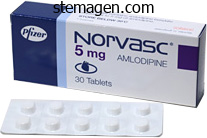
Buy generic norvasc 2.5mg online
Taken collectively blood pressure 9260 discount norvasc 2.5 mg on-line, the medical importance of security pharma cological and toxicological research could be very obvious blood pressure medication manufacturers effective norvasc 10 mg. With respect to reprogramming arterial insufficiency buy 10mg norvasc overnight delivery, main strides have been made in using nonintegrating and smallmoleculebased methods. Regardless of the strategies used, nonetheless, the overall reprogramming efficiency remains <1%, thus significantly growing the time, manpower, and prices associated with this process. The presence of karyotypic abnormality following longterm culture and repeated cell passaging is one other challenge. This heteroge neity corresponds to varied levels of improvement and maturity as corroborated by single cell transcriptional profiling, which is also suggestive of significant intercell line variability (Narsinh et al. A detailed discussion of maturation strategy is past the scope of this chapter, and interested readers are inspired to discuss with different evaluations (Matsa et al. This, coupled with improvement of simplified, lowcost chemically outlined cardiac differen tiation medium (Burridge et al. Kolossov, Dynamic monitoring of beating periodicity of stem cellderived cardio myocytes as a predictive software for preclinical security evaluation. Chakravarti, Understanding heart problems by way of the lens of genomewide association research. Wu, Production of de novo cardiomyocytes: human pluripotent stem cell differentiation and direct reprogramming. Sullivan, A scientific drug library display screen identifies astemizole as an antimalarial agent. Wu, Overview of high throughput sequencing applied sciences to elucidate molecular pathways in cardiovascular diseases. Brown, Blockade of a quantity of human cardiac potassium currents by the antihistamine terfenadine: possible mechanism for terfenadine associated cardiotoxicity. DesmondHellmann, the Cost of Creating a New Drug Now $5 Billion, Pushing Big Pharma to Change. Benghozi, Drug attrition during preclinical and medical growth: understanding and managing druginduced cardiotoxicity. Kolaja, Cardiotoxicity of kinase inhibitors: the prediction and translation of preclinical fashions to scientific outcomes. Daley, Induced pluripotent stem cells-opportunities for disease modelling and drug discovery. Jonsson, Lessons from the guts: mirroring electrophysiological charac teristics throughout cardiac improvement to in vitro differentiation of stem cell derived cardiomyocytes. Verkerk, Induced pluripotent stem cell derived cardiomyocytes as models for cardiac arrhythmias. Liu, Pluripotent stem cells induced from mouse somatic cells by smallmolecule compounds. Melton, Induction of pluripotent stem cells from major human fibroblasts with solely Oct4 and Sox2. Guideline on S7B, the nonclinical evaluation of the potential for delayed ventricular repolarization (Qt Interval Prolongation) by human pharmaceuticals. Koszka, A smallmolecule inhibitor of Tgf signaling replaces Sox2 in reprogramming by inducing Nanog. Glikson, Modeling of cate cholaminergic polymorphic ventricular tachycardia with patientspecific humaninduced pluripotent stem cells. Hulot, Small moleculemediated directed differentiation of human embryonic stem cells toward ventricular cardiomyocytes. Gepstein, Human embryonic stem cells can differentiate into myocytes with structural and functional properties of cardiomyocytes. Fischer, Controlling expansion and cardiomyogenic differentiation of human pluripotent stem cells in scalable suspension culture. Li, Developmental cues for the maturation of metabolic, electrophysiological and calcium handling properties of human pluripotent stem cellderived cardiomyocytes. Lanza, Generation of human induced pluripotent stem cells by direct supply of reprogram ming proteins. Swan, Cell model of catecholaminergic polymorphic ventric ular tachycardia reveals early and delayed afterdepolarizations. Sun, Abnormal calcium dealing with properties underlie familial hypertrophic cardiomyop athy pathology in patientspecific induced pluripotent stem cells. Schaffer, A totally outlined and scalable 3D tradition system for human pluripotent stem cell growth and differentiation. Wang, Drug screening using a library of human induced pluripotent stem cell�derived cardiomyocytes reveals diseasespecific patterns of cardiotoxic ity. Li, Mechanismbased facilitated maturation of human pluripotent stem cellderived cardiomyocytes. Plath, Generation of human induced pluripotent stem cells from dermal fibroblasts. January, High purity human induced pluripotent stem cellderived cardiomyocytes: electro physiological properties of action potentials and ionic currents. Class, Cardiotoxicity testing using pluripotent stem cellderived human cardiomyocytes and stateoftheart bioanalytics: a evaluate. Ebert, Exploiting pluripotent stem cell tech nology for drug discovery, screening, safety, and toxicology assessments. Olson, Toward transcriptional therapies for the failing coronary heart: chemical screens to modulate genes. Wu, A evaluation of human pluripotent stem cellderived cardiomyocytes for highthroughput drug discovery, cardiotoxicity screening, and publication stan dards. Kamp, Differentiation of human embryonic stem cells and induced pluripotent stem cells to cardiomyocytes a methods overview. Keller, Differentiation of embryonic stem cells to clinically relevant populations: lessons from embryonic improvement. Musunuru, Genome enhancing of human pluripotent stem cells to generate human cellular illness models. Rosenberg, Single cell transcriptional profiling reveals heterogeneity of human induced pluripotent stem cells. Takayasu, Development of faulty and protracted Sendai virus vector a novel gene delivery/expression system perfect for cell reprogram ming. Hsieh, Biowire: a platform for maturation of human pluripotent stem cellderived cardiomyocytes. Yamanaka, Generation of mouse induced pluripotent stem cells with out viral vectors. Bruening Wright, the action potential and comparative pharmacology of stem cellderived human cardiomyocytes. Vergely, Anthracyclines/trastuzumab: new features of cardiotoxicity and molecular mechanisms. Hoffman, Action potential prolongation and induction of abnormal automaticity by low quinidine concentra tions in canine Purkinje fibers. Wu, Finding the rhythm of sudden cardiac demise: new opportunities utilizing induced pluripotent stem cell�derived cardiomyocytes.
Diseases
- Chromosome 1, monosomy 1p22 p13
- Polycystic kidney disease, adult type
- Hereditary carnitine deficiency
- Hereditary resistance to anti-vitamin K
- Shellfish poisoning, neurotoxic (NSP)
- Linear hamartoma syndrome
- Follicular dendritic cell tumor
- Chromosome 3, monosomy 3q27
- Eosinophilic synovitis
Norvasc 5mg line
Cytologic Atypia Comedonecrosis (Left) Enteric-type adenocarcinoma with comedonecrosis could also be morphologically indistinguishable from colon cancer hypertension journal articles generic norvasc 10mg overnight delivery. In distinction arrhythmia young cheap 2.5 mg norvasc with visa, urothelial carcinoma with glandular differentiation has a urothelial morphology in some areas normal pulse pressure 60 year old 10 mg norvasc with mastercard. Exophytic and Invasive Histology 412 Invasive Adenocarcinoma Urinary Bladder Adenocarcinoma of Bladder Adenocarcinoma of Bladder (Left) Primary vesical adenocarcinoma (enteric and colloid type) is usually high stage and shows obvious destructive invasion of the muscularis propria. This characteristic could be very useful within the distinction from cystitis glandularis with mucin extravasation. Adenocarcinoma of Bladder Adenocarcinoma of Bladder (Left) Signet ring cell adenocarcinoma of the urinary bladder is rare but is morphologically indistinguishable from metastatic gastric signet ring cell adenocarcinoma. There could additionally be complete immunophenotypic overlap between these 2 lesions and adenocarcinomas arising in the urachus. The whole range of major mucosal-based vesical adenocarcinoma histology may be seen in urachal adenocarcinomas. Urachal Adenocarcinoma Urachal Adenocarcinoma (Left) Urachal adenocarcinomas are indistinguishable from major vesical adenocarcinoma. The presence of a muscularis propria-based tumor in the dome and absence of surface lesions favor a urachal main. Metastatic Colonic Adenocarcinoma to Bladder M�llerianosis of Bladder (Left) Lesions in the spectrum of m�llerianosis may also enter the differential prognosis of adenocarcinoma because of the presence of glands deep throughout the bladder wall, together with the muscularis propria. M�llerianosis of Bladder 414 Invasive Adenocarcinoma Urinary Bladder Prostatic Adenocarcinoma Prostatic Adenocarcinoma (Left) Prostatic adenocarcinoma could contain the lumina of the prostatic urethra and may extend superiorly to contain the bladder. The columnar cells of ductal carcinoma extra closely mimic enteric-type vesical adenocarcinoma. In comparability to vesical adenocarcinomas, prostatic carcinoma has extra rounded monomorphic nuclei. Urothelial Carcinoma With Tubular Features Cystitis Glandularis With Intestinal Metaplasia (Left) In this field, the blandlooking tubular structures invade deeply into the bladder wall and characterize a tubular component of in any other case classic urothelial carcinoma elsewhere in the tumor, effectively ruling out the prognosis of adenocarcinoma. Some glands have outstanding luminal cytoplasm, and others show intensive goblet cell metaplasia. Architectural diversity is the rule rather than an exception in clear cell adenocarcinoma. Clear Cell Adenocarcinoma of Bladder: Tubulocystic Architecture Clear Cell Adenocarcinoma of Bladder: Invasion and Myxoid Stromal Reaction (Left) A low-power photomicrograph reveals clear cell adenocarcinoma with a basic tubulocystic sample. The papillae are lined by a single layer of cuboidal cells, a function distinct from typical papillary urothelial carcinoma. There is subtle stalk invasion that, with haphazard growth when present, is diagnostic of adenocarcinoma. Clear Cell Adenocarcinoma of Bladder: Papillary Tufting and Stromal Invasion Clear Cell Adenocarcinoma of Bladder: Bland Morphologic Features (Left) the degree of cytologic atypia may be variable within a given clear cell adenocarcinoma. This papillary focus is comparatively bland, which may result in confusion with nephrogenic adenoma. Areas with more typical cytologic features of carcinoma ought to be sought and are usually recognized. Clear Cell Adenocarcinoma of Bladder: Nuclear Atypia and Prominent Nucleoli Clear Cell Adenocarcinoma of Bladder: Heterogeneous Tumor Patterns (Left) Multiple admixed architectural progress patterns are frequent in clear cell adenocarcinoma of the bladder. The neoplastic cells have a extra eosinophilic cytoplasm, which can be very putting in some circumstances. Clear Cell Adenocarcinoma of Bladder: Marked Eosinophilic Cytoplasm and Myxoid Stroma 418 Clear Cell Adenocarcinoma Urinary Bladder Clear Cell Adenocarcinoma of Bladder: High-Power View, Solid Growth Clear Cell Adenocarcinoma of Bladder: Round Uniform Cuboidal Cells (Left) the foci of stable intratubular growth may mimic a poorly differentiated urothelial carcinoma, especially if the cytoplasm is more eosinophilic, as on this example. Identification of more attribute patterns of clear cell adenocarcinoma is useful. Clear Cell Adenocarcinoma of Bladder: Short Papillae and Nuclear Hobnailing Clear Cell Adenocarcinoma of Bladder: Invasive With Minimal Stromal Reaction (Left) Clear cell adenocarcinoma with small papillary projections is shown. Tumor cells contain clear to eosinophilic cytoplasm with an occasional hobnail appearance. Clear Cell Adenocarcinoma of Bladder: Bland Features, Cystic and Flattened Tumor Nuclei Clear Cell Adenocarcinoma of Bladder: Tumor Heterogeneity, Cystic and Papillary Patterns (Left) this example of clear cell adenocarcinoma has dilated tubulocystic foci with very bland cytologic features. This sample might trigger diagnostic confusion with a cystic nephrogenic adenoma or other benign glandular lesions. There is a refined but distinct variation in nuclear measurement and shape and chromatin characteristics to render a malignant analysis. Clear Cell Adenocarcinoma of Bladder: Morphologic Similarity to Gynecologic Tract Tumors Clear Cell Adenocarcinoma of Bladder: Solid Growth and Clear Cytoplasm (Left) Solid development can also be seen in clear cell adenocarcinoma of the urinary bladder, which can mimic other poorly differentiated carcinomas, including renal cell carcinoma. The small rim of clear cytoplasm, the rounded nuclei, and the distinguished nucleoli are typical options of this tumor. Clear Cell Adenocarcinoma of Bladder: Clear Cytoplasm and Prominent Nucleoli Clear Cell Adenocarcinoma of Bladder: Clear Cytoplasm and Stromal Hyalinization (Left) Dense stromal hyalinization, admixed with nests and individual tumor cells, and the abundant clear cytoplasm are typical options of clear cell adenocarcinoma but is in all probability not at all times current in all cells. Such a discovering, along with the extra atypical cytologic features (nuclear pleomorphism, hyperchromasia, and markedly distinguished nucleoli), can additionally be helpful within the distinction from nephrogenic adenoma. The lack of serious cytologic atypia and the flattened lining epithelium are characteristic of nephrogenic adenoma. Other patterns of extra typical nephrogenic adenoma are normally present, and mitotic figures are often absent. Clear Cell Adenocarcinoma of Bladder: Superficial Biopsy Mimicking Nephrogenic Adenoma Urothelial Carcinoma With Clear Cell Features (Left) Superficial biopsy of a clear cell adenocarcinoma of the bladder resembles nephrogenic adenoma, in particular the inflammatory background and focally flattened cells. More atypical options, nevertheless, were additionally present in this area and elsewhere within the tumor. The focality of this glycogenrich appearance is typical and is distinct from clear cell carcinoma. Urothelial Carcinoma With Large Vacuolated Clear Cells Urothelial Carcinoma With Papillary Architecture (Left) that is one other instance of high-grade urothelial carcinoma with clear cells as a outcome of large cytoplasmic vacuoles. The presence of otherwise typical areas of urothelial carcinoma is useful in establishing the analysis. The presence of advanced micropapillary structures and multilayering of the urothelial lining ought to appropriately place this tumor into urothelial carcinoma prognosis (urothelial carcinoma with villoglandular features). When this is in depth, sampling of a quantity of areas is essential to explore the potential of an associated in situ neoplasia. Higherpower evaluation of cytologic options is critical to rule out in situ neoplasia. Squamous Metaplasia Squamous Papilloma (Left) Squamous papilloma of the bladder consists of hyperplastic, welldifferentiated squamous mucosa with distinct papillary formation. It is essential to look at such lesions at excessive magnification to rule out dysplastic or viral cytopathic changes. There is a distinct central vascular core surrounded by hyperplastic squamous mucosa.
Generic norvasc 2.5mg online
Immunoreactivity for histiocytic hypertension headaches symptoms buy 2.5 mg norvasc amex, however not epithelial blood pressure categories discount norvasc 2.5mg free shipping, markers will resolve most troublesome instances heart attack female cheap norvasc 10mg overnight delivery. Malakoplakia: Typical Pattern Malakoplakia: Typical Pattern (Left) Numerous intracytoplasmic inclusions (Michaelis-Guttman bodies) are present within the eosinophilic histiocytes of malakoplakia. Identification of those attribute inclusions help to exclude the risk of other histiocytic infiltrates. Malakoplakia: Typical Pattern Malakoplakia: Typical Pattern (Left) Under high-power examination with oil immersion, the lamellated nature of the intracytoplasmic Michaelis-Guttman our bodies is evident. Malakoplakia: von Kossa Stain 282 Malakoplakia Urinary Bladder Malakoplakia: Spindled Pattern Malakoplakia: Spindled Pattern (Left) this instance of malakoplakia has a extra distinguished spindled look and scant inflammatory infiltrate. Because the histiocytes appear spindled, lesions similar to reactive stromal proliferations or spindle cell neoplasms may be thought-about within the differential prognosis. Extranodal Rosai-Dorfman Extranodal Rosai-Dorfman (Left) Extranodal RosaiDorfman illness is well documented in the genitourinary tract and will closely resemble different histiocytic infiltrates, similar to malakoplakia. The histiocytes of Rosai-Dorfman illness usually contain more voluminous cytoplasm and emperipolesis could also be present. Extranodal Rosai-Dorfman: S100 Protein Granulomatous Inflammation: Fungal (Left) the histiocytes of RosaiDorfman disease, unlike malakoplakia, show diffuse cytoplasmic and nuclear reactivity for S100 protein. The sheet-like look of histiocytes in a fibrotic background with combined inflammatory cells mimics malakoplakia. The sheets of cells with amphophilic cytoplasm may provoke a differential that features malakoplakia, particularly since histiocytic markers may be positive. Leukemia in Bladder Prostatic Adenocarcinoma (Left) this instance of Gleason pattern 5 prostatic adenocarcinoma involving the bladder has eosinophilic cytoplasm which will prompt consideration of malakoplakia. Prostatic Adenocarcinoma Urothelial Carcinoma (Left) In this instance of invasive urothelial carcinoma, the sheet-like progress is related to more ample frothy and lightly eosinophilic cytoplasm. These cells are extra uniformly cohesive than usually anticipated in a histiocytic infiltrate, corresponding to malakoplakia. Urothelial Carcinoma 284 Malakoplakia Urinary Bladder Urothelial Carcinoma Urothelial Carcinoma (Left) this example of invasive urothelial carcinoma with an related combined inflammatory infiltrate has a morphology with putting resemblance to histiocytes. Demonstration of cytokeratin reactivity with a broad spectrum keratin is necessary in difficult instances. Mycobacterial Spindle Cell Pseudotumor Mycobacterial Spindle Cell Pseudotumor: Acid-Fast Stain (Left) Mycobacterial spindle cell pseudotumor has overlapping morphologic features with malakoplakia. Note the sheet-like aggregates of spindled histiocytes with an inflammatory infiltrate. Primary Signet Ring Cell Adenocarcinoma Plasmacytoid Urothelial Carcinoma (Left) Primary signet ring cell adenocarcinoma of the urinary bladder, as in different anatomic sites, could have morphologic overlap with histiocytes. Precursor lesions embrace in depth keratinizing metaplasia, dysplasia, and in situ carcinoma. Some nested carcinomas share similar features; therefore, appreciation of the structure is essential to distinguish von Brunn nests from delicate patterns of carcinoma. Predominant Pattern/Injury Type � Nests 289 von Brunn Nests Urinary Bladder von Brunn Nests (Left) von Brunn nests are cytologically benign invaginations of urothelium into the lamina propria. There is a close relationship, or generally a reference to the overlying floor. Carcinomas sometimes have a more random distribution and deeper extension into the lamina propria (or muscularis propria). The superficial location of the nests and welldelineated/lobular architecture, in addition to the clinical historical past, should assist on this distinction. The nests lack the aggregated, lobular association typical of von Brunn nests, that are more superficial with a pointy linear border on the base. Nested Urothelial Carcinoma Nested Urothelial Carcinoma (Left) Nested urothelial carcinomas present deeper invasion into the lamina propria with involvement of the muscularis mucosae. The cytology of the malignant cells is extraordinarily bland, which adds to the degree of histologic overlap with von Brunn nests. Nested Urothelial Carcinoma Pseudocarcinomatous Hyperplasia (Left) this florid reactive urothelial proliferation is from a affected person who underwent radiation remedy for prostate most cancers. Despite the back-toback nests, the cytology of the urothelial cells is bland, and at most is within the vary of reactive atypia. Pseudocarcinomatous Hyperplasia 292 von Brunn Nests Urinary Bladder Inverted Papilloma Inverted Papilloma (Left) Inverted papillomas also have an endophytic development but they usually present a more trabecular, anastomosing development pattern. The distinction in small lesions could also be arbitrary, although more established structure and expansile progress favors inverted papilloma. Carcinoid Tumor (Low-Grade Neuroendocrine Neoplasm) Carcinoid Tumor (Low-Grade Neuroendocrine Neoplasm) (Left) When seen on a superficial biopsy, the standard monomorphic cytology could trigger consideration of a benign lesion. The complicated cribriforming or ribbon-like progress should alert one to the potential of a carcinoid tumor. Paraganglia Paraganglia (Left) Paraganglionic tissue in a biopsy specimen reveals the lesion is well demarcated and unrelated to urothelium. Paraganglia are current within the lamina propria, not often in muscularis propria or deeper. The papillary cores are characteristically lined by a single layer of cuboidal epithelial cells. Nephrogenic Adenoma: Tubular Nephrogenic Adenoma: Tubular and Cystic (Left) this crowded glandular/tubular sample of nephrogenic adenoma is common. Not uncommonly, other patterns are admixed, such as the cystic sample seen in this instance. They are normally lined by eosinophilic cells with an atrophic or hobnail association. Nephrogenic Adenoma: Solid Cords Nephrogenic Adenoma: Papillary (Left) Small tubules of nephrogenic adenoma could have a pseudoinfiltrative appearance that carefully mimics prostatic adenocarcinoma. Although not all the time seen, the thin rim of basement membrane-like materials is attribute. Unlike papillary urothelial neoplasia, the papillae are lined by a single layer of cytologically bland cuboidal epithelial cells. Nephrogenic Adenoma: Flat Nephrogenic Adenoma: Solid Nests (Left) the recently described flat pattern of nephrogenic adenoma is difficult to distinguish from denuded urothelium with residual basal cells but is normally seen adjoining to other patterns. This histologic look might intently mimic prostatic adenocarcinoma because of the monomorphic nuclei. Other more typical patterns of nephrogenic adenoma are nearly always present to assist in analysis. Awareness of this rare sample is necessary as a end result of it could probably mimic myxoid mesenchymal neoplasms. Nephrogenic Adenoma: Fibromyxoid Nephrogenic Adenoma: Diffuse (Left) Solid progress in nephrogenic adenoma has been described because the diffuse sample, and it resembles renal cell carcinoma.
Order norvasc 10 mg free shipping
The tumor cells within and surrounding the tubules are comparable and have relatively uniform cells with distinguished nucleoli blood pressure of normal person buy norvasc 2.5mg online. Metastatic Prostate Carcinoma 896 Metastatic Tumors arrhythmia flutter discount 5 mg norvasc, Testis and Paratesticular Structures Testis and Paratesticular Structures Metastatic Renal Cell Carcinoma Metastatic Renal Cell Carcinoma (Left) this metastatic carcinoma is a high-grade clear cell renal carcinoma near the rete testis heart attack cpr purchase norvasc 2.5mg with amex. The ample sinusoidal vasculature and alveolar progress sample are options of clear cell renal cell carcinoma. It might mimic a seminoma but has a larger degree of nuclear pleomorphism and lacks a stromal lymphoid infiltrate. Metastatic Lung Carcinoma Metastatic Merkel Cell Carcinoma (Left) Metastatic poorly differentiated lung carcinoma with an interstitial development pattern is proven. Metastatic Melanoma Large B-Cell Lymphoma (Left) High-power image exhibits metastatic melanoma composed of large epithelioid cells with prominent nucleoli and melanin pigments. In case of an amelanotic melanoma, differential diagnoses would come with Leydig cell tumor, seminoma, embryonal carcinoma, and enormous B-cell lymphoma. M�llerian Papillary Serous Carcinoma (-) (+) (+) Mesothelioma (-) (-) (+) Prostatic Adenocarcinoma (+) (-) (-) Testicular Tumors vs. The atypical cells have ample clear cytoplasm and enlarged nuclei with distinguished nucleoli. Podoplanin is an antibody against oncofetal protein M2A, which also stains lymphatic endothelial cells and mesothelial cells. Mixed Cell Tumor Mixed Germ Cell Tumor (Left) this picture shows a combined germ cell tumor, i. When predominant, the morphologic differential diagnoses would come with seminoma, embryonal carcinoma, monophasic choriocarcinoma, and strong Leydig cell and Sertoli cell tumors. Other germ cell tumor elements are adverse for glypican-3 besides in syncytiotrophoblasts and barely in immature teratoma. It can additionally be a sensitive marker for traditional seminoma and germ cell neoplasia in situ. Condyloma, Low Magnification Condyloma, Architectural Features (Left) Medium magnification of this lesion exhibits papillomatosis with fibrovascular cores composed of unfastened connective tissue and small vascular spaces. Koilocytosis Giant Condyloma (Left) Papillomatosis with evident fibrovascular cores, floor koilocytosis, and absence of evident nuclear atypia are pathognomonic findings of ordinary condyloma acuminatum. Koilocytosis Koilocytosis (Left) In both condyloma acuminatum and large condyloma, koilocytes are easily discovered, subtle nuclear atypia could also be current in cells of the basal layer, especially within the latter, and a thin parakeratotic layer is often noticed. The cytoplasm is eosinophilic, mobile borders are distinctive, and binucleation is common. Philippou P et al: Genital lichen sclerosus/balanitis xerotica obliterans in males with penile carcinoma: a important analysis. An extraordinarily well-differentiated, nonverruciform neoplasm that preferentially impacts the foreskin and is frequently misdiagnosed: a report of 10 instances of a particular clinicopathologic entity. Flat Hyperplasia Cytologic Features (Left) Hyperkeratosis and acanthosis in flat squamous hyperplasia. Histological Subtypes � Flat Most widespread subtype Nonatypical acanthosis Hyperkeratosis with orthokeratosis 5. Verruciform Xanthoma Verruciform Lesion (Left) Verruciform xanthoma is characterized by prominent acanthosis, hyperkeratosis, papillomatosis, absence of cytologic atypia, and foamy cells current in lamina propria between rete ridges. In addition to histiocytes, on this case, the inflammatory response is also wealthy in lymphocytes. Lipogranuloma of Penis Sclerosing Lipogranuloma of Scrotum (Left) Numerous lipid vacuoles of different sizes are apparent in the dermis and dartos. This case exhibits outstanding sclerosis and histiocytic infiltrate with only a few lymphocytes. Scrotal Calcinosis Scrotal Calcinosis (Left) Homogeneous granular and globular pink and purple material is seen inside the dermis admixed with sclerosis and palisading histiocytic irritation. Shah V et al: Scrotal calcinosis outcomes from calcification of cysts derived from hair follicles: a sequence of 20 instances evaluating the spectrum of changes leading to scrotal calcinosis. Ito A et al: Dystrophic scrotal calcinosis originating from benign eccrine epithelial cysts. Polk P et al: Polypoid scrotal calcinosis: an uncommon variant of scrotal calcinosis. There is delicate spongiosis with abnormal maturation, keratin pearl formation (usually in deep rete ridges), and parakeratosis. Amin A et al: Penile intraepithelial neoplasia with pagetoid features: report of an uncommon variant mimicking Paget disease. There is also a thickened basement membrane and hyalinization of the upper lamina propria comparable to related lichen sclerosus. In spite of the apparently subtle cytologic adjustments, juxtaposition to carcinoma argues for its premalignant potential. Note the elongation of the rete ridges and delicate abnormal maturation of the epithelium. Irregular nuclear contours, biand multinucleation, and numerous mitoses are easily recognized. The decrease a half of the epidermis is replaced by small basaloid cells, and the higher part is changed by larger cells exhibiting koilocytotic changes. Warty-Basaloid Penile Intraepithelial Neoplasia Warty-Basaloid Penile Intraepithelial Neoplasia (Left) Spiky surface shows parakeratosis and atypical koilocytosis. Differentiated Penile Intraepithelial Neoplasia With Lichen Sclerosus Squamous Hyperplasia and Lichen Sclerosus (Left) Hyperplasia of the squamous epithelium with atypia in the basal layer is diagnostic of this lesion. Verruciform Growth Pattern Verruciform Growth Pattern (Left) Penile verruciform tumors embrace warty, papillary, and verrucous carcinoma and are characterised by an exophytic papillomatous growth pattern, infiltration as much as the lamina propria, or corpus spongiosum, and are associated with a comparatively favorable prognosis. The tumor deeply infiltrates the corpora cavernosa and perforates the tunica albuginea. Squamous Cell Carcinoma: Grade 3 934 General Concepts, Squamous Cell Carcinoma Penis and Scrotum Warty Carcinoma Warty Carcinoma (Left) Warty (condylomatous) carcinoma is a verruciform tumor characterized by acanthosis, papillomatosis, and hyperkeratosis, with straight and spiky parakeratotic papillae, outstanding and fixed fibrovascular cores, and irregular and jagged tumor base. Warty Carcinoma Papillary Carcinoma (Left) Verrucous carcinoma is characterized by papillomatosis, reasonable to marked acanthosis, minimal cytological atypia, distinguished hyperkeratosis with absent or inconspicuous fibrovascular cores, and a broad, pushing tumor base. Carcinoma Cuniculatum Basaloid Carcinoma (Left) Carcinoma cuniculatum, a rare verruciform tumor, reveals outstanding acanthosis, papillomatosis and hyperkeratosis, broad and pushing tumor base, and a deeply infiltrating endophytic sample of growth. Perineural Invasion Vascular Invasion (Left) Perineural invasion, an necessary pathological prognostic issue, is precisely outlined as invasion of perineural space by neoplastic cells and never simply extension of the tumor alongside nerve bundles (nerve entrapment). The impression is of a poorly to undifferentiated, high-grade carcinoma with minimal proof of squamous differentiation. This carcinoma is incessantly related to vertical growth pattern of penile carcinoma. Basaloid Nests Mitotic Activity (Left) Neoplastic cells of basaloid carcinomas present average nuclear pleomorphism, vague cellular borders, and nonkeratinized cytoplasm. There is an abundance of mitotic figures, which on low power might impart a starry-sky appearance.
Khulakhudi (Gotu Kola). Norvasc.
- Preventing blood clots in the legs while flying.
- Decreased return of blood from the feet and legs back to the heart called venous insufficiency.
- Are there any interactions with medications?
- Fatigue, anxiety, increasing circulation in people with diabetes, atherosclerosis, stretch marks associated with pregnancy, common cold and flu, sunstroke, tonsillitis, urinary tract infection (UTI), schistosomiasis, hepatitis, jaundice, diarrhea, indigestion, improving wound healing when applied to the skin, a skin condition called psoriasis, and other conditions.
- What is Gotu Kola?
- Is Gotu Kola effective?
- Are there safety concerns?
- How does Gotu Kola work?
Source: http://www.rxlist.com/script/main/art.asp?articlekey=96735
Discount norvasc 2.5mg on-line
SteidlNichols J hypertension 5 mg discount 2.5 mg norvasc amex, Bhatt S arteria pharyngea ascendens generic norvasc 10mg line, Hemkens M heart attack 86 years old order norvasc 2.5 mg overnight delivery, Heyen J, Marshall C, Li D, Flynn D, Wisialowski T, Northcott C (2014). Task Force of the Working Group on Arrhythmias of the European Society of Cardiology (1991). A new strategy to the classification of antiarrhythmic drugs based mostly on their actions on arrhythmogenic mechanisms. The European Agency for the Evaluation of Medicinal Products Human Medicines Evaluation Unit. A new homogeneous highthroughput screening assay for profiling compound activity on the human etheragogorelated gene channel. Effects of calcium, sodium and potassium ions on contractility of isolated atria and their responses to noradrenaline. Functional assessments in nonhuman primate toxicology research to support drug improvement. The electromechanical window: a danger marker for Torsade de Pointes in a canine mannequin of drug induced arrhythmias. Scientific evaluation and suggestions on preclinical cardiovascular security analysis of biologics. Nonclinical strategy concerns for safety pharmacology: evaluation of biopharmaceuticals. The impact of single cell voltage clamp on the understanding of the cardiac ventricular motion potential. Techniques and methodologies to research the ryanodine receptor at the molecular, subcellular and cellular stage. Preclinical evaluation of druginduced proarrhythmias: function of the arterially perfused rabbit left ventricular wedge preparation. Analysis of the direct and indirect results of adenosine on atrial and ventricular cardiac muscle. Emerging trends in ion channelbased assays for predicting the cardiac safety of drugs. Cardiovascular problems of cancer therapy: incidence, pathogenesis, analysis and management. In phrases of operate, the renal epithelium is uncovered to any chemical in the blood by advantage of both glomerular filtration and secretion from the nearby renal circulation. A critical incontrovertible truth that emphasizes the unique physiology of the kidneys is that they typically comprise <1% of total physique weight yet obtain 20�25% of cardiac output. Glomerular filtration leads to accumulation of blood borne chemical substances within the tubular lumen, whereas secretion from the renal circulation initially ends in mobile accu mulation of these chemical compounds. Another facet of renal perform that will significantly contribute to the kidneys as a goal organ in drug or chemical exposures is the urinary concentrating mechanism that serves to reabsorb vitamins such as hexoses and amino acids; excrete waste products such as ammonia, creatinine, and other nitrogenous compounds; and regulate water and electrolyte steadiness. This can outcome in very high concentra tions of chemical compounds in renal cells, lumens, or interstitial area. The urinary concentrating mechanism additionally supplies an illustration of structure serving function. In this case, the renal vasculature and epithelium exist in parallel such that renal capillaries are in shut proximity to reabsorptive and secretory surfaces of the tubular epithelium. Finally, regardless of their major perform being considered as the site for reabsorption and excretion of medication and chemical substances, the kidneys are also essential websites of metabolism for so much of chemical substances (Lohr et al. Besides these four "susceptibility components," one must consider the heterogeneous structure of the kidneys, as a outcome of many medication and chemicals act at particular sites within the tissue or specific websites are extra susceptible than others to harm as a end result of their intrinsic properties. At the extent of the whole organ, the kidneys are subdivided into three areas: cortex, outer medulla, and inner medulla. The useful unit of the kidneys is the nephron, which is a sequence of epithelial cells of varying types that form a tube of onelayer thick cells enclosing a luminal compartment. Mammalian kidneys contain hundreds of thousands of nephrons, every of which leads into a typical amassing duct. Nephrons are categorized as both superficial, midcortical, or juxtamedullary, depending on the location of their glomeruli and loop of Henle. The renal cortex accommodates glomeruli, proximal convoluted, and a few proximal straight tubules, macula densa, cortical thick ascending limbs, distal convoluted tubules, connecting tubules, initial amassing ducts, interlobular arteries, and afferent and efferent capillary networks. The outer stripe of the outer medulla is situated slightly below or inside the cortex and incorporates proximal straight tubules, medullary thick ascending limbs, and outer medullary accumulating ducts. The inside stripe of the outer medulla is situated under or contained in the outer stripe and incorporates skinny descending limbs, thick ascending limbs, and outer medullary amassing ducts. The inner medulla incorporates skinny descending limbs, thin ascending limbs, and internal medullary amassing ducts. Differences exist amongst cell types in morphology, membrane structure, energetics, and the composition of transporters and drug metabolism enzymes. As a consequence of this functional heterogeneity, exposure of the kidneys, both in vivo or in vitro, to numerous nephrotoxicants or to pathological situations similar to ischemia or hypoxia produces distinct patterns of mobile harm. Thus, certain cell populations are both specific targets of or are notably vulnerable to one form of injury or one other. In some instances, the specificity is as a result of of the selective presence in a given cell population of a membrane transport system or a bioactivation or detoxication enzyme. In other instances, nevertheless, susceptibility is defined by the fundamental biochemical perform of the cell. Hence, when char acterizing chemically induced toxicity or screening drug candidates, data of the positioning of action or websites inside the nephron which are doubtlessly extra vulnerable than others is important in achieving the targets of the study. Although in vivo fashions, similar to rodents, are critical in establishing goal organ specificity for investigating effects involving systemic processes, similar to immunemediated toxicity, and for performing invasive studies in vivo that might obviously not be potential in humans, they suffer from many limitations, not the least of which is expense and the high utilization of animals. In distinction, in vitro fashions derived from the kidneys of each laboratory animals and humans have been developed and optimized over the previous few many years to extra accurately replicate processes that occur in vivo. Despite these advances, nevertheless, there are significant limitations in the use of in vitro rodent fashions to assess relevant results for humans and to make correct predictions for responses in humans (Steinberg and DeSesso, 1993; Pelekis and Krishnan, 1997). Due the existence of each quantitative and qualitative speciesdependent variations, use of animal models for human well being threat assessment or drug screening involving the kidneys as a target organ can be limited by important uncertainties. Accordingly, in vitro fashions have been developed which are either derived from human kidneys or that extra carefully mimic human kidneys. One potential strategy that has been instructed to higher approximate occasions that happen in people is to use nonhuman primates or in vitro fashions derived from nonhuman primate kidneys. Caution is needed with such research, however, as a result of while responses of nonhuman primates or renal cells derived from nonhuman primates might differ from these of rodents or renal cells derived from rodents, respectively, they might nonetheless additionally differ from those of people or human derived renal cells. This means that species differ ences in drug susceptibility and mechanisms of motion also can exist amongst different primate species. Advantages of in vitro models embody the flexibility to fastidiously modulate exposure situations and dissect mechanisms of action. Unlike research in laboratory animals, in vitro models enable using paired controls and treated samples from the same set of incubations. Another advantage particularly for research of renal operate and nephrotoxicity is that in vitro models can be constructed that derive from particular nephron segments, thereby allowing research of effects at the particular sites within the kidneys where they occur.
Generic 5 mg norvasc visa
Ultrasound is the first-line imaging study performed in patients with acute scrotum blood pressure reading chart buy cheap norvasc 5mg line. Knowledge of the normal and pathologic sonographic look of the scrotum and an understanding of the right technique are essential for accurate analysis of an acute scrotum blood pressure juicing recipes generic norvasc 2.5 mg visa. High-frequency transducer sonography combined with shade and power Doppler sonography supplies info essential to reach a particular prognosis in sufferers with testicular torsion blood pressure medication nightmares cheap norvasc 5mg without a prescription, epididymoorchitis, and scrotal trauma. History and physical examination are important in making the correct analysis together with the sonographic appearance of the scrotum. Approach to Scrotal Sonography Diagnoses: Scrotum (Left) Transverse grayscale ultrasound of the scrotum shows an irregular axis of the left testis in comparability with the right. It is necessary to study each testes using the identical shade scale and similar colour Doppler gain settings. This affected person exhibits decreased flow on the best side, surgically confirmed to be right-sided partial testicular torsion. Comparison of both sides using the same scale is crucial to detect asymmetry in dimension and vascularity. In a patient with acute right-sided pain, findings counsel acute epididymoorchitis. Note the compressed and near-complete alternative of regular testicular parenchyma. A few scattered microliths are also seen in the noninvolved portion of the testicle. Pathology revealed a mixed germ cell tumor with 95% embryonal component and 5% teratoma. Testicular Germ Cell Tumors Diagnoses: Scrotum (Left) Longitudinal grayscale ultrasound of the testis in a affected person with a cystic teratoma exhibits a posh heterogeneous mass with cystic areas of varying sizes and small echogenic foci. Pathology revealed a blended germ cell tumor with choriocarcinoma and mature teratoma. Pathology after orchiectomy confirmed it to be a benign intercourse cord stromal tumor (unclassified type). Miliaras D et al: Adult kind granulosa cell tumor: a really uncommon case of sex-cord tumor of the testis with evaluation of the literature. Testicular Metastases, Lymphoma, Leukemia � Often multiple; in any other case indistinguishable Intratesticular Hematoma � Scrotal trauma, no inner colour move in hematoma 7. The gross discovering is different from Leydig cell tumor, which often is tanbrown to yellow because of high lipid content. These findings are consistent with tubular ectasia of the rete testis with associated spermatocele. Annual screening ultrasound is indicated as a outcome of elevated risk for cancer in an atrophic testicle with microlithiasis. This must be annually screened by ultrasound due to elevated danger for cancer. The patient had a history of right orchiectomy for germ cell tumor and was being screened yearly for increased risk of cancer. The affected person had a historical past of proper orchiectomy for germ cell tumor and was being followed-up by ultrasound on an annual foundation. This limits enough evaluation of the testicular parenchyma for tumor, therefore these must be referred to specialist facilities for alternate strategies of future screening. Patients with any intratesticular calcification should be considered to be at higher danger of a testicular malignancy. Demographics � Epidemiology Infant & adolescent boys most frequently affected 702 Testicular Torsion/Infarction Diagnoses: Scrotum (Left) Transverse color Doppler ultrasound of the left testis in a male with an acute painful scrotum for 2 hours exhibits complete absence of inner blood circulate. Pathology confirmed it to be a hemorrhagic infarction of the testis with torsion > 360 levels. An undescended testis could also be positioned wherever from the kidney to the inguinal canal. Testicular microlithiasis in a cryptorchid testis is related to increased threat of development of testicular carcinoma. The heterogenous echogenicity and lack of shade move within this testis is consistent with testicular torsion. Abaci A et al: Epidemiology, classification and management of undescended testes: does treatment have worth in its therapy The findings are consistent with epididymitis complicated by an epididymal abscess. In instances of focal intratesticular hematomas, follow-up to resolution is really helpful to exclude underlying neoplasm. Rafailidis V et al: Sonography of the scrotum: from appendages to scrotolithiasis. Note that the belly probe somewhat than the high-frequency linear probe was wanted to seize this huge lesion. This ovoid, well-circumscribed mass within the tail of the epididymis is oechoic to testis and exhibits minimal internal vascularity. Note the very totally different echogenicity in comparability with the in any other case comparable affected person shown previously. Blood flow within the varicocele is slow and could also be detected only with low Doppler settings. Cantoro U et al: Reassessing the position of subclinical varicocele in infertile males with impaired semen quality: a potential examine. Most commonly, the uterus lies in the midline with the fundus pointing in direction of the anterior belly wall and the external os pointing in direction of the rectum (anteversion and anteflexion). The opposite configuration (retroversion and retroflexion) is also generally encountered and typically the uterus strikes during the ultrasound examination. Both variants of uterine position are well imaged at endovaginal sonography as a result of the transducer could be positioned within the anterior or posterior vaginal fornices and near the uterine serosa, allowing for high-frequency and high-resolution imaging. The uterus may be in a impartial position, which is commonly the most troublesome to picture as a outcome of the fundus factors superiorly, away from the transducer. In this position, the fundus is at quite some depth from the vaginal fornix, making it difficult to penetrate with the higher frequencies of endovaginal transducers. In this example, the affected person or sonographer can apply stress to the anterior belly wall to try to transfer the uterus to a good position. Ovaries the position of the ovaries can be variable and could be affected by the bladder filling, parity, uterine place, and pathology. The ovaries are typically discovered alongside the pelvic sidewall, adjacent to the external iliac vessels. The broad ligament, fallopian tube, and mesosalpinx are found between the ovary and uterus.
Discount norvasc 2.5mg free shipping
Alternatively blood pressure 5545 order norvasc 5mg without prescription, goal organ toxicity is identified during efficacy heart attack blues buy norvasc 2.5mg otc, pharmacokinetic arrhythmia 10 year old order norvasc 10 mg amex, or pilot toxicity studies. Common themes in investigative approaches embody determination of ontarget versus off course effects and mechanisms, cross species translation, and reversibility assessments. With regard to ontarget versus offtarget effects, "software" molecules that are structurally distinct and/or structurally related molecules that lack pharmacological activity are often compared to the offending molecule; likewise, the evaluation of genetically engineered rodent fashions is commonly evaluated. When goal organ toxicity is identified in vivo, gaining an understanding of the potential translation to people is a significant focus but is a significant problem. Such an investigation may rely upon the presence of the target organ toxicity in two species, as nicely as a cross species susceptibility evaluation for the goal organ in vitro to assess the potential translation to people. Often in vitro studies make the most of a cell kind identified histologically in the goal organ to assess species translation and to investigate underlying mobile drivers of toxicity. This information can be used to decide a attainable structure�activity relationship or to identify danger factors for sufferers based mostly on the mobile mechanisms, for example, mitochondrial toxin. Human druginduced liver injury severity is extremely related to twin inhibition of mitochondrial perform and bile salt export pump. An in vitro assay to assess transporter based mostly cholestatic hepatotoxicity utilizing sandwichcultured rat hepatocytes. Predicting security toleration of pharmaceutical chemical leads: cytotoxicity correlations to exploratory toxicity research. The final goal is to determine a lead growth candidate that has the most superior safety characteristics and, for those safety issues identified, a wellcharacterized danger assessment to scale back scientific attrition because of toxicity and inform the clinical development plan. Highthroughput multiparameter profiling of electrophysiological drug results in human embryonic stem cell derived cardiomyocytes utilizing multielectrode arrays. Pharmacokinetic drivers of toxicity for fundamental molecules: strategy to decrease pKa ends in decreased tissue publicity and toxicity for a small molecule Met inhibitor. Structural and functional screening in human inducedpluripotent stem cellderived cardiomyocytes accurately identifies cardiotoxicity of multiple drug varieties. Role of electrostatic potential in the in silico prediction of molecular bioactivation and mutagenesis. Comparison of responses of basespecific Salmonella tester strains with the standard strains for figuring out mutagens: the outcomes of a validation study. Using in vitro cytotoxicity assay to aid in compound choice for in vivo security studies. Comparative analysis of in silico techniques for ames check mutagenicity prediction: scope and limitations. Guidance on nonclinical security research for the conduct of human scientific trials and advertising authorization for prescribed drugs M3(R2). Guidance on genotoxicity testing and information interpretation for prescribed drugs intended for human use S2(R1). Underlying mitochondrial dysfunction triggers flutamide induced oxidative liver harm in a mouse model of idiosyncratic drug toxicity. In vitro approaches to develop weight of evidence (WoE) and mode of action (MoA) discussions with optimistic in vitro genotoxicity results. A core in vitro genotoxicity battery comprising the Ames check plus the in vitro micronucleus check is adequate to detect rodent carcinogens and in vivo genotoxins. The utility of discovery toxicology and pathology in path of the design of security pharmaceutical lead candidates. How can we improve our understanding of cardiovascular safety liabilities to develop safety medicines Human hepatocytes: isolation, cryopreservation and applications in drug improvement. Building a tiered method to in vitro predictive toxicity screening: a focus on assays with in vivo relevance. Interference with bile salt export pump operate is a susceptibility issue for human liver damage in drug development. A multifactorial strategy to hepatobiliary transporter assess ment enables improved therapeutic compound development. Assessment of testing methods for druginduced repolarization delay and arrhythmias in an ips cellderived cardiomyocyte sheet: multisite validation study. A zone classification system for danger evaluation of idiosyncratic drug toxicity utilizing daily dose and covalent binding. Troglitazoneinduced hepatic necrosis in an animal model of silent genetic mitochondrial abnormalities. Application of an in silico strategy to predict intrinsic in vitro cytotoxicity for compounds in main human hepatocytes throughout preclinical growth. Human stem cellderived cardiomyocytes in cellular impedance assays: bringing cardiotoxicity screening to the entrance line. An algorithm for evaluating potential tissue drug distribution in toxicology research from readily available pharmacokinetic parameters. Rechanneling the cardiac proarrhythmia safety paradigm: a meeting report from the Cardiac Safety Research Consortium. Preclinical strategy to cut back medical hepatotoxicity utilizing in vitro bioactivation knowledge for >200 compounds. Testing paradigm for prediction of developmentlimiting barriers and human drug toxicity. Relating molecular properties and in vitro assay outcomes to in vivo drug disposition and toxicity outcomes. The nationwide institutes of health microphysiological techniques program focuses on a important problem within the drug discovery pipeline. Preclinical fashions of nicotinamide phosphoribosyltransferase inhibitormediated hematotoxicity and mitigation by cotreatment with nicotinic acid. Risk evaluation and mitigation methods for reactive metabolites in drug discovery and growth. In vitro safety pharmacology profiling: an essential tool for profitable drug development. The relevance and potential roles of micro physiological methods in biology and medication. Mitochondrial toxicity evaluation in industry-a decade of technology improvement and insight. Influence of lipophilicity and lysosomal accumulation on tissue distribution kinetics of fundamental medicine: a physiologically primarily based pharmacokinetic model. Retinal toxicity, in vivo and in vitro, related to inhibition of nicotinamide phos phoribosyltransferase. Proceedings of the National Academy of Sciences of the United States of America, 98, 12671�12676.
Purchase 5mg norvasc overnight delivery
Motallebnejad M blood pressure chart app purchase norvasc 10mg line, Akram S blood pressure medications with the least side effects discount norvasc 5mg overnight delivery, Moghadamina A blood pressure very low buy 5 mg norvasc, et al: the effect of topical software of pure honey on radiation-induced mucositis: a randomized clinical trial. Starmer H, Yang W, Raval R, et al: Effect of gabapentin on swallowing during and after chemoradiation for oropharyngeal squamous cell cancer. Show how disorders of esophageal origin may affect other elements of the swallowing chain. Discuss attainable therapy approaches for swallowing disorders of esophageal and pharyngoesophageal origin. In most instances sufferers must be taking their treatment each day, so reinforcing this point or reviewing correct dietary restrictions could also be wanted to keep away from a rise in reflux events. In these circumstances data of other professional roles and methods of analysis and treatment may be helpful to improve patient compliance and answer any questions sufferers might need about their dysphagia. His oral peripheral examination was normal, and a modified barium swallow with liquids and solids was carried out. Because he was an outpatient and was capable of stand, the bolus was adopted by the radiologist from the mouth to the region of the stomach. For this cause he recommended a full examination of the esophagus with the patient within the supine place to more absolutely examine this impression. Why is it essential to study the esophagus with the affected person within the supine or side-lying place A change in the structure of the esophagus could also be caused by a luminal stenosis or narrowing or by a luminal deformity similar to one other construction compressing it, thereby limiting its ability to open. Esophageal Stenosis Esophageal stenosis is conceptually the easiest mechanism of dysphagia to understand. In addition, the kind of stable materials ingested often is necessary for symptom manufacturing. For instance, dysphagia of esophageal origin is extra probably when solids are tough or fibrous. Softer, extra simply chewed meals are a lot much less prone to trigger symptoms of esophageal dysphagia. An exception to this robust food�soft meals dichotomy is that many patients even have particular hassle with gentle, absorbent foods corresponding to bread or pasta, which swell when blended with saliva throughout mastication. Once bolus impaction occurs, the affected person could have problem with liquids as properly, obscuring the characteristic solids-only nature of esophageal stenosis. However, a careful history often reveals that liquid dysphagia begins with ingestion of solids (see Differential Diagnosis later in this chapter). The frequent wisdom that patients accurately localize symptoms to the site of obstruction is often inaccurate. In truth, approximately one third of patients with obstructing lesions of the distal esophagus level to the neck as the location of obstruction. Between these extremes (20 and 10 mm), symptoms range both in frequency and severity depending on the presence of related motor dysfunction and the selection and preparation of food. Stenosis is handled by opening or removing the narrowed segment, relying on the specific trigger. This is normally completed with Maloney (bougie) dilators or with balloon dilatation. Common intrinsic structural abnormalities that narrow the esophagus embody mucosal rings, benign strictures, and malignant tumors. Rings and Webs the esophagus may be narrowed by a band of tissue composed of mucosa and submucosa. By custom, this type of lesion is identified as a ring when positioned at the esophagogastric junction and an internet when located elsewhere in the esophagus or hypopharynx. A suspected net at the cervical degree additionally may be seen in Video 5-1 on the Evolve web site accompanying this text. Patients often report that signs are intermittent and fewer likely if they choose their meals correctly and chew fastidiously (see the part on Differential Diagnosis). Conversely, signs are extra likely if the patient eats away from house or carries on a dialog while eating; in these conditions the choice of food is more restricted and correct preparation of food before swallowing is tougher. Once the food is dislodged, the affected person often can return to the meal with out additional issue. The extent to which attention to the mechanics of slicing and chewing controls signs is limited. When the lumen is severely compromised, the affected person may find it inconceivable to maintain the level of attention required to stay symptom free with out avoiding solids totally. The patient might describe signs without any apparent development in frequency or severity that date again for a couple of years. Radiographically, rings and webs seem as thin (2 to four mm) bands that kind shelflike constrictions anyplace along the esophagus. Although radiologists often check with thicker lesions as webs or rings, these are probably brief strictures or abnormal muscular contractions. Treatment of webs or rings entails dilatation or rupture of the ring by any considered one of quite so much of esophageal dilator techniques. Dilatation may present everlasting aid, although a big proportion of sufferers need periodic redilatation at variable intervals. The majority of benign esophageal strictures are acquired in adulthood as a consequence of esophagitis. In a circular construction such as the esophagus, edema resulting from ongoing inflammation and fibrosis as part of the healing course of occurs on the expense of luminal diameter. However, dysphagia is progressive, with episodes changing into extra frequent and severe over a interval of months or years. As luminal narrowing will increase, the patient reports hassle swallowing meals that beforehand brought on no issue. Stenosis often can become so extreme that even thick liquids trigger dysphagia. Even then, however, dysphagia is just about at all times higher for solids than liquids. Esophagitis could range in severity from microscopic inflammation to mucosal edema to erosion, ulcerations, and stricture. Gastroesophageal reflux Infections (Candida, viral) Trauma (prolonged nasogastric intubation) Acute chemical ingestion (lye, industrial acids) Drug-induced esophagitis (tetracycline, iron, potassium, quinidine, nonsteroidal antiinflammatory drugs) 6. Skin conditions (pemphigus, cicatricial pemphigoid, epidermolysis bullosa dystrophica, lichen planus, poisonous epidermal necrolysis, Stevens-Johnson syndrome) 8. Because sudden onset of a swallowing dysfunction is uncommon in younger individuals, the only potential cause that got here to thoughts was pillinduced esophagitis. When I requested whether he was taking treatment, she reported that he had just began taking tetracycline for his zits and the day earlier than he had forgotten to take his treatment at home using the conventional quantity of water.
Order norvasc 2.5 mg line
As famous above quercetin and blood pressure medication buy norvasc 5mg overnight delivery, the more challenging dimension of early rodent screening is identifying potential non-hybridization primarily based pre hypertension nursing diagnosis generic norvasc 5mg on line, sequencespecific toxicities-the general incidence of which tends to be comparatively uncommon for modified oligonucleotides (and not essentially identified more or less frequently in mouse vs heart attack music video quality 2.5 mg norvasc. There are current revealed reviews from singlestranded oligonucleotides that debate the association of specific short sequence motifs with pronounced 4. The underlying hypothesis is that there are short sequence motifs that may promote binding to intracellular proteins and/or sample recognition receptors concerned in activating acute antiviraltype responses that will finally be concerned in the pathophysiology of liver harm. It follows then that given a large sufficient "gene walk" of oligonucleotides, one would encounter uncommon "bad actors" by likelihood inclusion of some offending sequence motif. The fact that this has been demonstrated for singlestranded antisense molecules is not to indicate that this class is more or less topic to uncommon sequencespecific effects. Rather, the fact that nearly all of singlestrand antisense oligonucleotides make use of mounted chemical modification patterns simply makes them more amenable to this sort of retrospective evaluation. Across large screening units, most of these libraries are extra amenable to the identification of structure�activity relationships, such as brief sequence pattern recognition. Given these complexities, the subacute rodent toxicity screening (with standard endpoints) becomes an effective hazard identification display, albeit not a very effective mechanistic tool. The actual extent and price at which this occurs will depend on numerous factors together with cell kind, subcellular localization, nuclease exercise, chemical modifications, and positioning and would need to be decided experimentally during conventional metabolite profiling studies. Nonetheless, the use of non-naturally occurring chemical modifications and subsequent liberation of those monomers may warrant considerations much like antiviral or anticancer nucleoside analog medication. Since the identical modification chemistry could also be utilized in modular trend throughout totally different sequences, any in vitro analysis of particular person modified nucleotides want solely be performed once in help of the oligonucleotide platform at giant. For chemically modified nucleotides, developing semiquantitative or quantitative bioanalytical strategies for monomeric varieties in organic matrices is technically challenging, as monomers are very small polar molecules with plenty more likely to be practically identical to the comparatively abundant endogenous bases current within the background. In addition, nucleotides are usually in a dynamic state of flux between mono, di, and triphosphate forms, as ruled by intracellular kinases and phosphatases, and the kinetics for each nucleotide (natural or nonnatural) will likely be completely different. Despite these challenges, figuring out the abundance of modified monomeric metabolites relative to the endogenous nucleotide type offers a context for relative exposures. Determination of this relative ratio helps to contextualize other ex vivo/in vitro evaluations. Similar to nucleoside analog medication, the potential for direct inhibitory results on key nuclear and/or mitochondrial polymerases might be determined using routine in vitro enzyme kinetic assays. Chemical modifications on the 2hydroxyl place, for example, could alter steric conformations and alter affinities for polymerases (incorporation, chain elongation) and/or exonucleases (repair processes). This is especially evident in the presence of endogenous nucleotides (preferential substrates). Cellfree recombinant transcription/translation kits are commercially obtainable (bacterial or mammalian origin) to allow the characterization of particular person monomeric metabolites. Consistent with the event of nucleoside analog medication, the cell varieties must be representative of tissues of parent drug distribution (likely liver based for oligonucleotides). In addition, as mitochondrial toxicity is often associated with nucleoside analog medication, mitochondrialbased toxicity endpoints could be included as part of the in vitro evaluations. The chemical modifications (backbone, sugar) used in the oligo design would have minimal potential for interplay with endogenous polymerases and minimal potential to trigger mitochondrial toxicity in monomeric varieties. Intracellular publicity to the fulllength parent drug product has been demonstrated in all three assay methods (thereby relevant across different sequences). The current scientific growth landscape is dominated by hepatic targets, but therapies focusing on different tissues are actively being pursued and hold promise to convey differentiated medicines to sufferers in need. Clinical professional panel on monitoring potential lung toxicity of inhaled oligonucleotides: consensus points and proposals. The role of surface carbohydrates within the hepatic recognition and transport of circulating glycoproteins. Galactose and Nacetylgalactosaminespecific endocytosis of glycopeptides by isolated rat hepatocytes. Sequence motifs related to hepatotoxicity of locked nucleic acidmodified antisense oligonucleotides. Considerations for assessment of reproductive and developmental toxicity of oligonucleotidebased therapeutics. Speciesspecific inflammatory responses as a main part for the development of glomerular lesions in mice and monkeys following persistent administration of a secondgeneration antisense oligonucleotide. Hepatotoxic potential of therapeutic oligonucleotides can be predicted from their sequence and modification pattern. Oligo security working group exaggerated pharmacology subcommittee consensus doc. Difficulties in the quantitation of asialoglycoprotein receptors on the rat hepatocyte. Targeting hepatocytes for drug and gene delivery: rising novel approaches and purposes. Proteins are associated with the cell membrane to present signaling to the cell inside, preserve the intracellular homeostasis, or transport nutrients and different substances. Cells are linked to one another by junction proteins which have various levels of tightness. Small molecules can diffuse by way of these junctions that resemble an aqueous pore. Paracellular diffu sion is necessary for small, nonlipophilic (see following text) watersoluble drug molecules within the higher gastrointestinal tract and for most molecules to move from the vasculature to the extravascular space (water). Most drugs transfer by way of the cellular phospholipid/lipid bilayers in a course of termed lipoidal diffusion. This passive bidirectional flux is correlated positively with lipophilicity and negatively with the hydrogen bonding potential of a molecule. The relationship between lipoidal permeability and lipophilicity is due to the alkyl chain inside of the bilayer. Hydrogen bonding teams have to undergo desolvation when they transfer from an aqueous extracellular environment to the lipophilic interior of membranes. Hydrogen bonding and the removal of solvent thus characterize a big part of the power value concerned in membrane permeability. The most used measure of lipophilicity is offered by octanol/buffer partitioning. Octanol is chosen to represent one of the best compromise of solvent to represent a organic membrane. The ratio of partitioning is normally expressed on a log scale because of the wide range of ratios for medication or other compounds. In common the unionized molecule is that which is considered to cross the bilayer. For such compounds the diploma of ionization at a specific pH is expounded to the pKa (a measure of the energy of the ionizable moiety).
References
- Wijns W, Vatner SF, Camici PG: Hibernating myocardium, N Engl J Med 339:173-181, 1998.
- Giannantoni A, Di Stasi SM, Nardicchi V, et al: Botulinum-A toxin injections into the detrusor muscle decrease nerve growth factor bladder tissue levels in patients with neurogenic detrusor overactivity, J Urol 175(6):2341, 2006.
- Hall HD, Werther JR. Conventional alveolar cleft bone grafting. Oral Maxillofac Clin North Am 1991;3:609-616.
- Steiner MS: Review of peptide growth factors in benign prostatic hyperplasia and urological malignancy, J Urol 153(4):1085n1096, 1995.

