Erectafil
Peter M. Pollak, MD
- Internal Medicine Resident, Department of Medicine, University of
- Virginia, Charlottesville, VA, USA
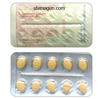
Erectafil dosages: 20 mg
Erectafil packs: 10 pills, 20 pills, 30 pills, 60 pills, 90 pills, 120 pills, 180 pills, 270 pills, 360 pills

Cheap erectafil 20mg with mastercard
This loss of myelin ends in an interruption of the propagation of the motion potential down these axons impotence synonym purchase erectafil 20 mg with visa. The specific array of motor impotence natural supplements safe 20mg erectafil, visual erectile dysfunction caused by fatigue generic erectafil 20 mg mastercard, or general sensory losses in a affected person with multiple sclerosis displays the places of the demyelinating lesions. Metabolic activation of microglia may be much more necessary to their functioning than the dramatic shape changes noticed pathologically. As immune cells, microglia can be stimulated to secrete cytokines, similar to interleukins and tumor necrosis factor-, and different immune mediators, corresponding to arachidonic acid derivatives prostaglandin E2 and plateletactivating issue. Like macrophages, in addition they secrete development components, for instance, brain-derived neurotrophic factor. The broad range of microglial products thus contains probably neurotoxic and neuroprotective mediators of irritation and tissue restore. The internet results of microglial activation can be beneficial and protecting, as in the case of synaptic stripping. This term is applied to a scenario during which the facial nerve (a motor nerve) has been minimize peripherally. Microglia then encompass the motor neuron cell our bodies positioned within the brainstem and displace or take away all synapses from the floor of the neurons. Once the peripheral axon has grown back and reconnected with its goal muscle tissue, new synapses kind around the neurons. When researchers and physicians prevented this secondary irritation by administering steroids before giving penicillin, microglial cytokine secretion was inhibited, and the survival rate for bacterial meningitis in kids vastly improved. In cases of trauma or extreme tissue damage, Primary mind tumors arise from the cells that make up the structure of the brain and spinal cord as properly as its coverings. Individual cells belonging to any of the cell populations found in mind tissue or the leptomeninges can provide rise to a brain tumor, provided genetic and environmental stimuli favor cell proliferation. Glia-Derived Tumors Glial cells are a frequent supply of major brain tumors in adults and children, and of those, astrocytomas are the glial tumors encountered most often. An analysis of astrocytomas is traditionally made on the idea of how intently or how little the neoplastic cells resemble nonneoplastic astrocytes (degree of differentiation), and this helps predict the end result for the affected person. Grade 1: the density of astrocytes, cells with vesicular chromatin and stubby processes, is increased in white matter in contrast with that in gray matter (A, evaluate upper right with lower left). These enlarged astrocytes have homogeneous nuclei (E, arrows), nucleoli, conspicuous cytoplasmic bodies, and stellate processes (E). Grade 2: Astrocyte nuclei vary in shape; the staining intensity and cell density are elevated (B). Grade three: Heterogeneity of astrocyte size and form (pleomorphism) is more obvious (C). Cells with enlarged nuclei or with two nuclei (binucleate astrocytes) are current. Abnormal tripolar mitotic figures and mitotic figures on spindles (C, inset) signify a heightened stage of cell proliferation. Grade four: Spindleshaped, oval, elongated, and curved nuclei are abundant, indicating the extreme pleomorphism of this grade, which is also called glioblastoma multiforme (D). Large, complicated vascular structures with clusters of cells surrounding the lumina (D, arrows) are characteristic of glioblastoma. Another feature is the sharp border and the transitional zone between the stay tumor and the necrotic zone (F). On occasion, protoplasmic astrocytes, denizens of gray matter with fewer processes, might kind tumors that include fluid-filled cysts. However, if they recur after surgery, these astrocytomas could turn into extra aggressive, remodeling into a higher grade. Grade 3 astrocytomas have nuclei which may be often enlarged, with elevated density of chromatin. The Cell Biology of Neurons and Glia 31 Grade four astrocytomas are extremely malignant tumors. The astrocytes of these tumors may be spindled, and the elongated nuclei may have many mitotic figures. They can invade the leptomeninges, spreading from one contiguous gyrus to its neighbor. Unfortunately, this "high-grade" tumor is the most common astrocytoma encountered in middle-aged and aged grownup patients. These cells can produce slow-growing tumors, situated within the lobes of the mind somewhat than within the diencephalon or in the basal nuclei. The oligodendroglia in these tumors have dark, spherical nuclei centered inside clear cytoplasm, very like the yolk of a fried egg embedded in egg white. Tumor cells kind sheets which are subdivided into geometric items by capillary twigs. Hallmarks of oligodendrogliomas are enlarged clusters around neurons (satellitosis) and nodules of tumor cells beneath the pia. The ventricular spaces of the brain and the central canal of the spinal twine are lined by an epithelium of ependymal cells. Tumors that stem from the final member of the glial household, microglia, have been first depicted as forming mobile aggregates around tiny arterioles. The fashionable interpretation is that these tumors are lymphomas, a large family of neoplasms consisting of bone marrow�derived cells (B lymphocytes) or thymusderived cells (T lymphocytes). Unlike the lymph nodes during which these tumors predominate, the brain has no lymphatics. In addition, genetic footprints of one other virus (Epstein-Barr virus) can incessantly be detected when molecular genetic strategies are applied to microscopic sections of lymphomas. Benign Primary Brain Tumors Benign main mind tumors are sometimes lined by a fibrous, vascularized capsule that discretely demarcates the tumor from surrounding normal mind. As a benign tumor enlarges, it pushes towards brain tissue, quite than extending finger-like projections that invade white and gray matter for great distances. Benign main mind tumors trigger issues by compressing regular tissue as they grow. Frequently encountered benign major tumors of the mind embrace meningioma (see Chapter 7) and schwannoma. Metastatic Brain Tumors Metastatic brain tumors arise from malignant cells that originate exterior the nervous system. Microscopic clumps of malignant cells break free from the preliminary growths and travel by way of the bloodstream to the mind. These cell aggregates turn out to be lodged at tiny arteriolar department points, regularly positioned on the junction of gray and white matter. Using intracellular enzymes that dissolve basement membranes, the malignant cells escape from the confines of the vasculature and begin to grow in the brain. Some of essentially the most prevalent malignant tumors that affect men and women frequently metastasize to the mind. Prostate carcinoma can unfold to the spinal wire by way of veins of the Batson venous plexus. Medulloblastomas come up in the cerebellar hemispheres of kids and consist of primitive "blue cells" that are able to growing along several pathways. The preliminary cells can mature into members of the glial family or into neurons, all of which may be found in the identical tumor.
Cheap 20mg erectafil free shipping
Collectively erectile dysfunction doctor kolkata cheap erectafil 20mg with visa, this group of language disorders are termed pericentral aphasias and occur with harm outside of language centers that have an result on projections from language centers to different areas of the mind erectile dysfunction is caused by generic 20mg erectafil with visa. The language deficits for pericentral aphasias are often comparable but less extreme than for central aphasias latest news erectile dysfunction treatment safe 20 mg erectafil. And not like central aphasias, where repetition capability is usually misplaced, with pericentral aphasias, repetition capability is typically maintained. Parietal Association Cortex: Space and Attention Conduction Aphasia and Global Aphasia the severity and length of the aphasia rely upon the severity of the related mind injury. In gentle instances, only one or two A fully totally different set of mental capabilities is mediated within the parietal association cortex of the nondominant hemisphere. Much of our knowledge of the useful properties of different regions of the cerebral cortex has been gained from neurologic case research of patients with cortical harm produced by stroke or head trauma. In this respect, the 2 nice wars of the primary half of the twentieth century led, inadvertently, to great progress in our understanding of the consequences of mind injuries. One of the 478 Systems Neurobiology the cat ran up the tree to catch a squirrel for his lunch. For example, the affected person could additionally be requested to learn a short passage and check off every word within the course of. Another method to demonstrate contralateral neglect is to draw a circle and ask the affected person to draw in the numbers of a clock face. A patient with contralateral neglect is probably not conscious of individuals standing to the left, may stumble upon large stationary objects on the left, and should not reply to sounds or phrases coming from the left. In excessive instances, the patient may not even recognize the left side of his or her own physique (asomatognosia). For instance, the patient may ignore the left aspect when dressing or grooming (dressing apraxia) or, if in a hospital, could even demand that the workers get this "different individual" (the left facet of his or her personal body) out of the mattress. Another characteristic group of signs of right parietal lobe lesions concerns the power to function efficiently within the spatial environment. Yet another problem is an incapability to efficiently manipulate objects in area. In addition, disorders of affect are frequent, including a decreased capability to understand and respect humor, a lack of the power to appreciate the prosody of speech, and infrequently an inappropriate cheerfulness and lack of concern for and even awareness of the implications of the illness. This lack of concern may be noted even when the deficit is as critical as complete left hemiplegia. Apraxia and Agnosia Damage in many areas of affiliation cortex can produce greater stage disorders of habits. Apraxia is a disorder of motor management which will occur after harm in parietal affiliation cortex, premotor cortex, or supplementary motor cortex. Nevertheless, the affected person is unable to coordinate his or her muscular tissues to execute complex habits. Apraxia of speech is a separate disorder from aphasia (described earlier), by which the internal processing of the symbols of language is impaired, thus affecting the understanding and manufacturing of spoken language, written language, and signal languages. Agnosia is a general time period used to describe a big group of higher-level issues of sensory notion. The term "agnosia" is derived from two Greek terms that imply a "lack of knowledge. For instance, an lack of ability to recognize a well-known object by sight is visual agnosia, whereas the shortcoming to recognize noises or sounds is auditory agnosia. This sort of deficit additionally extends to the sense of scent (olfactory agnosia), the sense of taste (gustatory agnosia), and even the inability to establish colours (color agnosia). They are generally produced by harm in modality-specific areas of the sensory association cortex. John Harlow, one of many physicians who attended Gage, perceived the importance of this case with respect to the localization of intellectual capabilities in the mind. The equilibrium or balance, so to communicate, between his mental faculties and animal propensities seems to have been destroyed. In this regard his mind was radically changed, so decidedly that his associates and acquaintances stated that he was "not Gage. Patients with significant bilateral damage to the prefrontal cortex have a constellation of deficits that might be summarized as follows. Fourth, the patient with prefrontal harm displays a profound lack of ambition, a loss of the sense of accountability, and a loss of a way of social propriety. The first and third signs (distractibility versus perseveration) are obviously in conflict. The individual is as an alternative imprisoned in a chaotic world, along with his or her actions governed by randomly altering whims. It was this set of symptoms that prompted the Portuguese neurosurgeon Egas Moniz to develop the prefrontal lobotomy procedure in the late 1930s to deal with a variety of extreme, intractable mental issues. At that point, psychological hospitals ("insane asylums") all round the world contained many sufferers who were so immobilized by nervousness that they may not even care for their very own bodily wants. The discovery that a neurosurgical process may alleviate the anxiousness to the extent that the patients could lead a somewhat more normal existence (albeit nonetheless within the confines of a psychological institution) was hailed as a great breakthrough. In these determined patients, the signs as described before appeared a justifiable price to pay for freedom from the crushing nervousness that had immobilized them. The discovery of tranquilizers in the late Nineteen Fifties provided a more effective technique of remedy, having fewer undesirable unwanted facet effects, and prefrontal lobotomy was quickly deserted as a way of therapy. Prefrontal Cortex and Plans for Future Operation the opposite main area of multimodal affiliation cortex is the massive expanse anterior to the primary motor and premotor cortices, the prefrontal affiliation cortex. This region has historically been connected with a few of the most distinctly human mental traits, such as judgment, foresight, a sense of function, a sense of responsibility, and a way of social propriety. One of the earliest accounts of the impact of brain harm on greater intellectual capabilities described a sequence of events that started on September thirteen, 1848. A crew of railroad building workers was blasting a right-of-way by way of the rugged granite mountains of Vermont. The well-liked younger foreman of the crew, Phineas Gage, was in cost of placing a black powder charge in a deep gap drilled in the rock, adding a fuse, covering the powder with sand, and at last tamping the sand and powder down firmly with an iron rod before lighting the fuse and running for cover. On this present day, one thing apparently distracted Gage, and he started to tamp down a cost before the sand had been added. The rod struck Gage just beneath the left eye and exited by way of the highest of his head, destroying most of his prefrontal cortex. Amazingly, Gage was not killed immediately, and much more extremely, he survived the inevitable serious wound infection that adopted. The return of Phineas Gage: clues in regards to the brain from the skull of a famous affected person. Varieties and distribution of non-pyramidal cells in the somatic sensory cortex of the squirrel monkey. Corticocortical networks and corticosubcortical loops for the higher management of eye motion. Elementary processes in chosen thalamic and cortical subsystems-the structural substrates. Now that many aspects of practical systems neurobiology have been mastered, the chance to apply this data is at hand. The neurologic examination is an excellent instance of how basic neuroscience can apply directly to occasions (both normal and abnormal) encountered within the medical setting.

Generic 20mg erectafil free shipping
For example erectile dysfunction doctor tampa discount erectafil 20 mg mastercard, an X cell may have a yellow-responsive middle and a blue-responsive encompass erectile dysfunction types purchase erectafil 20 mg with mastercard. These cells are also known as P cells because in humans and different primates erectile dysfunction 21 years old discount erectafil 20 mg free shipping, they consistently hook up with other, smaller cells within the parvocellular layers in the lateral geniculate nucleus. All ganglion cells ignored of the preceding two classes are classified anatomically as gamma, delta, and epsilon cells and physiologically as W cells. These cells tend to have smaller cell bodies and axons, and they show quite so much of receptive field sizes and physiologic responses. The superior colliculus, in flip, initiatives to the pulvinar, the largest nucleus of the thalamus. The pulvinar receives enter from the superior colliculus, pretectum, and visible cortex (see later) and sends data to visible affiliation areas. This temporary overview illustrates the purpose that the visible system influences a variety of areas of the brain. Lesions of the retina or to the optic nerve lead to visible deficits attribute of their location. Scotoma could additionally be manifested as a randomly formed lesion in a single or each visible fields in which imaginative and prescient could additionally be decreased or absent surrounded by areas of normal imaginative and prescient. A scotoma may be symmetric or asymmetric and unilateral or bilateral; it may be manifested in a extensive variety of shapes (annular, ring, sickle) and locations (central, peripheral, paracentral) throughout the retina. Causes of scotoma are varied and include publicity to toxins, retinal hemorrhage, trauma, and tumors that impinge on the eyeball. The most common deficits related to injury to the optic nerve are partial or full blindness in that eye and the potential lack of the direct and consensual pupillary mild reflex when gentle is shined in that eye. The blind eye may have a consensual response when a light is shined within the reverse eye as a end result of the efferent limb of the reflex is unbroken in the blind eye. Among the targets are the suprachiasmatic nucleus, a hypothalamic area that controls diurnal rhythms (see Chapter 30); the accent optic and olivary Most retinal ganglion cells send their axons to the lateral geniculate nucleus by method of the optic nerve, chiasm, and tract. A commonly used various time period for this pathway is the geniculocalcarine radiations. In this pathway, an the Visual System 295 orderly map of visual space must be maintained. This orderly illustration of the visual world on the retina known as a retinotopic map. It consists of a binocular zone-a broad central region seen by both eyes-and proper and left monocular zones (or monocular crescents) seen only by the corresponding eye. The light ray is bent (refracted) by the cornea and lens so that the image is targeted on the retina. These patterns are important to understanding regular vision and the defects in visible fields seen in sufferers with lesions within the visual pathways. However, as they cross via the sclera, they turn into ensheathed with myelin fashioned by oligodendrocytes. Thus the subarachnoid house extends along the optic nerve, which is bathed in cerebrospinal fluid. For this cause, will increase in intracranial pressure could additionally be transmitted alongside the optic nerves and can trigger blockage of axoplasmic circulate at the optic nerve head. Terminal branches of the central retinal artery, a branch of the ophthalmic artery, issue from the optic disc and radiate over the retina. Changes in the configuration of the retinal vessels or in the dimension or form of the optic disc could indicate illnesses of the retina, the vascular system, or the central nervous system. Just rostral to the pituitary stalk, the optic nerves come collectively to type the optic chiasm, from which the optic tracts diverge as they move caudally. Visual acuity is sharpest within the fovea (20/20) and drops precipitously within the outer parts of the retina (to 20/600). This drop correlates with a lower density of photoreceptors and ganglion cells in the peripheral regions of the retina. A detached retina in the lower part of the attention ends in an irregular defect within the higher visible field (left), whereas an irregular lesion of the macula or compression of the optic nerve produces a central scotoma (area of reduced vision) in the center of the visible field (right). D, Increased intracranial stress could produce a "choked disc" (papilledema in the right eye), which is swelling of the optic nerve head visible via an ophthalmoscope. Blood vessels emerge from the optic disc, the sunshine area in the middle of the photograph. A lesion that damages the lateral a part of the chiasm might interrupt solely fibers conveying information from the nasal visible subject on the same facet, although in apply this situation is uncommon. It is extra probably that this kind of defect (a proper or left hemianopia) may actually be the results of a central scotoma; an ophthalmologic examination would make clear this issue. Extending caudolaterally from the chiasm, the axons of retinal ganglion cells continue as a compact bundle, the optic tract. Some of those radiations course directly to the visible cortex; others loop rostrally adjacent to tip of the temporal horn (the Meyer-Archambault loop), then flip caudally to terminate in the visible cortex. The optic chiasm receives blood from the small anteromedial branches of the anterior speaking artery and A1 phase of the anterior cerebral artery. The optic nerve receives its blood provide from small branches of the ophthalmic artery traveling parallel to the nerve. As noted beforehand, the optic nerve head and retina are supplied by the central artery of the retina. The observer draws the diagrams as in the event that they have been on the wall the patient is taking a glance at. Representative visible system lesions and the corresponding visual area deficits ensuing from such lesions illustrate these concepts. The gray-shaded area within the visible area of the left eye (A) signifies the extent of the visible deficit. The lesion transects crossing retinal axons from both the left and proper optic nerves (C) and ends in temporal visible field deficits in each eyes (D). The visible loss is clear within the temporal half of the visual subject in every eye and leaves the nasal visual fields of every eye unaffected. In this case, as a result of the deficit entails the temporal one-half of the field of every eye, the deficit is called bitemporal hemianopia. The ensuing visual subject hemideficits are indicated by the gray-shaded areas of the visual area of the left and right eye. Notice that in this case the deficits involve the best half of the visible area of every eye. Because the same (right) or corresponding halves of the visible area of each eye are affected, the deficit is known as a homonymous hemianopia. Note that a dorsal view of the mind and visible pathways is used on this illustration.


Order 20 mg erectafil fast delivery
Adherens junctions contribute to cell motility and form by way of hyperlinks to inside cytoskeletal elements such as actin erectile dysfunction caused by steroids cheap 20 mg erectafil free shipping. Important components of those constructions within the keratinocyte include the transmembrane hyperlink drugs for erectile dysfunction ppt order 20mg erectafil overnight delivery, E-cadherin erectile dysfunction drugs lloyds generic erectafil 20 mg free shipping, and the intracellular actin-binding -catenin. Gap junctions are fashioned from aggregation of connexins into connexons that be part of adjoining cells to allow intercellular transport of ions and other small molecules. The keratinocyte life cycle as described earlier ranges from 5 to 30 days, relying on the anatomic web site and state of well being of the skin. A few specialised cell types current in the pores and skin deserve special point out with respect to perform of the skin as an organ. Melanocytes the melanocyte offers pigmentation to the pores and skin or fur needed for behavioral features of survival similar to camouflage in sure species. In addition to their presence within the epidermis, melanocytes are also found in the eye, cochlea of the ear, the brain/meninges, and the heart. Melanin can even bind certain potentially dangerous compounds similar to cations and metals. Each melanocyte "serves" about 30�40 keratinocytes within the basal layers of the epidermis. Melanosomes are organelles derived from the endoplasmic reticulum of melanocytes and serve as packages for switch of melanin. Although the number of melanocytes stays constant, the manufacturing and transfer of melanosomes to keratinocytes may be up- or downregulated. In healthy pores and skin, these cells are comparatively inactive and even act to attenuate inflammatory response. They are able to preferentially reply to particular or extreme international antigens, thereby stopping continual upregulation of inflammatory mediators whenever overseas antigens are sensed, which is almost fixed in the pores and skin. The dermis offers the pores and skin tensile power through collagen, contributes to motion by allowing stretch by way of elastic fibers, has immune regulatory capabilities, accommodates vascular and neurologic parts necessary for communication with the dermis and surroundings, and varieties the matrix for adnexa. In species missing epidermal rete ridges, these subregions are normally simply described as superficial and deep, respectively. The main parts of the dermis are collagens and elastins, conferring tensile energy, and proteoglycans, glycosaminoglycans, and hyaluronans, which help dissipate stress forces. They represent a cell type unique to the pores and skin and oral mucosa, able to appearing as mechanoreceptors with dense granules containing neurotransmitter-like mediators. The dendrites of the Merkel cells contact unmyelinated axons in the epidermis, where they function collectively as a unit (tylotrich pads) to signal adnexal secretions (sweat), adjustments in blood circulate, tactile sensation, and probably serve in a paracrine function together with different cells of the skin. Motor innervation is from sympathetic autonomic nerves and responds to either cholinergic or adrenergic stimuli. Adrenergic responses embody vasoconstriction, apocrine gland secretion, and hair follicle positioning. Eccrine sweat glands are underneath cholinergic management; however, these glands are largely absent in most areas of nonhuman mammalian pores and skin. Superficial, center, and deep vasculature plexuses provide corresponding layers of the pores and skin. Arteriovenous anastomoses are common in the pores and skin of the extremities particularly, essential for adaptation to frequent modifications in temperature and blood pressure. These junctions reply to vasoconstrictors and vasodilators corresponding to epinephrine and histamine. Many vessels of the skin have comparatively thick walls in comparison with those of comparable measurement in inside organs, a necessary adaptation for protection against the shear and strain forces to which the pores and skin is regularly subjected. Also current within the dermis are the fibroblast-like veil cells that define spaces for vessels within the dermis, surrounding microvasculature, and making a perivascular area. Cutaneous lymphatics arise as capillaries within the superficial dermis, however under the level of superficial blood capillaries. Gaps throughout the lymphatic vessels form channels by linking with the dermal matrix, directing extra interstitial fluids from the dermis into the lymphatic system. Lacking smooth muscle and pericytes, lymphatics of the dermis rely on subcutaneous muscle contraction, pressure of the surrounding matrix, and associated blood vessel movements to initiate circulate of immunologic, waste, and even degraded pathogenic and xenobiotic supplies away from the dermal interstitium. In the deeper subcutis, lymphatics have smooth muscle walls and are actively contractile directly directing move of lymph to and from the skin. The Adnexa the cutaneous adnexal unit refers to a hair follicle, the related arrector pili muscle, and related glands. The hair shaft consists of an internal medulla, a surrounding cortex, and outer cuticle. The hair follicle has subanatomic regions often known as the infundibulum (from the floor epidermis to the sebaceous gland duct entrance to the hair follicle), the isthmus (just beneath the infundibulum, from sebaceous duct to the arrector pili muscle insertion), and the inferior section from the isthmus deep to the dermal papilla. The outer root sheath that accommodates the hair is contiguous with the surface dermis. The inner root sheath is attached to the cuticle of the hair and is distinguishable in tissue sections by the presence of eosinophilic cytoplasmic trichohyalin granules. In people and mice, a structure known as "the bulge" is current and connected to the outer root sheath, close to the insertion of the arrector pili muscle. The bulge supplies the adnexal unit and, in some cases the surface epidermis. The bulge area may be absent in different species, with stem cells being present in infundibular and isthmus areas of the follicle itself, though some analysis does help bulge cell presence within the dog. Hair growth is split into the following levels: anagen (active growth), catagen (transition phase), telogen (resting stage), and exogen (shedding). During hair progress, an ordered array of keratinized cells is progressively pushed upward within the form of hair shafts. These cells give rise to hair by a strategy of terminal differentiation, analogous to , however extra complicated than, the method described for epidermal cornification. Keratinization of hair follicle cells is of 4 morphologic subtypes: infundibular (like the floor dermis, that includes keratohyalin), trichilemmal (important in identification of catagen hairs, with dense eosinophilic keratin "flames"), trichogenic/matrical ("ghost cell" keratinization), and medullary/inner root sheath (with deeply eosinophilic trichohyalin granules). The Subcutis the subcutis is the adipose-rich tissue beneath the dermis answerable for attachment to underlying muscle, fascia, or periosteum. Connective tissue septa present throughout the subcutis facilitate movement and assist dense vessel and nerve networks in the tissue. This layer additionally serves to take up shock to underlying structures, form the exterior options of the organism, and regulate temperature. The rich triglyceride stores of the subcutis can be utilized as an vitality store and also serve to shield underlying tissues from temperature extremes. Visceral and subcutaneous adipose stores are also important in the secretion and concentrating on of assorted hormones and cytokines. Zone of dividing cells of the hair matrix, comparable with the stratum basale of the epidermis.

Buy 20mg erectafil mastercard
Synthesis of keratin in any of its types is an almost unique characteristic of epithelial cells impotence 25 years old buy 20 mg erectafil otc. Desmosomes are formed largely from desmogleins and desmocollins and characterize the primary technique of cell�cell adhesion in nucleated epidermal layers erectile dysfunction medications causes symptoms purchase 20 mg erectafil visa. Layers of the dermis embrace the stratum corneum (sc) impotence group purchase 20mg erectafil, stratum granulosum (sg), stratum spinosum (ss), and stratum basale (sb) consisting of basal cells (bc) adherent to the basement membrane (bm). In the dermis, fibroblasts (f), dermal vessels (v), perivascular mast cells (pmc), and dendritic cells (dendrocytes, dc) are pictured. This zone incorporates melanocytes that give shade to the hair by passing melanin to the matrix cells. Eccrine glands participate in thermoregulation by secreting water and salts directly to the epidermal surface. Abundant all through the skin in nice apes and humans, in home and laboratory animals, the eccrine sweat glands are largely restricted to the foot pads in canine, and the nasal planum and carpus of pigs. These are merocrine glands composed of a long-coiled secretory tubule and a connecting lengthy excretory duct that ends within the epidermis separate from the hair follicle; the epithelium lining each is straightforward columnar. While distributed typically over the skin of most species, apocrine glands in humans are restricted to armpits, groin, and nipples. They kind sebum, comprised of wax esters, squalene, free fatty acids, and triglycerides. Evaluation of Toxicity Physiologic and Morphologic Safety Evaluation Strategies and Techniques Evaluation of cutaneous toxicity is crucial for any therapeutic agents intended for use by topical administration. In addition, evaluation of potential adverse effects on the skin is necessary for therapeutic compounds that unintentionally come into contact with the pores and skin. These guidances also extend to the various excipients used in preparation of formulations. In vivo topical toxicity testing methods have focused on assessing skin irritation, cutaneous sensitization, ocular toxicity, and photosafety testing in various animal species and strains. The animal mannequin chosen and kind of protocol used will depend upon the target of toxicity take a look at. Unlike many different main organs, there are very distinct variations in pores and skin construction amongst laboratory animal species and between these species and humans. Consequently, comparative analysis of pores and skin toxicity could be difficult to accomplish. The rabbit has been used for many years as the animal mannequin of choice for evaluation of topical irritation potential. Although an intensive historical database exists for dermal irritation within the rabbit, this animal model has been proven to be more delicate to major irritants than human skin. Most species used commonly in toxicity evaluations (mice, rabbits, rats, guinea pigs, dogs, and nonhuman primates) have comparatively dense fur overlaying much of their bodies, and as such serve as poor comparators to human skin. The minipig has turn out to be the animal mannequin of choice for assessing dermal irritation and tolerability of topical compounds on the idea of the higher similarity of morphologic and physiologic traits of pig pores and skin to human pores and skin. There are a number of breeds of laboratory minipig, but some of the commonly utilized for regulatory toxicology is the Gottingen, based on its � small measurement (about 45 kg as an adult). Both humans and pigs have a relatively thick dermis with a big elastic fiber element. Also much like people, minipig pores and skin thickens and has increased permeability with lowered effectiveness at wound healing with age. Enzymatic properties and drug metabolism within the dermis and some adnexa also are comparable, as are lipid composition of the dermis and sebum. There are even spontaneous fashions of human pores and skin disease corresponding to melanoma and bullous pemphigoid that exist in minipig strains (the Sinclair and Yucatan, respectively). There are additionally some differences in the enzymatic profile of the skin, within the distribution of eccrine and apocrine sweat glands, and in regulation of exogen as properly. Still the minipig seems to be the species best suited to comparative toxicology of the pores and skin. In addition, other porcine fashions, in particular the Duroc/Yorkshire mannequin, are considered one of the best animal models for recreating human wounds. However, for sure evaluations, traditional laboratory animal models are nonetheless necessary. For example, the guinea pig is the animal mannequin of selection for assessing allergic contact dermatitis potential of chemical compounds. The route of administration chosen in assessing the security of topically applied compounds will depend on the tip point of the evaluation. Similar to therapeutic brokers intended for oral or parenteral administration, in vivo toxicity testing must be evaluated in each rodent and nonrodent species. The rat is the rodent species of selection for analysis of potential systemic toxicity of topical compounds, whereas the minipig is recommended because the nonrodent species. For compounds supposed for topical software, there have been quite a few initiatives to scale back animal use in topical toxicity testing. Assessment of Cutaneous Irritation Evaluation of the irritation potential of topically utilized compounds has utilized animal fashions of skin irritation. The Draize scale approach has been generally utilized for quantitatively figuring out the degree of irritation caused by topical software of compounds and for providing a quantitative measure of comparability and differentiation of the irritation potential of varied compounds. Human pores and skin irritancy evaluation is often required to complement animal irritancy testing in order to extra precisely understand human threat (Table 24. Photoactivation of a chemical may end in adverse effects termed photosensitivity reactions. Photosafety testing is intended to determine agents with photosensitivity potential. Both in vitro and in vivo assays have been developed to assess the photosensitivity potential of photoreactive chemical compounds. Key issues in the evaluation of photosensitivity potential in these guidances are: photoirritation, photoallergenicity, photogenotoxicity, photocarcinogenicity, and photococarcinogenicity. In vivo phototoxicity testing could also be carried out in guinea pigs, rabbits, hairless mice, or hairless guinea pigs. After a time period to enable for absorption of the check article, the Draize scale is used to decide the phototoxic response by grading of irritation potential. In a review of concordance of toxicity of prescribed drugs in humans and animals, phototoxicity response in guinea pigs correlated well with that in humans. The Buehler Guinea Pig Sensitization assay (with publicity to simulated light) is most well-liked over the Guinea Pig Maximization Assay, which though thought of extra delicate than the Buehler assay, is related to subcutaneous reactions attributed to the utilization of adjuvant in this assay. However, photocarcinogenicity testing is most likely not needed for compounds that are photoirritants if a warning is provided in patient information. Photococarcinogenic potential should even be considered for chemical compounds that is most likely not photoreactive however might affect carcinogenicity through immunosuppressive results.
Buy erectafil 20 mg otc
Immune responses to gene therapy vectors: influence on vector operate and effector mechanisms impotence grounds for annulment philippines generic erectafil 20mg. Cellular innate immunity and restriction of viral infection: implications for lentiviral gene remedy in human hematopoietic cells impotence urban dictionary order 20mg erectafil visa. The inflammasome: a molecular platform triggering activation of inflammatory caspases and processing of proIl-beta erectile dysfunction gabapentin discount erectafil 20 mg with amex. The evolution of adenoviral vectors through genetic and chemical surface modifications. Bone morphogenetic protein-2 nonviral gene remedy in a goat iliac crest model for bone formation. In vivo endochondral bone formation using a bone morphogenetic protein 2 adenoviral vector. Adenovirus-mediated direct gene remedy with bone morphogenetic protein-2 produces bone. Human bone morphogenetic protein 2-transduced mesenchymal stem cells improve bone regeneration in a model of mandible distraction surgery. Rapid and reliable healing of critical size bone defects with genetically modified sheep muscle. Bone morphogenetic proteins four and 2/7 induce osteogenic differentiation of mouse skin derived fibroblast and dermal papilla cells. Regeneration of bone- and tendon/ligament-like tissues induced by gene transfer of bone morphogenetic protein-12 in a rat bone defect. Cellular and molecular mechanisms of accelerated fracture healing by CoX2 gene remedy: research in a mouse model of multiple fractures. Microarray evaluation of gene expression reveals that cyclo-oxygenase-2 gene remedy up-regulates hematopoiesis and down-regulates inflammation during endochondral bone fracture healing. Marrow stromal cell-based cyclooxygenase 2 ex vivo genetransfer strategy surprisingly lacks bone-regeneration effects and suppresses the bone-regeneration motion of bone morphogenetic protein 4 in a mouse critical-sized calvarial defect model. Repair of critical-sized bone defects with anti-miR-31-expressing bone marrow stromal stem cells and poly(glycerol sebacate) scaffolds. Adeno-associated virus-mediated osteoprotegerin gene transfer protects against particulate polyethylene-induced osteolysis in a murine mannequin. Adenoviral vector-mediated overexpression of osteoprotegerin accelerates osteointegration of titanium implants in ovariectomized rats. Role of sclerostin in bone and cartilage and its potential as a therapeutic goal in bone ailments. Validation in mesenchymal progenitor cells of a mutation-independent ex vivo strategy to gene therapy for osteogenesis imperfecta. Marrow stromal cells as a source of progenitor cells for nonhematopoietic tissues in transgenic mice with a phenotype of osteogenesis imperfecta. Treatment of osteoarthritis using a helper-dependent adenoviral vector retargeted to chondrocytes. Demineralized bone matrix combined bone marrow mesenchymal stem cells, bone morphogenetic protein-2 and transforming growth factor-beta3 gene promoted pig cartilage defect restore. Acceleration of articular cartilage repair by mixed gene transfer of human insulin-like growth factor I and fibroblast progress factor-2 in vivo. Nanoparticle delivery of the bone morphogenetic protein 4 gene to adipose-derived stem cells promotes articular cartilage repair in vitro and in vivo. Direct bone morphogenetic protein 2 and Indian hedgehog gene transfer for articular cartilage repair using bone marrow coagulates. Adenovirus-mediated gene switch of insulin-like development factor 1 stimulates proteoglycan synthesis in rabbit joints. Transformation from a neuroprotective to a neurotoxic microglial phenotype in a mouse mannequin of Als. Disease-regulated local Il-10 gene therapy diminishes synovitis and cartilage proteoglycan depletion in experimental arthritis. Intraarticular expression of biologically active interleukin 1-receptor-antagonist protein by ex vivo gene transfer. Il-37 alleviates rheumatoid arthritis by suppressing Il-17 and Il-17-triggering cytokine manufacturing and limiting Th17 cell proliferation. Adeno-associated virus-mediated osteoprotegerin gene switch protects against joint destruction in a collagen-induced arthritis rat model. Direct lentiviral-cyclooxygenase 2 application to the tendon-bone interface promotes osteointegration and enhances return of the pull-out tensile strength of the tendon graft in a rat model of biceps tenodesis. Bone marrow-derived mesenchymal stem cells transduced with scleraxis improve rotator cuff healing in a rat model. Co-transfection of adeno-associated virus-mediated human vascular endothelial progress factor165 and transforming development factor-1 into annulus fibrosus cells of rabbit degenerative intervertebral discs. The disease impacts up to 40% postmenopausal women and 15% elderly men of Caucasian background. Osteoporosis provides an interesting case for the appliance of genetics in threat prediction, because the susceptibility to the illness is set by genetic Genetics of Bone Biology and Skeletal Disease. Moreover, osteoporosis is a condition characterized by a quantity of phenotypes, including worsened bone energy and structure, and in the end fragility fracture. Currently, there are multiple remedies and combination of remedies obtainable for lowering bone loss and decreasing fracture threat. The efficacy and clinical outcome of current therapies are extremely variable among patients. Pharmacogenetics and pharmacogenomics provide the possibility to individualize fracture prognosis and remedy. Pharmacogenetics refers to the science of how genetic components have an effect on the interindividual variation in drug efficacy and security. In actuality, most pharmacogenetic studies give consideration to single genes and their associations with interindividual variations in drug behaviors, while pharmacogenomics is concerned with the genomic interactions amongst genes within the overall variation in drug metabolism and response. Both pharmacogenetics and pharmacogenomics have important applications in studies of the pathogenesis and therapy of osteoporosis. Fracture is a direct consequence of bone fragility and is therefore a key component of an osteoporosis phenotype. However, fracture is a dichotomous occasion, resulted from cumulative deterioration in a quantity of tissues, that are in flip linked to genetic and nongenetic elements. Moreover, fracture is age-dependent such that the risk of fracture increases exponentially with advancing age. Theoretically, if the life expectancy of a population had been infinite, then the lifetime danger of fracture could be 100 percent. Fracture itself is associated with a number of threat factors, a few of which may be causal elements. Pooling the results from all research (women and men) and for all fracture websites, the risk of subsequent fracture among these with a previous fracture at any web site is 2. Biochemical markers of bone remodeling can additionally be thought-about phenotypes of osteoporosis. Bone mass is the net results of two counteracting processes of bone resorption and bone formation, typically referred to as bone remodeling. Bone remodeling is a normal, pure course of that maintains skeletal power, allows restore of bone injury, and is essential for calcium homeostasis.
Cheap erectafil 20mg on line
Reduced inhibition of this excitatory projection results in elevated cardiac output and increased resistance in vascular beds of skeletal muscle and stomach visceral organs however not of the skin erectile dysfunction vasectomy cheap erectafil 20mg otc, coronary heart herbal erectile dysfunction pills review purchase erectafil 20 mg visa, and mind erectile dysfunction treatment new zealand generic erectafil 20 mg without a prescription. Some of this solitary input to vasopressor neurons may be relayed through vasodepressor cell groups within the caudal ventrolateral medulla. Thus when a person strikes from a reclining to a standing posture, the resultant pooling of blood in the decrease half of the body is quickly countered by increased vascular tone and increased cardiac Visceral Motor Pathways 441 output. Without this reflex, movement to a standing place leads to dizziness or fainting (reflex syncope) because of decreased blood flow to the brain. The chemoreceptor reflex maintains homeostasis of blood gasoline composition by adjusting respiration, cardiac output, and peripheral blood flow. Decreased partial strain of oxygen (Po2) and elevated partial stress of carbon dioxide (Pco2), detected by receptors within the carotid and aortic bodies, are signaled by glossopharyngeal and vagal afferents that terminate in the solitary nucleus. Within the medulla, the reflex pathway for cardiovascular effects parallels that for the baroreceptor reflex. A lower in blood Po2 activates this reflex and promotes increased heart fee and vascular tone. These adjustments result in a decreased blood circulate to skeletal muscular tissues and viscera, whereas blood move to the brain is maintained. Thus proportionately more oxygenated blood is available to the brain than to skeletal muscle and viscera. The cardiovascular component of the chemoreceptor reflex is carefully coordinated with respiration, a somatic motor perform coordinated by other neurons of the brainstem reticular formation. For instance, if breathing is suspended (as in diving), heart price is slowed (bradycardia) quite than accelerated (tachycardia). Contraction of the detrusor and inhibition of the inner sphincter are mediated by parasympathetic outflow. The exterior urethral sphincter, which is subject to each reflex and voluntary management, is provided by alpha motor neurons in spinal cord segments S3 and S4. The low exercise of the sensory neurons leads to (1) low exercise of parasympathetic excitatory innervation to the detrusor and inhibitory innervation of the interior sphincter, (2) tonic activity of sympathetic neurons that inhibit both the parasympathetic ganglion cells in the bladder wall and the detrusor muscle directly, and (3) tonic exercise of sacral somatic motor neurons mediating constriction of the external sphincter. As urine accumulates, stress on the bladder wall prompts rigidity receptors until bladder afferent exercise rises to a threshold degree. This increased exercise of bladder afferents induces micturition by way of both spinal and brainstem reflexes that lead to inhibition of sympathetic outflow, activation of parasympathetic outflow, and inhibition of somatic motor neurons supplying the external sphincter muscle. Dysfunction of the bladder is a common consequence of dysautonomias and may manifest variably as urgency, incontinence, or retention. Autonomic issues that promote urinary retention are related to elevated incidence of urinary tract infections, which may trigger irritation of the afferent innervation of the bladder lining and consequent activation of the micturition reflex. Thus the contraction of the detrusor muscle can induce a robust urge to void the bladder even when urine quantity is low. Causalgia and reflex sympathetic dystrophy: does the sympathetic nervous system contribute to the technology of ache Neuropeptides within the sympathetic system: presence, plasticity, modulation, and implications. Cardiovascular penalties of loss of supraspinal management of the sympathetic nervous system after spinal wire damage. Axon transport and neuropathy: related views on the etiopathogenesis of familial dysautonomia. Physiology and pathophysiology of the interstitial cell of Cajal: from bench to bedside: I. Perkins brain volume) is dwarfed in size by the the rest of the mind (weighing roughly 1300 to 1400 g). The lamina terminalis separates the hypothalamus from the more rostrally located septal nuclei. Medially, the hypothalamus is bordered by the inferior portion of the third ventricle. Externally, the boundary between the hypothalamus and the midbrain is represented by the caudal edge of the mammillary physique. This is an especially good landmark to use when viewing a sagittal magnetic resonance picture within the prognosis of hypothalamic lesions. In addition to its role in regulating visceromotor functions, the hypothalamus, through a wide range of circuits, influences circadian rhythms, neurohormones, reproductive functions, common homeostasis, and behavior. Although its major position is within the upkeep of homeostasis, the hypothalamus partially regulates numerous features, including water and electrolyte stability, meals intake, temperature, blood pressure, possibly the sleep-waking mechanism, circadian rhythmicity, and common physique metabolism. The infundibulum is positioned immediately caudal to the optic chiasm, is somewhat funnel formed (hence its name), and incorporates a small portion of the third ventricle, the infundibular recess. The smaller parts of the pituitary, the tuberal half (pars tuberalis) and the intermediate half (pars intermedia), also originate in affiliation with the anterior lobe. In addition, the extension of the hypophysial stalk and infundibulum via the diaphragma sellae is a susceptible relationship. For instance, trauma to the top could lead to a shearing of the stalk and the eventual growth of diabetes insipidus. The preoptic space is a transition region that extends rostrally, by passing laterally to the lamina terminalis, to form a continuation with constructions in the basal forebrain. The thin periventricular zone is the most medial and is subjacent to the ependymal cells that line the third ventricle. Cross sections of the hypothalamus via preoptic (B), tuberal (C), and mammillary (D) regions. The myelin-stained sections in B, C, and D correspond to the comparably labeled strains in A. Preoptic Area the preoptic space, although functionally a half of the hypothalamus (and diencephalon), is embryologically derived from the telencephalon. Because gonadotropin release is steady in males and cyclic in females, the medial preoptic nucleus of males tends to be more lively and consequently bigger than that of females. Accordingly, the medial Inferior side of developing diencephalon Infundibulum Tuber cinereum Infundibular recess Optic chiasm Infundibulum Pars tuberalis Hypophysial stalk Posterior lobe Pars intermedia Anterior lobe Rathke pouch Stomodeum preoptic nucleus is often referred to because the sexually dimorphic nucleus of the preoptic space. The medial preoptic nucleus additionally influences behaviors which may be related to consuming, reproductive activities, and locomotion. However, by way of its connections with the ventral pallidum, it might operate partially in locomotor regulation. Some investigators think about the nuclei of the preoptic area to be part of the supraoptic region of the medial hypothalamic zone. This diffuse bundle of fibers traverses the lateral hypothalamic zone and interconnects the hypothalamus with rostral areas, such as the septal nuclei, and with caudal areas, such as the brainstem reticular formation. The lateral hypothalamic zone contains a large, diffuse population of neurons commonly known as the lateral hypothalamic area as well as smaller condensations of cells located in its anterior (ventral) parts. The latter cell teams are the lateral hypothalamic nucleus and the tuberal nuclei. The lateral hypothalamic nucleus is a unfastened aggregation of relatively giant cells that extends all through the rostrocaudal extent of the lateral hypothalamic zone. The tuberal nuclei consist of small clusters of neurons, each containing small, pale, multipolar cells.

Effective 20mg erectafil
It serves to coordinate the exercise of individual muscle teams to produce easy impotence exercises purchase erectafil 20 mg line, purposeful erectile dysfunction vacuum pump order 20 mg erectafil with mastercard, synergistic actions erectile dysfunction herbs generic erectafil 20mg fast delivery. This part of the mind is, fairly actually, the link between the brainstem and the forebrain. Ascending or descending pathways to or from the forebrain must traverse the midbrain. Other midbrain centers are involved with visual and auditory reflex pathways, motor function, transmission of ache, and visceral capabilities. Thalamus Pons and Cerebellum the pons and cerebellum originate embryologically from the same segment of the developing neural tube. We shall see later that the thalamus actually consists of several regions-for example, the hypothalamus, subthalamus, epithalamus, and dorsal thalamus. The thalamus is also generally known as the diencephalon, a time period that reflects its embryologic origin. The thalamus is rostral to the midbrain and almost utterly surrounded by components of the cerebral hemisphere. Individual elements of the thalamus could be seen in detail only when the mind is minimize in coronal or axial planes. This layer of cells is thrown into elevations or peaks called gyri (singular, gyrus) separated by valleys called sulci (singular, sulcus). The second main a part of the hemisphere is the subcortical white matter, which is made up of myelinated axons that carry info to or from the cerebral cortex. This bundle accommodates fibers passing to and from the cerebral cortex, corresponding to corticospinal and thalamocortical fibers. The third main part of the hemisphere is a distinguished group of neuronal cell our bodies collectively referred to as the basal nuclei. Parkinson disease, a neurologic dysfunction related to the basal nuclei, is characterized by a progressive impairment of actions and in many circumstances eventual dementia. The gyri and sulci that make up the cerebral cortex are named, and lots of are associated with particular functions. Some gyri obtain sensory input from thalamic relay nuclei, whereas descending fibers from these gyri could affect centers within the brainstem or spinal twine. The cerebral cortex additionally contains affiliation areas that are important for evaluation and cognitive thought. A practical system is a set of neurons linked together to convey a specific block of data or to accomplish a selected task. In this respect, systems and pathways, in some instances, could additionally be comparable, and their meanings may frequently overlap. The research of their structure and function, referred to as regional neurobiology, is the major target of the second part of this book. The system of neurons and axons that allows you to feel the sting of this page, for instance, crosses every area of the nervous system between your fingers and the somatosensory cortex of the cerebral hemisphere. The research of functional techniques, known as methods neurobiology, is the focus of the third section of this text. It is important to do not forget that the useful traits of regions coexist with these of techniques. Let us contemplate an instance of how the interrelation of systems and regions could be essential clinically. The signals that influence movements of the hand originate within the cerebral cortex. Neurons in the hand area of the motor cortex ship their axons to cervical ranges of the spinal wire, the place they influence spinal motor neurons that innervate the muscles of the higher extremity. These are called corticospinal fibers as a outcome of their cell bodies are in the cerebral cortex (cortico-) and their axons end within the spinal twine (-spinal). These fibers move by way of the subcortical white matter, the whole brainstem, and the upper levels of the cervical spinal cord. In the midbrain, for example, they move close to fibers of the oculomotor nerve, which originate in the midbrain and management sure extraocular muscular tissues. In the medulla, they cross close to fibers that originate in the medulla and innervate the musculature of the tongue. An damage to the midbrain may due to this fact cause motor issues in the hand (systems damage) combined with partial paralysis of eye motion (regional damage). In comparable trend, an injury to the medulla might trigger the identical hand downside but now in affiliation with partial paralysis of the tongue rather than of eye actions. With the exception of olfaction (sense of smell), all sensory info that eventually reaches the cerebral cortex should synapse within the thalamus. One perform of the thalamus, due to this fact, is to obtain sensory info of many types and to distribute it to the particular regions in the cerebral cortex which may be specialised to decode it. Other areas of the thalamus receive enter from pathways conveying information on, for example, place sense or the stress in a tendon or muscle. This enter is relayed to areas of the cerebral cortex that operate to generate smooth, purposeful actions. Through descending connections, the hypothalamus influences visceral centers within the brainstem and spinal twine. Fibers of the motor system that control hand movement descend from the motor cortex to the cervical spinal wire. In the twine, these fibers affect motor neurons that control hand and forearm muscle tissue. Injury at any level alongside the method in which can injury fibers of the system and structures particular to the region. For example, injury to the midbrain may harm both fibers to the hand and fibers to the eye muscular tissues, whereas harm to the medulla may harm both fibers to the hand and fibers to the tongue musculature. In circumstances of damage to a long tract (motor or sensory) and to a cranial nerve (root or nucleus), the cranial nerve deficits are usually the most effective localizing indicators. First, deficits (motor or sensory) positioned on the same aspect of the head and body incessantly signify lesions within the cerebral hemisphere. Second, deficits on one side of the pinnacle and on the alternative facet of the body typically point out a lesion within the brainstem. Although there are exceptions to these general guidelines, we will see that they hold true in many clinical conditions. Brain harm that results in only a weak spot or paralysis of the upper extremity generally localizes the lesion solely to one cerebral hemisphere or maybe to one aspect of the brainstem. However, if the paralysis of the upper extremity is coupled with a partial paralysis of eye motion, the lesion may be specifically localized to the midbrain. Another basic concept of localization states that sure mixtures of neurologic deficits might point out involvement the terms afferent and efferent are used to describe a big selection of constructions in the human body, corresponding to nerve fibers, small vessels, and lymphatics. Afferent refers to conduction (of an impulse on a nerve or fluid in a vessel) toward a structure; that is an incoming bit of data. Efferent refers to conduction (of an impulse or fluid) away from a construction; that is an outgoing bit of data.
References
- Shen, C.H., Cheng, M.C., Lin, C.T. et al. Innovative metal dilators for percutanous nephrostomy tract: report on 546 cases. Urology 2007;70:418-422.
- Boyd SD, Schiff WM, Skinner DG, et al: Prospective study of metabolic abnormalities in patient with continent Kock pouch urinary diversion, Urology 33:85n88, 1989.
- Saramaki OR, Harjula AE, Martikainen PM, et al: TMPRSS2:ERG fusion identifies a subgroup of prostate cancers with a favorable prognosis, Clin Cancer Res 14:3395n3400, 2008.
- Banik R, Lubach D: Skin tags: localization and frequencies according to sex and age, Dermatologica 174:180n183, 1987.
- Xu W, Shi J, Li X, et al. Endoscopic ultrasound elastography for differentiation of benign and malignant pancreatic masses: a systemic review and meta-analysis. Eur J Gastroenterol Hepatol. 2013;25:218-224.
- Rehman J, et al: Total intracorporeal robot-assisted laparoscopic ileal conduit (Bricker) urinary diversion: technique and outcomes, Can J Urol 18:5548n5556, 2011.
- Kuyper LM, Pare PD, Hogg JC, et al. Characterization of airway plugging in fatal asthma.AmJMed2003;115(1):6-11.
- Roussi JH, Houbouyan LL, Goguel AF: Use of low-molecular-weight heparin in heparin-induced thrombocytopenia with thrombotic complications, Lancet 1:1183, 1984.

