

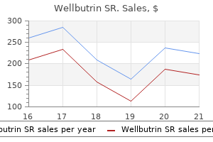
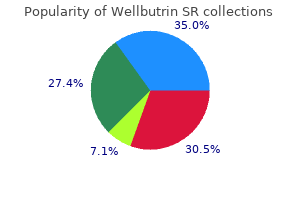
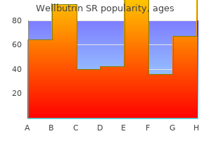
Wellbutrin SR
Clarisse M. Machado, M.D.
- Virology Laboratory
- S?o Paulo Institute of Tropical Medicine
- University of S?o Paulo
- S?o Paulo, Brazil
Wellbutrin SR dosages: 150 mg
Wellbutrin SR packs: 30 pills, 60 pills, 90 pills, 120 pills, 180 pills, 270 pills, 360 pills

Wellbutrin sr 150 mg with visa
The impulses inhibit the release of prolactin-inhibiting issue depression hereditary buy cheap wellbutrin sr 150 mg on line, and prolactin is then launched from the adenohypophysis depression symptoms without sadness 150mg wellbutrin sr otc. Oxytocin stimulates the myoepithelial cells that encompass the bottom of the alveolar secretory cells and the base of the cells in the larger ducts depression symptoms acronym buy wellbutrin sr 150 mg low cost, inflicting them to contract and eject the milk from the alveoli and the ducts. Ovulation normally resumes after 6 months or earlier with a lower in suckling frequency. In cultures by which breast-feeding may continue for 2 to 3 years, lactational amenorrhea is the principal technique of contraception. In the absence of suckling, secretion of milk ceases, and the mammary glands start to regress and atrophy. Female Reproductive System Involution of the Mammary Gland the mammary gland atrophies or its specialized stroma involutes after menopause. The connective tissue also demonstrates degenerative modifications, marked by a lower in the variety of fibroblasts and collagen fibers, and loss of elastic fibers. Branches of the vessels cross primarily along the path of the alveolar ducts as they attain capillary beds surrounding the alveoli. Veins basically comply with the path of the arteries as they return to the axillary and internal thoracic veins. Lymphatic capillaries are located in the connective tissue surrounding the alveoli. The bigger lymphatic vessels drain into axillary, supraclavicular, or parasternal lymph nodes. Innervation the nerves that offer the breast are anterior and lateral cutaneous branches from the second to sixth intercostal nerves. The secretory function is primarily underneath hormonal control, however afferent impulses associated with suckling are concerned in the reflex secretion of prolactin and oxytocin. Ovaries have a medulla in their middle that accommodates unfastened A growing follicle that accommodates a single fluid cavity (antrum) is called the secondary (antral) follicle. It nonetheless connective tissue, nerves, blood and lymphatic vessels, and a cortex on their periphery that incorporates a giant number of ovarian follicles that present a microenvironment for creating oocytes. The floor of the ovary is roofed by germinal epithelium, which is a single cuboidal epithelium that overlies a dense layer of connective tissue known as tunica albuginea. There are three fundamental developmental stages of an ovarian follicle: primordial, growing (both major and secondary), and mature (Graafian) follicle. After puberty following cyclic hormonal adjustments, a particular cohort of main follicles develops into rising follicles. Follicular cells surrounding the oocyte become cuboidal and endure additional stratification to form the first follicle. As granulosa cells proliferate, they become concerned in steroid hormone metabolism (conversion of androgens produced by theca interna into estrogens) and are actively secreting follicular fluid that accumulates in cavities between the granulosa cells. The mature (Graafian) follicle has a big antrum and a outstanding, steroid-producing theca interna layer. All other follicles in the creating cohort endure follicular atresia, a strategy of degeneration involving apoptosis. During ovulation, a secondary oocyte is launched from the ruptured Graafian follicle. At ovulation, the follicular wall, composed of the remaining granulosa and thecal cells, is remodeled into the corpus luteum. The corpus luteum of menstruation is formed within the absence of fertilization; it degenerates 10 to 12 days after ovulation to turn out to be the corpus albicans. During capacitation, the mature spermatozoa acquire the power to fertilize the oocyte within the female reproductive tract. After capacitation, the spermatozoa bind to the zona pellucida receptors, which set off the acrosome response. Enzymes released from the acrosome enable a single spermatozoon to penetrate the zona pellucida and impregnate the oocyte. During impregnation, the entire spermatozoon, except for the tail cytoplasm, becomes incorporated into the ooplasm, which triggers resumption of the second meiotic division (transforms the secondary oocyte into a mature oocyte). The sperm head within the oocyte cytoplasm undergoes modifications to type the male pronucleus, which fuses with the feminine pronucleus to form a diploid zygote. Each uterine tube has four segments: infundibulum (a funnel-shaped finish surrounded by fimbriae adjoining to the ovary), ampulla (common site of fertilization), isthmus (narrow segment adjacent to the uterus), and intramural half (traversing the uterine wall). The uterine tube wall consists of three layers: external serosa, thick muscularis, and highly folded mucosa. The mucosal lining is simple columnar epithelium composed of two cell sorts: ciliated and nonciliated (peg) cells. The oocyte (and zygote after fertilization) is propelled into the uterine cavity by a coordinated motion of cilia on the floor of mucosa and peristaltic muscular contractions of the uterine tube. The uterine wall consists of endometrium (lining mucosa of the uterus), myometrium (smooth muscular layer), and perimetrium (a serous layer of visceral peritoneum). The endometrium is lined by easy columnar epithelium that invaginates into the underlying lamina propria (endometrial stroma), forming uterine glands. The endometrium consists of stratum basale and stratum functionale, which undergoes cyclic modifications as a end result of fluctuat- ing levels of estrogens and progesterone in the course of the menstrual cycle. The thickness of the endometrium, its glandular activity, and its vascular pattern are distinctive for each of the three phases (proliferative, secretory, and menstrual) of the menstrual cycle, which lasts a median of 28 days. If the embryo implants successfully, the endometrium undergoes decidualization (the process of conversion to decidua) and together with the trophoblastic cells from the embryo provoke growth of the placenta. The part of the cervix projecting into the vagina has a metamorphosis zone where easy columnar epithelium of the cervix modifications abruptly into stratified squamous epithelium of the vagina. Fetal and maternal blood is separated by the placental barrier, which develops in the tertiary chorionic villi (projections of chorion containing syncytiotrophoblast, cytotrophoblast, mesenchymal connective tissue, and fetal blood vessels). Villi are immersed within the maternal blood that fills vascular areas in the placenta (cotyledons). The placenta is a major endocrine organ that helps improvement of the fetus; it produces each steroid hormones (mainly progesterone) and protein hormones. Female exterior genitalia (vulva) encompass the mons pubis (formed by underlying adipose tissue), labia majora (longitudinal folds of skin containing adipose tissue, a thin layer of clean muscle, and sebaceous and sweat glands), labia minora (core of connective tissue devoid of adipose tissue but contains massive sebaceous glands), clitoris (erectile tissue homologous to the penis), and vestibule (lined with stratified squamous epithelium with quite a few small mucous glands). The morphology of the secretory portion of the inactive mammary gland varies with the menstrual cycle. Mammary glands bear dramatic proliferation and development during being pregnant in preparation for lactation under the influence of estrogen (proliferation of duct components) and progesterone (growth of alveoli). The protein part of milk is released by alveolar cells using merocrine secretion, whereas the lipid part of milk is launched by apocrine secretion. On one side is a hilum for the transit of neurovascular constructions; on this same facet is a mesovarium that joins the ovary to the broad ligament. The capabilities of the ovary are the production of ova and the synthesis and secretion of estrogen and progesterone. In the cortex are numerous primordial follicles which are present on the time of delivery and that remain unchanged until sexual maturation. The oogonia in these follicles are arrested in prophase of the primary meiotic division. At puberty, beneath the influence of pituitary gonadotropins, the ovaries start to bear the cyclical adjustments designated the ovarian cycle.
Buy generic wellbutrin sr 150 mg line
All three agents are comparatively selective for 2-adrenoceptors and theoretically are able to producing bronchodilation with minimal cardiac stimulation anxiety young living essential oils proven wellbutrin sr 150 mg. However depression definition economic wellbutrin sr 150mg with visa, the time period 2-selectivity is a pharmacological classification based mostly primarily on the relative efficiency of a person adrenomimetic to stimulate the pulmonary or the cardiovascular system anxiety uncertainty theory wellbutrin sr 150mg otc. Indeed, 2agonists invariably produce a degree of tachycardia at massive doses, either by activating sympathetic reflex pathways as a consequence of systemic vasodilation or by directly stimulating cardiac 1-adrenoceptors. In addition, a significant number of 2-adrenoceptors are present in the human coronary heart, and stimulation of these receptors could contribute to the cardiac effects of 2adrenoceptor agonists. Inhaled salmeterol has a pharmacological half-life in extra of 12 hours, much longer than both albuterol or terbutaline. The likely foundation for this long half-life is that the long lipophilic tail of salmeterol promotes retention of the molecule in the cell membrane. Its lengthy length of motion makes salmeterol particularly appropriate for prophylactic use, corresponding to in stopping nocturnal signs of bronchial asthma. This course of is triggered by the interaction of the adrenomimetics with 2-adrenoceptors on airway smooth muscle. Isoproterenol is used principally by inhalation for the management of bronchospasm. Terbutaline, albuterol, salmeterol and different 2adrenoceptor agonists are used primarily in the administration of bronchial asthma. Terbutaline and albuterol have very rapid onset of action and are indicated for acute symptom relief. Salmeterol, in distinction, has a slow onset of action however a protracted duration of motion. Salmeterol is thus used as prophylactic therapy only, to not reverse acute signs. In addition to its use as a bronchodilator, terbutaline is used extensively to control premature labor, since contractions of uterine easy muscle are abolished by adrenomimetics (see Chapter 62). Adverse Effects Patients treated with recommended dosages of epinephrine will complain of feeling nervous or anxious. Some will have tremor of the hand or upper extremity and many will complain of palpitations. Epinephrine is harmful if beneficial dosages are exceeded or if the drug is used in patients with coronary artery disease, arrhythmias, or hypertension. The inappropriate use of epinephrine has resulted in excessive hypertension and cerebrovascular accidents, pulmonary edema, angina, and ventricular arrhythmias, together with ventricular fibrillation. At beneficial dosages, opposed effects from inhaled isoproterenol are rare and not critical. When extreme dosages are used, tachycardia, dizziness, and nervousness could happen, and some sufferers might have arrhythmias. The limiting facet impact associated with orally administered 2-adrenoceptor agonists is muscle tremor, which results from a direct stimulation of 2-adrenoceptors in skeletal muscle. This impact is most notable on the initiation of therapy and steadily improves on continued use. When used by intravenous infusion for premature labor, 2-agonists have been re- 39 Drugs Used in Asthma 463 ported to produce tachycardia and pulmonary edema within the mom and hypoglycemia in the child. A few epidemiological studies suggest that the overuse of -adrenoceptor agonists is associated with an general deterioration in disease control and a slight enhance in asthma mortality. This obvious trend may be brought on by a number of elements, the more than likely of which is that sufferers rely too heavily on bronchodilator therapy to control acute symptoms on the expense of antiinflammatory remedy to management the underlying disease course of. Adverse Effects, Drug Interactions, and Contraindications Theophylline has a slim therapeutic index and produces unwanted aspect effects that may be extreme, even life threatening. In one study, the oral dosage of theophylline required to produce therapeutic plasma levels. Heterogeneity amongst people in the price at which they metabolize theophylline appears to be the principal issue liable for the variability in plasma ranges. Such circumstances as heart failure, liver illness, and extreme respiratory obstruction will gradual the metabolism of theophylline. The most frequent complaints of sufferers taking theophylline are nausea and vomiting, which occur most incessantly in sufferers receiving theophylline for the first time and when the plasma degree approaches 20 g/mL however hardly ever happen at plasma concentrations under 15 g/mL. A speedy intravenous injection of theophylline may cause arrhythmias, hypotension, and cardiac arrest. Consequently, plasma concentrations of theophylline ought to be determined when a affected person begins therapy after which at common intervals of 6 to 12 months thereafter. Theophylline ought to be used with warning in sufferers with myocardial illness, liver disease, and acute myocardial infarction. The half-life of theophylline is prolonged in patients with congestive coronary heart failure. Because of its slim margin of security, extreme warning is warranted when coadministering drugs, such as cimetidine or zileuton, that will intrude with the metabolism of theophylline. It can additionally be prudent to be careful when utilizing theophylline in patients with a history of seizures. Theophylline Twenty years in the past theophylline (Theo-Dur, Slo-bid, Uniphyl, Theo-24) and its extra soluble ethylenediamine salt, aminophylline, had been the bronchodilators of alternative within the United States. Although the 2-adrenoceptor agonists now fill this main function, theophylline continues to have an necessary place within the remedy of bronchial asthma as a result of it appears to have antiinflammatory as well as bronchodilator exercise. The motion of theophylline on the respiratory system is easily seen within the asthmatic by the resolution of obstruction and improvement in pulmonary perform. Other mechanisms that will contribute to the action of theophylline in bronchial asthma embody antagonism of adenosine, inhibition of mediator release, increased sympathetic activity, alteration in immune cell perform, and discount in respiratory muscle fatigue. Theophylline also may exert an antiinflammatory impact by way of its capacity to modulate inflammatory mediator release and immune cell function. Inhibition of cyclic nucleotide phosphodiesterases is broadly accepted because the predominant mechanism by which theophylline produces bronchodilation. It can additionally be used to deal with the reversible component of airway obstruction related to persistent obstructive pulmonary illness and to relieve dyspnea related to pulmonary edema that develops from congestive heart failure. Anticholinergics the parasympathetic cholinergic pathway emanating from the vagus nerve exerts the primary neuronal management in human airways. Stimulation of these nerve fibers, with the resultant release of acetylcholine and activation of muscarinic cholinoreceptors, elicits bronchoconstriction, mucous secretion, and bronchial vasodilation. Thus, the cholinergic pathways play a key position within the maintenance of the caliber of the airways and contribute to the airway obstruction in each bronchial asthma and persistent obstructive pulmonary illness. Ipratropium has larger effectiveness than 2-adrenoceptor agonists in two settings: in psychogenic bronchial asthma and in patients taking 2-adrenoceptor antagonists. A mounted mixture of ipratropium and albuterol (Combivent) is permitted to be used in continual obstructive pulmonary illness. The most prevalent peripheral unwanted effects are dry mouth, headache, nervousness, dizziness, nausea, and cough. Basic Pharmacology the airway results of launched acetylcholine are mediated through activation of three distinct muscarinic receptor subtypes: M1, in parasympathetic ganglia, mucous glands and alveolar partitions; autoinhibitory M2, in parasympathetic nerve terminals; and M3, in airway clean muscle, mucus glands, and airway epithelium. To improve the scientific utility of anticholinergics, quaternary amine derivatives of atropine had been developed.
Discount wellbutrin sr 150 mg amex
The tear film accommodates proteins (tear albumins volcanic depression definition buy wellbutrin sr 150 mg with visa, lactoferrin) depression kanji generic 150 mg wellbutrin sr with amex, enzymes (lysozyme) depression quest buy wellbutrin sr 150mg with mastercard, lipids, metabolites, electrolytes, and drugs, the latter secreted during remedy. The tear cationic protein lactoferrin increases the exercise of assorted antimicrobial agents corresponding to lysozyme. The eye is moved throughout the orbit by coordinated contraction of extraocular muscular tissues. Six muscular tissues of the eyeball (also referred to as extraocular or extrinsic muscles) connect to every eye. These are the medial, lateral, superior, and inferior rectus muscle tissue and the superior and inferior oblique muscles. The combined, precisely managed action of those muscle tissue permits vertical, lateral, and rotational motion of the eye. Normally, the actions of the muscles of both eyes are coordinated in order that the eyes move in parallel (called conjugate gaze). The tissues of the eye are derived from neuroectoderm (retina), floor ectoderm (lens, corneal epithelium), and mesoderm (sclera, corneal stroma, vascular coat). The eyeball consists of three structural layers: the outer corneoscleral (fibrous) coat consisting of the clear cornea and the white opaque sclera; the center vascular coat consisting of the choroid, ciliary body, and iris; and the internal layer, the retina. The layers of the eye and the lens function boundaries for 3 chambers: the anterior chamber and posterior chamber, that are full of aqueous humor, and the vitreous chamber, which is occupied by a transparent gel, the vitreous physique. It communicates with the cornea on the corneoscleral limbus, which contains corneolimbal stem cells. The iris arises from the ciliary physique and its opening (pupil) is managed by smooth muscle fibers of the sphincter pupillae muscle and the myoepithelial cell layer of the dilator pupillae muscle. It incorporates ciliary processes that secrete aqueous humor, anchors zonular fibers that suspend the lens, and accommodates ciliary muscle that alters the form of the lens throughout lens accommodation. Major cells within the retina embody photoreceptors (rods and cones), conducting neurons (bipolar neurons and ganglion cells), association neurons, and supporting cells. Retinal pigment epithelium (layer 1) is the outermost layer of the retina and absorbs scattered mild, contributes to the blood�retina barrier, restores photosensitivity to visual pigments, and phagocytoses membranous discs from the rods and cones. Rods (layer 2) are most quite a few (120 million) within the retina and detect gentle intensity with their cylindrical outer segments. Cones (layer 2) are much less quite a few (7 million) and with their conical outer segment detect three completely different wavelengths of light comparable to primary colours: blue, green, and pink. Rods comprise the visual pigment rhodopsin that consists of opsin and a small light-absorbing compound, retinal. Conversion of sunshine into nerve impulses within the photoreceptors is known as visual processing. It includes a photochemical response based on the conversion of 11-cis-retinal into alltrans-retinal within the rhodopsin. The outer nuclear layer (layer 4) contains the nuclei of rods and cones, and the outer plexiform layer (layer 5) incorporates their processes, which synapse with the horizontal, amacrine, and bipolar cells (the nuclei of which reside within the internal nuclear layer [layer 6]). Axons from cells within the outer plexiform layer synapse within the internal plexiform layer (layer 7) with ganglion cells, the cell our bodies of which reside within the ganglion cell layer (layer 8). These cells send axons to the layer of optic nerve fibers (layer 9), which types the optic nerve. It lines the area between the inner surface of the eyelid and the anterior floor of the eye lateral to the cornea. The tarsal glands (Meibomian glands) are long sebaceous glands embedded in the tarsal plates of the upper and decrease eyelids. The lacrimal gland produces tears that moisten the cornea and move to the nasolacrimal duct and into the nasal cavity. The wall of the eye consists of three concentric layers or coats: the retina, the inner layer; the uvea, the middle or vascular layer; and the corneosclera, the outer fibrous layer. The eye is usually compared to a simple digicam with a lens to capture and focus mild, a diaphragm to regulate the quantity of sunshine, and film to report the image. The iris, positioned between the cornea and lens, regulates the dimensions of the pupil by way of which light enters the attention. It is suspended within the bony orbit by six extrinsic striated muscle tissue that management its movement. The extraocular muscle tissue are coordinated in order that both eyes transfer synchronously, with every transferring symmetrically round its personal central axis. The receptor parts of the retina are situated in the posterior three-fifths of the eyeball. This anterior nonreceptor extension of the inner layer is extremely pigmented, and the pigment (melanin) is obvious because the black inner border of those buildings. The uvea, the middle layer of the eyeball, consists of the choroid, the ciliary physique, and the iris. On this basis, the choroid (Ch) is identified as being just exterior to the pigmented layer of the retina. It can be extremely pigmented; the choroidal pigment is clear as a discrete layer in several parts of the section. This incorporates the ciliary muscle (see Plate 106), which brings about adjustments of the lens to focus mild. The ciliary body additionally incorporates processes to which the zonular fibers are attached. The iris (I) is the most anterior element of the uvea and incorporates a central opening, the pupil. The outermost layer of the eyeball, the fibrous layer, consists of the sclera (S) and the cornea (C). Both of these contain collagenous fibers as their primary structural element; nonetheless, the cornea is clear, and the sclera is opaque. The extrinsic muscles of the attention insert into the sclera and effect movements of the eyeball. Just posterior to the lens is the massive cavity of the eye, the vitreous cavity (V), which is crammed with a thick jelly-like materials, the vitreous humor or physique. The fibrous cowl of the optic nerve is an extension of the meninges of the mind. The internal limiting membrane is the basal lamina of these cells; the exterior limiting membrane is actually a line formed by the junctional complexes between processes of these cells and the photoreceptor cells. The neurons of the retina are arranged sequentially in three layers: (1) a deep layer of rods and cones; (2) an intermediate layer of bipolar, horizontal, and amacrine cells; and (3) a superficial layer of ganglion cells. Nerve impulses originating within the rods and cones are transmitted to the intermediate layer after which to the ganglion cells. Synaptic connections happen within the outer plexiform layer (between the rods and cones and the intermediate neuronal layer) and the internal plexiform layer (between the intermediate layer and the ganglion cells), resulting in summation and neuronal integration. Finally, the ganglion cells send their axons to the mind as parts of the optic nerve. The fibers that give rise to the optic nerve originate within the retina, extra specifically, within the ganglion cell layer (see below).
Buy generic wellbutrin sr 150mg
Clinical examination contains checking for previous scars in the abdomen and for intact hernial orifices bipolar depression 4 months purchase 150 mg wellbutrin sr mastercard, including within the femoral canal bipolar depression symptoms mania buy 150 mg wellbutrin sr fast delivery. Complications of unrelieved intestinal obstruction are ischaemia of the proximal phase depression internet test purchase wellbutrin sr 150mg without a prescription, perforation of the bowel wall, peritonitis, sepsis, and even maternal death. Clinical features of complications include fever, tachycardia, and indicators of peritonism on stomach palpation. A single erect anteroposterior X-ray of the abdomen is useful in making the prognosis, with features of air-fluid ranges in dilated small bowel loops seen in 82 per cent of circumstances. These loops of small bowel could additionally be seen in the periphery of the film and are due to displacement of the bowel by the gravid uterus. In early being pregnant the prognosis would be much like that of a non-pregnant female. However, as being pregnant advances, any belly mass could additionally be displaced upwards and laterally, a proven reality that have to be borne in mind when making a analysis. Furthermore, indicators of peritonitis in stomach swellings of an inflammatory nature can be markedly altered, probably leading to misdiagnosis with potential critical consequences. Abdominal swellings could additionally be categorised according to the anatomical layer of the stomach, which contains the: anterior belly wall; peritoneal cavity; retroperitoneal area. The overlying skin can be made to transfer independently of a lipoma, and asking the woman to tense her stomach muscle tissue will make the lump more distinguished. Swellings can additionally be due to herniation of abdominal contents through areas of potential weak point of the stomach wall, the most typical being the umbilicus. Like all herniae, these plenty have an expansile cough impulse and are usually reducible on mendacity supine. As the neck of these paraumbilical herniae is normally extensive, problems of irreducibility and strangulation are comparatively uncommon. Herniation can even occur through earlier incisions, including those made for caesarean sections, and often happen on the lateral edge of the Pfannensteil scar. A situation that occurs particularly with repeated pregnancy is divarification of the recti. This is a defect of the median raphe and is palpable below the extent of the umbilicus. Abdominal swellings arising from the anterior belly wall the layers of the belly wall which will give rise to belly wall swellings is shown in Box 1. Box 1 the layers of the belly wall that will give rise to abdominal wall swellings Skin and appendages Subcutaneous tissue Herniation of intra-abdominal contents by way of the wall Lumps can come up from the skin and its appendages. A punctum could also be seen in sebaceous cysts, which may be tender and erythematous in the occasion that they turn into infected. Diagnostic confusion could occur as pigmented naevi may change during pregnancy owing to a rise in junctional activity with the altering hormonal levels. In the later levels of being pregnant, these circumstances may be suspected when the abdominal enlargement is larger than could be anticipated for the gestational age. A symphysiofundal height larger than that anticipated for the gestational age could also be because of uterine fibroids that are making the uterus bigger, extra amniotic fluid, a large child, or the upward displacement of a gravid uterus by an ovarian cyst. An appropriate symphysiofundal top is found if the generalised abdominal distension is secondary to faeces or flatus, the place a historical past of constipation plus or minus vomiting is elicited. The advances in ultrasound permit for correct fibroid mapping in the gravid uterus. Cervical fibroids are specific necessary as they could affect the mode of supply. Fibroids, however, are susceptible to undergo pink degeneration (where the fibroid outgrows the blood supply and haemorrhagic necrosis occurs) at any time during the being pregnant and puerperium. Ovarian cysts that trigger generalised abdominal distension are usually mucinous cystadenomas. Ultrasound features embrace the presence of septae making the cyst multiloculated. There are now simple guidelines utilized for ultrasound options to verify whether or not the cyst is benign (B) or malignant (M). If a benign cyst is identified early in pregnancy, then ovarian cystectomy may be carried out laparoscopically, ideally within the early second trimester. This can occur by way of an obstruction of the cystic duct or of the common bile duct. The commonest cause is calculi, although it might be as a end result of carcinoma of the top of the pancreas, which is extremely uncommon in pregnancy. This is predicated on the idea that persistent irritation secondary to calculi causes fibrosis of the gallbladder, thereby making it tough to distend and current as an abdominal swelling. Mass in the right hypochondrium the potential causes of a mass in the proper hypochondrium are proven in Box 2. Mass within the epigastrium the possible causes of a mass in the epigastrium are given in Box three. Generalised enlargement may be due to infections, cirrhosis, chronic energetic hepatitis, cirrhosis, or myeloproliferative issues. If the surface of the liver is irregular, polycystic disease and carcinoma have to be excluded. Liver enlargement could additionally be accompanied by jaundice in infective hepatitis, Localised enlargements of the left lobe of the liver can present with a mass in the epigastrium. Epigastric ache in the presentation in a girl with severe pre-eclampsia could be because of pressure on the liver capsule, which might very rarely rupture with fatal penalties. This mass is often hard and irregular, and is pre-dated by signs of anorexia and weight loss. Investigations that may assist with the differential prognosis embody a full blood count, a blood image, thick and skinny blood movie for malarial parasites, and tissue biopsy of lymph nodes or the liver. Pancreatic pseudocysts are normally very troublesome to really feel, because the abdomen is anterior to it, thereby making it troublesome to delineate and resonant to percussion. Masses within the left hypochondrium the buildings that can enlarge to give rise to a mass within the left hypochondrium are shown in Box 4. Masses in the proper and left lumbar areas the anatomical origins of plenty within the loin are shown in Box 5. Box four the anatomical origins of masses within the left hypochondrium Box 5 the anatomical origins of plenty in the loin Enlargement of the spleen Extension of lots from the epigastrium (stomach and pancreas) Enlargement of the kidney Extension of lots from the best hypochondrium the spleen has to enlarge considerably to turn into palpable beneath the left costal margin. Small enlargements could also be felt by tilting the affected person in the direction of the examiner, lifting the lower ribs forwards and asking the affected person to breathe deeply. Depending on the trigger of splenic enlargement the sting may be gentle or agency and a splenic notch may be palpable. Splenomegaly happens in the following situations: Infection: splenomegaly in pregnant ladies is common in areas endemic for malaria. There is an increase in measurement in the first trimester owing to a rise in parasitaemia.
Purchase 150 mg wellbutrin sr
The base of the cell tapers into an axonal course of that enters the lamina propria and joins axons from other receptor cells to form the olfactory nerve definition of depression in geography order wellbutrin sr 150 mg with mastercard. Large depression symptoms bupa purchase wellbutrin sr 150mg without prescription, cuboidal Schwann cells are a distinguished characteristic of these axons anxiety coach wellbutrin sr 150mg with amex, giving the nerve an uncommon look. They connect to the receptor cells by way of adhering junctions and provide mechanical and metabolic assist to the olfactory cells. Basal cells are stem cells from which olfactory and supporting cells differentiate. Brush cells are the identical cell sort that happens in nonsensory respiratory epithelium. These are tubuloalveolar serous glands whose watery secretion serves as a entice and solvent for odorant substances and continuously washes the olfactory surface. This low-magnification orientation micrograph exhibits a half of the wall of the nasal cavity. The olfactory mucosa is immediately connected to the bone tissue; no submucosa is current. In this specimen, nevertheless, the mucosa is separated from the bone tissue due to shrinkage, a incessantly encountered artifact. Note that the adjoining respiratory mucosa lacks the nerves and displays a relative paucity of glands. The supporting cell has a cylindrical form and extends from the basement membrane via the total thickness of the epithelium. Careful examination of the nuclei of these bipolar neuronal cells reveals that they include more euchromatin than the nuclei of the supporting cells and often exhibit a number of nucleoli. Note that the olfactory mucosa in distinction to respiratory mucosa lacks goblet cells. The duct elements extend from the secretory portion of the gland beginning in shut proximity to the overlying epithelium (arrowhead) and cross instantly via the epithelium to ship their secretions on the floor. The nuclei present within the olfactory nerves characterize Schwann cell nuclei (ScC). It consists of a cartilaginous framework to which both extrinsic and intrinsic muscle tissue are attached and a mucosal surface that varies in character from pseudostratified to stratified squamous in areas subject to abrasion by the air stream. The muscles transfer certain cartilages with respect to others, thus growing or decreasing the opening of the rima glottis and increasing or decreasing the strain on the vocal folds (cords). In this way, vibrations of various wavelengths are generated in the passing air, and sound is produced. The vocal folds are ridge-like structures that are oriented in an anteroposterior (ventral-dorsal) course. Just above each vocal fold is an elongated recess called the ventricle (V), and above the ventricle is one other ridge called the ventricular fold (VnF) or, generally, the false vocal fold. It lies in an anteroposterior course within the substance of the vocal fold and performs an essential position in phonation. The surfaces of a vocal fold and the dealing with ventricular fold within rectangle 1 in high figure are turned 90� clockwise and shown at higher magnification on this determine. This area of the larynx beneath the ventricles and rima glottidis communicates with trachea and is identified as the infraglottic cavity. The lamina propria consists of loose connective tissue by which glands (Gl) are current. Note the cylinders of cytoplasm that clearly indicate the columnar nature of the surface cells. This distinction is tough to make from the examination of a single pattern such as that proven here, and different data is required to make the evaluation. The extra data is the presence of cilia on the pseudostratified columnar epithelium; this epithelium is typically ciliated. Although not evident within the photomicrographs, observe that stratified columnar epithelium has a very restricted distribution, usually occurring between stratified squamous epithelium and another epithelial sorts. The lamina propria is a free cellular connective tissue, and it also reveals some glands (Gl). It extends from the larynx to in regards to the middle of the thorax, the place it divides into the two main bronchi (primary bronchi). The lumen of the trachea is held open by a collection of C-shaped hyaline cartilages that are stacked on each other to form a supporting construction. Fibroelastic tissue and smooth muscle (the trachealis muscle) bridge the gap between the free ends of the cartilages at the posterior border of the trachea, adjoining to the esophagus. Typical respiratory (ciliated pseudostratified columnar) epithelium lines the trachea and primary bronchi. On entering the lungs, the primary bronchi branch instantly to give rise to the lobar bronchi (secondary bronchi) that offer the 2 lobes of the left lung and the three lobes of the best lung. Within the lung, the C-shaped cartilages are changed by an investment of (sometimes overlapping) cartilaginous plates that fully surround the bronchi. The basement membrane, which consists of tightly packed, nice collagen fibers, is actually an unusually thick and dense reticular layer and is, thus, a part of the lamina propria. It is particularly distinct within the human trachea and should thicken with continual irritation, as in smokers. Adipose tissue (Ad) can also be present within the submucosa between the esophagus and trachea. A important quantity of adipose tissue (Ad) is found within the connective tissue between the trachealis muscle and the wall of the esophagus (not shown in this figure). The thickness and the density of the basement membrane (Bm) are extra easily seen right here than within the lower magnification views in the different figures. Portions of the seromucous glands (Gl) are simply seen on the backside edge of the determine. As the bronchi turn into smaller, some parts of the wall are lost or decreased in quantity. The features that characterize the bronchiole are the absence of cartilage, lack of submucosal glands, and gradual disappearance of goblet cells. The epithelium changes from pseudostratified columnar to simple ciliated columnar, and a few columnar cells even lack cilia. Smooth muscle occupies a comparatively larger portion of the bronchiolar wall than of the bronchial wall. The smallest diameter conducting bronchioles, the terminal bronchioles, are lined with easy ciliated cuboidal epithelium in which Clara cells, cells that secrete a surface-active agent that prevents luminal adhesion of bronchiolar partitions throughout expiration, are found among the many ciliated cells. Respiratory bronchioles are the first a half of the bronchial tree that enables gas trade to happen. Respiratory bronchioles represent a transition zone in which each air conduction and fuel trade happen. Scattered, thin-walled evaginations of the lumen of the respiratory bronchiole are referred to as alveoli; these are the structures in which gasoline change between the air passages and the blood capillaries happens. Surrounding the bronchiole, comprising many of the lung substance, are the air areas or alveoli of the lung. The last portion of a bronchiole that leads into respiratory bronchioles is called a terminal bronchiole.
Syndromes
- Failure to gain weight
- Infections that occur in the womb or after birth
- Surgery to fix a winged scapula
- Gets worse when you change positions or when you bend, strain, or cough
- Nausea and vomiting
- Sweating
- Choriocarcinoma (rare)
- Have your blood pressure checked every year.
- Altered medical status or thinking ability
- Fever

150mg wellbutrin sr sale
Enteroendocrine cells and Paneth cells are also derived from the stem cells at the base of the intestinal gland anxiety disorder treatment discount 150 mg wellbutrin sr visa. They live for about 4 weeks and are then changed by differentiation of a nearby "dedicated" cell within the intestinal gland depression symptoms in kittens cheap 150 mg wellbutrin sr. As talked about in the chapter on epithelial tissue (page 146) depression laboratory test cheap 150 mg wellbutrin sr free shipping, expression of the transcription factor Math1 appears to decide the destiny of differentiating cells in the intestinal stem cell niche. The colon is additional subdivided on the idea of its anatomic location into ascending colon, transverse colon, descending colon, and sigmoid colon. Local contractions displace intestinal contents both proximally and distally; this kind of contraction known as segmentation. They serve to flow into the chyme locally, mixing it with digestive juices and transferring it into contact with the mucosa for absorption. Peristalsis, the second kind of contraction, entails coordinated motion of each round and longitudinal muscle layers and strikes the intestinal contents distally. Haustra coli which may be visible sacculations between the teniae coli on the exterior surface of the cecum and colon. Omental appendices which are small fatty projections of the serosa, observed on the outer floor of the colon. Serosa the serosa of the elements of the small intestine that are situated intraperitoneally in the abdominal cavity corresponds to the general description initially of the chapter. Mucosa the mucosa of the large gut has a "smooth" surface; neither plicae circulares nor villi are present. The glands consist of simple columnar epithelium, as does the intestinal surface from which they invaginate. Examination of the luminal floor of the big intestine at the microscopic stage reveals the openings of the glands, that are arranged in an orderly sample. The morphology of absorptive cells is actually equivalent to that of the enterocytes of the small intestine. Elimination of semisolid to strong waste supplies is facilitated by the big amounts of mucus secreted by the numerous goblet cells of the intestinal glands. Goblet cells are extra numerous in the large intestine than within the small gut. The mucosal epithelium of the big gut accommodates the identical cell types as the small intestine except Paneth cells, which are normally absent in people. This photograph shows the outer (serosal) surface (left) and inside (mucosal) surface (right) of the transverse colon. The easy mucosal floor shows semilunar folds (arrows) fashioned in response to contractions of the muscularis externa. The ratio decreases, nonetheless, approaching 1:1, near the rectum, where the number of goblet cells increases. This photomicrograph of an H&E preparation reveals the mucosa and a half of the submucosa. The surface epithelium is continuous with the straight, unbranched, tubular intestinal glands (crypts of Lieberk�hn). As the absorptive cells are adopted into the glands, they turn out to be fewer in number, whereas the goblet cells improve in quantity. The highly mobile lamina propria accommodates numerous lymphocytes and different cells of the immune system. Comparative electron-microscopic features of normal, hyperplastic, and adenomatous human colonic epithelium. Goblet cells may mature deep in the intestinal gland, even within the replicative zone. They secrete mucus continuously, even to the purpose where they attain the luminal surface. Here, at the surface, the secretion fee exceeds the synthesis fee, and "exhausted" goblet cells seem in the epithelium. These cells are tall and thin and have a small variety of mucinogen granules in the central apical cytoplasm. An occasionally noticed cell sort, the caveolated "tuft" cell, has additionally been described within the colonic epithelium; nevertheless, this cell could also be a type of exhausted goblet cell. The turnover times of the epithelial cells of the massive intestine are similar to these of the small intestine. Senile epithelial cells that attain the mucosal surface undergo apoptosis and are shed into the lumen at the midpoint between two adjoining intestinal glands. Lamina Propria Although the lamina propria of the massive intestine incorporates the same primary elements as the rest of the digestive tract, it demonstrates some extra structural features and larger growth of some others. These include the following: � Collagen desk, which represents a thick layer of col- Epithelial Cell Renewal within the Large Intestine All intestinal epithelial cells in the large intestine derive from a single stem cell population. As in the small gut, all the mucosal epithelial cells of the large intestine arise from stem cells positioned on the backside of the intestinal gland. The intermediate cell sorts discovered in the � lagen and proteoglycans that lies between the basal lamina of the epithelium and that of the fenestrated absorptive venous capillaries. This layer is as much as 5 m thick within the normal human colon and may be as a lot as three times that thickness in human hyperplastic colonic polyps. The collagen table participates in regulation of water and electrolyte transport from the intercellular compartment of the epithelium to the vascular compartment. Pericryptal fibroblast sheath, which constitutes a well-developed fibroblast inhabitants of often replicating cells. Some proof means that the macrophages of the core of the lamina propria within the large gut could arise as a terminal differentiation of the pericryptal fibroblasts. The intensive development of the immune system in the colon probably displays the big quantity and number of microorganisms and noxious end merchandise of metabolism usually present within the lumen. Only recently, using new very selective markers for lymphatic epithelium, researchers have discovered occasional small lymphatic vessels at the bases of the intestinal glands. These lymphatic vessels drain into the lymphatic community within the muscularis mucosae. The next step in lymph drainage occurs within the lymphatic plexuses within the submucosa and muscularis externa earlier than lymph leaves the wall of the big intestine and drains into the regional lymph nodes. To perceive the clinical significance of the lymphatic pattern in the giant gut, see Folder 17. In the rectum, anal canal, and vermiform appendix, the outer longitudinal layer of easy muscle is a uniformly thick layer, as within the small intestine. Bundles of muscle from the teniae coli penetrate the internal, round layer of muscle at irregular intervals alongside the length and circumference of the colon. These apparent discontinuities within the muscularis externa permit segments of the colon to contract independently, leading to the formation of haustra colli, sacculations of the colon wall. The muscularis externa of the massive gut produces two major kinds of contraction: segmentation and peristalsis. Mass peristaltic actions happen sometimes; in wholesome persons, they normally happen as quickly as a day to empty the distal colon. The fibroblasts then differentiate and migrate upward in parallel and synchrony with the epithelial cells.
Buy cheap wellbutrin sr 150mg line
Efferent fibers are thought to affect management of auditory and vestibular input to the central nervous system depression vines buy wellbutrin sr 150mg with mastercard, presumably by enhancing some afferent signals while suppressing other alerts depression meaning purchase wellbutrin sr 150mg with visa. Damage to the organ of Corti depression letters wellbutrin sr 150mg without a prescription, cochlear nerve, nerve pathways, or auditory cortex is responsible for sensorineural hearing loss (see Folder 25. Blood Vessels of the Membranous Labyrinth Arterial blood is provided to the membranous labyrinth by the labyrinthine artery; venous blood drainage is to the venous dural sinuses. The blood provide to the exterior ear, center ear, and bony labyrinth of the internal ear is derived from vessels related to the exterior carotid arteries. The arterial blood supply to tissues of the membranous labyrinth of the interior ear is derived intracranially from the labyrinthine artery, a standard department of the anterior inferior cerebellar or basilar artery. The labyrinthine artery is a terminal artery: It has no anastomoses with different surrounding arteries. Branches of this artery precisely parallel the distribution of the superior and inferior components of the vestibular nerve. Venous drainage from the cochlear labyrinth is by way of the posterior and anterior spiral modiolar veins that type the widespread modiolar vein. The common modiolar vein and the vestibulocochlear vein type the vein of the cochlear aqueduct, which empties into the inferior petrosal sinus. Venous drainage from the vestibular labyrinth is via vestibular veins that join the vein of the cochlear aqueduct and by the vein of vestibular aqueduct, which drains into the sigmoid sinus. Tissues of the ear are derived from surface ectoderm (epithelia lining of the membranous labyrinth) and elements of the first pharyngeal pouch (auditory tube and middle ear cavity), first pharyngeal groove (external acoustic meatus), first pharyngeal arch (malleus, incus, and anterior a half of the auricle), and second pharyngeal arch (stapes and posterior part of the auricle). The inside ear has three fluid-filled areas: the endolymphatic house within the membranous labyrinth (has a high K and a low Na concentration), the perilymphatic space between the wall of the bony and membranous laby rinth (has a low K and a excessive Na concentration), and the cortilymphatic area that lies within the tunnels of the organ of Corti of the cochlea. The bony labyrinth consists of three connected spaces: semicircular canals, vestibule, and cochlea, each containing totally different components of the membranous labyrinth. The membranous labyrinth consists of a collection of communicating sacs (utricle, saccule, and endolymphatic sac) and ducts (three semicircular ducts, cochlear duct, utriculosaccular duct, endolymphatic duct, and ductus reuniens) that contain endolymph. Specialized sensory cells are situated in six areas within the membranous labyrinth: three cristae ampullaris in the ampullae of semicircular ducts (receptors for angular acceleration of the head), two maculae within the utricle and saccule (receptors for position of the top and its linear movements), and the spiral organ of Corti (receptors for sound). Utricle and saccule maculae contain hair cells which are epithelial mechanoreceptors. These hair cells contain hair bundles on their apical surfaces (formed by rows of stereocilia with a single kinocilium) and are overlaid with a gelatin-like otolithic membrane that contains otoliths (otoconia). Movement of the otoliths is detected by the hair bundles, which activate the mechanically gated ion channels to generate action potential. Movement of the cupula stimulates the mechanically gated ion channels to generate motion potential. The cochlear canal is split into three parallel compartments: scala media or cochlear duct (the center compartment full of endolymph that incorporates the spiral organ of Corti), scala vestibuli, and scala tympani (both containing perilymph). The scala media is a triangular space with its decrease wall forming the basilar membrane on which the spiral organ of Corti resides. The upper wall (vestibular membrane) separates the scala media from scala vestibuli, and the lateral wall incorporates the stria vascularis that produces endolymph. The spiral organ of Corti consists of hair cells (arranged in internal and outer rows), supportive phalangeal cells, and pillar cells. Movement of the stereocilia on hair cells throughout interplay with the overlying tectorial membrane generates electrical impulses which would possibly be transmitted to the cochlear nerve. Sound waves are transmitted from the vibrating tympanic membrane by the ossicles to the oval window, the place they produce movement (waves) of the perilymph in the scala vestibule. This movement deflects the basilar membrane and spiral organ of Corti to generate electrical nerve impulses, that are perceived by the brain as sounds. Nerve impulses from the cristae ampullaris and maculae travel with the vestibular nerve, and the impulses from the spiral organ of Corti journey with the cochlear nerve. It is lined by skin that accommodates hair follicles in addition to sebaceous and ceruminous glands (which produce cerumen, or earwax). The tympanic membrane is composed of pores and skin of the external auditory meatus, a thin core of connective tissue, and mucous membrane of the middle ear. The auditory ossicles (malleus, incus, and stapes) cross the house of the middle ear in sequence and join the tympanic membrane to the oval window. Movement of the ossicles is modulated by the tensor tympani muscle that inserts to the malleus and the stapedius muscle that inserts to the stapes. These are referred to , respectively, because the bony labyrinth and membranous labyrinth. Within the area lined by the membranous labyrinth is a watery fluid referred to as endolymph. The bony labyrinth is split into three components: cochlea, semicircular canals, and vestibule. The cochlea and semicircular canals contain membranous counterparts of the same shape; nonetheless, the membranous components of the vestibule are extra complicated in form, being composed of ducts and two chambers, the utricle and saccule. In this part by way of the interior ear, bone surrounds the complete inner ear cavity. Because of its labyrinthine character, sections of the inner ear seem as a selection of separate chambers and ducts. These, however, are all interconnected (except that the perilymphatic and endolymphatic spaces stay separate). The communication of the vestibule with one of the semicircular canals is marked by the white arrow. Because of the airplane of section and the spiral arrangement of the cochlear tunnel, the tunnel is cut crosswise in seven places (note 3� turns). A more detailed examination of the cochlea and the organ of Corti is provided in Plate 109. The receptor for motion, the crista ampullaris (note its relationships in determine above), is current in every of the semicircular canals. A gelatinous mass, the cupula (Cu), surmounts the epithelium of the crista ampullaris. Each receptor cell sends a hair-like projection deep into the substance of the cupula. Hair cells are epithelial cells that possess numerous stereocilia, modified microvilli also known as sensory hairs. All hair cells have a typical basis of receptor cell operate that involves bending or flexing of their stereocilia. The particular means by which the stereocilia are bent varies from receptor to receptor, but in each case, stretching of the plasma membrane attributable to the bending of the stereocilia generates transmembrane potential changes which are transmitted to the afferent nerve endings associated with every cell. The most important practical element of the cochlea is the organ of Corti, enclosed by the rectangle and shown at larger magnification in determine under. Both the scala vestibuli and the scala tympani are perilymphatic spaces; these talk at the apex of the cochlea.

Generic 150 mg wellbutrin sr visa
On the other hand depression symptoms early morning cheap 150mg wellbutrin sr otc, the muscularis externa is organized in a distinctive manner anxiety 34 weeks pregnant wellbutrin sr 150 mg low cost, and this is evident within the photomicrograph depression articles purchase 150 mg wellbutrin sr with mastercard. The three teniae coli prolong along the length of the large gut as far as, however not into, the rectum. Two rectangles mark areas of the mucosa which are examined at larger magnification in figures beneath. Just above these cross-sectioned smooth muscle cells are others which were minimize longitudinally; they show elongate nuclei and elongate strands of eosinophilic cytoplasm. The arrows establish the openings of a number of the Lamina propria, colon, monkey, H&E 525. As the absorptive cells are followed into the glands, they turn out to be fewer, whereas the goblet cells enhance in number. Other cells within the gland are enteroendocrine cells, not easily recognized in routine H&E�stained paraffin sections, and, in the deep part of the gland, undifferentiated cells of the replicative zone, derived from the stem cells within the base of the crypt. It arises from the cecum (the first phase of the large intestine; the others, so as, are the ascending, transverse, and descending colon; the sigmoid colon; the rectum; and the anal canal) and types a blind-ending tube starting from 2. The wall of the appendix is much like that of the small gut, having a complete longitudinal layer of muscularis externa, nevertheless it lacks each plicae circulares and villi. Even this resemblance is often obliterated, nevertheless, by the massive quantity and size of the lymphatic nodules that normally fuse and prolong into the submucosa. In many adults, the traditional construction is misplaced, and the appendage is filled with fibrous scar tissue. Cross-section of an appendix from a preadolescent, exhibiting the various constructions composing its wall. It reveals the straight tubular glands (Gl) that extend to the muscularis mucosae. The more superficial a part of the submucosa blends and merges with the mucosal lamina propria due to the quite a few lymphocytes in these two websites. Note that the epithelium of the glands within the appendix is similar to that of the big intestine. Most of the epithelial cells comprise mucin, therefore the light look of the apical cytoplasm. The lamina propria, as famous, is heavily infiltrated with lymphocytes, and the muscularis mucosae on the base of the glands is difficult to recognize (arrows). At the identical degree, the round layer of the muscularis externa thickens to turn into the inner anal sphincter. The external anal sphincter is shaped by the striated muscular tissues of the pelvic flooring. Mucosa characteristic of the massive intestine (colorectal zone) is seen on the higher left of the micrograph. This area is the upper a part of the anal canal, and the intestinal glands are the same as those present within the colon. This area known as the anal transitional zone is examined at larger magnification in the backside left determine. The proper rectangular space consists of the stratified squamous epithelium (StS) of the pores and skin within the squamous zone of the anal canal and is examined at higher magnification in the bottom right determine. Between the 2 diamonds within the rectangular areas proven is epithelium of the decrease part of the anal canal. Characteristically, the lamina propria contains giant numbers of lymphocytes (Lym), particularly so in the area marked. A higher magnification of the stratified columnar epithelium (StCol) and stratified cuboidal epithelium (StC) found within the transition zone is shown within the inset. The final change in epithelial kind that happens on the squamous zone of the anal canal is proven here. Again, numerous lymphocytes (Lym) are in the underlying connective tissue, and tons of have migrated into the epithelium within the nonkeratinized space. It is located within the upper proper and partially in the higher left quadrants of the belly cavity, protected by the rib cage. The liver is anatomically divided by deep grooves into two giant lobes (the right and left lobes) and two smaller lobes (the quadrate and caudate lobes;. This anatomic division has only topographic significance as a end result of it relates lobes of the liver to different abdominal organs. Division into useful or surgical segments that correspond to the blood supply and bile drainage is more clinically important. In the embryo, the liver develops as an endodermal evagination from the wall of the foregut (specifically the location that may turn out to be the duodenum) to kind the hepatic diverticulum. The diverticulum proliferates, giving rise to the hepatocytes, which turn into organized in mobile (liver) cords, thus forming the parenchyma of the liver. An outgrowth from the frequent bile duct types the cystic diverticulum that provides rise to the gallbladder and cystic duct. Liver Physiology Many circulating plasma proteins are produced and secreted by the liver. The liver performs an essential role in the uptake, storage, and distribution of each nutrients and nutritional vitamins from the bloodstream. In addition, the liver degrades or conjugates quite a few toxic substances and drugs, however it can be overwhelmed by such substances and broken. The liver can be an exocrine organ; it produces a bile secretion that incorporates bile salts, phospholipids, and ldl cholesterol. Several vitamins are taken up from the bloodstream and are then stored or biochemically modified by the liver. They embody: � falciform ligament ligamentum teres terminal hepatic venule (central vein) vitamin A (retinol), an essential vitamin in vision. This diagram shows the gross view of the diaphragmatic and visceral surfaces of the liver, with labeled anatomic landmarks discovered on each surfaces. The enlarged cross-sectional area of the liver (bottom) exhibits the general microscopic group of the liver into lobules. Note the presence of hepatic portal triads at the periphery of every lobule, with the terminal hepatic venule (central vein) within the middle of the lobule. The circulating plasma proteins produced by the liver embody: � � albumins, which are involved in regulating plasma quantity and tissue fluid steadiness by sustaining the plasma colloid osmotic pressure. The liver additionally produces small amounts of other plasma lipoproteins, corresponding to low-density Vitamin A is the precursor of retinal, which is required for the synthesis of rhodopsin within the eye. The liver performs a major role in the uptake, storage, and upkeep of circulating levels of vitamin A. When the vitamin A levels within the blood decrease, the liver mobilizes its storage websites within the hepatic stellate cells (see pages 634�635). Vitamin D is acquired from dietary vitamin D3 and can also be produced within the pores and skin throughout publicity to ultraviolet light by conversion of 7-dehydrocholesterol. The liver performs an essential position in vitamin D metabolism by converting vitamin D3 to 25-hydroxycholecalciferol, the predominant form of circulating vitamin D.

Quality 150 mg wellbutrin sr
The vitreous physique (partially removed) occupies considerable space within the eyeball bipolar depression treatments 150 mg wellbutrin sr with amex. The retina depression job loss purchase 150 mg wellbutrin sr mastercard, the internal layer anxiety questionnaire for adults buy cheap wellbutrin sr 150 mg online, includes an outer pigment epithelium, the inner neural retina, and the epithelium of the ciliary physique and iris. The neural retina is continuous with the central nervous system via the optic nerve. The corneoscleral coat consists of the clear cornea and the white opaque sclera. The sclera is composed of dense fibrous connective tissue that gives attachment for the extrinsic muscular tissues of the eye. The uvea consists principally of the choroid, the vascular layer that provides nutrients to the retina. The ciliary body is a ring-like thickening that extends inward simply posterior to the level of the corneoscleral junction. Contraction of the ciliary muscle adjustments the shape of the lens, which allows it to bring gentle rays from different distances to focus on the retina. The iris is a contractile diaphragm that extends over the anterior floor of the lens. It additionally incorporates smooth muscle and melanin-containing pigment cells scattered within the connective tissue. It appears black because one seems via the lens towards the closely pigmented again of the eye. In the method of adaptation, the pupil changes in measurement to control the quantity of light that passes through the lens to reach the retina. The pigment absorbs scattered and reflected gentle to decrease glare inside the eye. The choroid incorporates many venous plexuses and layers of capillaries and is firmly attached to the retina. The anterior rim of the uveal layer continues forward, where it varieties the stroma of the ciliary physique and iris. The neural retina is an inside layer that accommodates lightsensitive receptors and complicated neuronal networks. The neural retina consists largely of photoreceptor cells, known as retinal rods and cones, and interneurons. Visual data encoded by the rods and cones is distributed to the mind through impulses conveyed alongside the optic nerve. The posterior chamber is the house between the posterior floor of the iris and the anterior floor of the lens. The vitreous chamber is the space between the posterior surface of the lens and the neural retina. The cornea, the anterior and posterior chambers, and their contents constitute the anterior phase of the eye. The vitreous physique consists of a transparent gel substance that fills the vitreous chamber. It acts as a "shock absorber" that protects the delicate retina throughout speedy eye movement and helps to preserve the form of the attention. The vitreous body is nearly 99% water with soluble proteins, hyaluronan, glycoproteins, extensively dispersed collagen fibrils, and traces of different insoluble proteins. The refractile media elements of the eye alter the light path to focus it on the retina. Four clear components of the eye, known as the refractile (or dioptric) media, alter the trail of the sunshine rays: the cornea is the chief refractive component of the attention. Because of its elasticity, the shape of the lens can endure slight modifications in response to the strain of the ciliary muscle. However, the aqueous humor plays an important position in providing nutrients to two avascular constructions, the lens and cornea. The tissues of the eye are derived from neuroectoderm, floor ectoderm, and mesoderm. The aqueous humor is the watery fluid situated within the anterior and posterior chambers. The lens is a transparent, crystalline, biconcave structure suspended from the inside floor of the ciliary physique by a ring of radially oriented fibers, the zonule of Zinn. By the twenty second day of development, the eyes are evident as shallow grooves-the optic sulci or grooves-in the neural folds at the cranial finish of the human embryo. This diagram reveals the relationship between the layers of the eye and inside buildings. Note the posterior chamber of the attention, which is a slim house between the anterior floor of the lens and posterior surface of the iris. The giant cavity posterior to the lens, the vitreous chamber is stuffed by the clear jelly-like substance referred to as the vitreous body. In this figure, most of the vitreous body has been removed to illustrate the distribution of the central retinal vessels on the floor of the retina. The other layers of the eyeball and the attachment of two of the extraocular muscular tissues to the sclera are additionally proven. The invagination of the optic vesicle leads to the formation of a double-layered optic cup. Invagination of the central area of every lens placode ends in the formation of the lens vesicles. By the fifth week of improvement, the lens vesicle loses contact with the floor ectoderm and involves lie within the mouth of the optic cup. After the lens vesicle detaches from the floor ectoderm, this similar website once more thickens to kind the corneal epithelium. Mesenchymal cells from the periphery then give rise to the corneal endothelium and the corneal stroma. Grooves containing blood vessels derived from mesenchyme develop alongside the inferior surface of each optic cup and stalk. Called the choroid fissures, the grooves enable the hyaloid artery to attain the internal chamber of the attention. This artery and its branches supply the internal chamber of the optic cup, lens vesicle, and mesenchyme within the optic cup. The distal parts of the hyaloid vessels degenerate, however the proximal parts remain because the central retinal artery and central retinal vein. By the tip of the seventh week, the perimeters of the choroid fissure fuse, and a spherical opening, the lengthy run pupil, varieties over the lens vesicle. The internal layer undergoes a fancy differentiation into the nine layers of the neural retina. The photoreceptor cells (rods and cones) as nicely as the bipolar, amacrine, and ganglion cells and nerve fibers are current by the seventh month. During the third month, development of the optic cup provides rise to the ciliary body and the lengthy run iris, which forms a double row of epithelium in entrance of the lens.
Generic 150 mg wellbutrin sr otc
Fringed extensions anxiety quotes and sayings order 150mg wellbutrin sr, or fimbriae anxiety 10 weeks pregnant buy wellbutrin sr 150mg without prescription, lengthen from the mouth of the infundibulum toward the ovary anxiety hypnosis wellbutrin sr 150 mg sale. The ampulla is the longest segment of the tube, constituting about two-thirds of the entire size, and is the location of fertilization. The isthmus is the slim, medial section of the uterine tube adjacent to the uterus. The uterine or intramural part, measuring about 1 cm in size, lies inside the uterine wall and opens into the cavity of the uterus. The uterine tube wall resembles the wall of other hollow viscera, consisting of an exterior serosal layer, an intermediate muscular layer, and an inside mucosal layer. Autonomic nerve fibers that supply the ovary are conveyed mainly by the ovarian plexus. The muscularis, all through most of its length, is organized into an internal, relatively thick circular layer and an outer, thinner longitudinal layer. The mucosal lining is easy columnar epithelium composed of two kinds of cells-ciliated and nonciliated. Nonciliated, peg cells are secretory cells that produce the fluid that gives nutritive material for the ovum. The epithelial cells bear cyclic hypertrophy during the follicular phase and atrophy in the course of the luteal phase in response to modifications in hormonal levels, significantly estrogens. Also, the ratio of ciliated to nonciliated cells changes in the course of the hormonal cycle. Estrogen stimulates ciliogenesis, and progesterone increases the number of secretory cells. At in regards to the time of ovulation, the epithelium reaches a height of about 30 m and is then reduced to about one-half that height simply earlier than the onset of menstruation. The uterine tube demonstrates active actions simply earlier than ovulation as the fimbriae become intently apposed to the ovary and localize over the area of the ovarian floor the place rupture will happen. As the oocyte is launched, the ciliated cells within the infundibulum sweep it towards the opening of the uterine tube and thus stop it from getting into the peritoneal cavity. Research suggests that both ciliary movements and peristaltic muscular exercise are concerned in the movements of the oocyte. The motion of the spermatozoa is far too speedy, nevertheless, to be accounted for by intrinsic motility. The ovum remains within the uterine tube for about three days before it enters the uterine cavity. The majority of ectopic pregnancies (98%) occur in the uterine tube (tubal pregnancies); remaining websites for the implantation of the blastocyst in ectopic pregnancies are the peritoneal cavity, ovaries, and cervix. The mucosa is thrown into extensive folds that project into the lumen of the tube. The muscularis is composed of a thick inner layer of circularly organized fibers and an outer layer of longitudinal fibers. The lumen of the tube is lined by a simple columnar epithelium composed of ciliated cells (above the purpose of the arrow) and nonciliated cells (below the purpose of the arrow). All subsequent embryonic and fetal growth happens throughout the uterus, which undergoes dramatic increases in size and improvement. The human uterus is a hollow, pear-shaped organ positioned in the pelvis between the bladder and rectum. Its lumen, which is also flattened, is steady with the uterine tubes and the vagina. Anatomically, the uterus is divided into two regions: endometrium Female Reproductive System � � the body is the large upper portion of the uterus. The higher, rounded a half of the body that expands above the attachment of the uterine tubes is termed the fundus. The cervix is the decrease, barrel-shaped a part of the uterus separated from the physique by the isthmus. The lumen of the cervix, the cervical canal, has a constricted opening at each finish. The internal os communicates with the cavity of the uterus; the exterior os with the vagina. The perimetrium, the outer serous layer or visceral peritoneal covering of the uterus, is continuous with the pelvic and stomach peritoneum and consists of a mesothelium and a thin layer of free connective tissue. The perimetrium covers the entire posterior floor of the uterus but only part of the anterior surface. The remaining a part of the anterior floor consists of connective tissue or adventitia. This part shows the three layers of the uterine wall: the endometrium, the innermost layer that traces the uterine cavity; the myometrium, the center layer of easy muscle; and the perimetrium, the very thin layer of peritoneum that covers the exterior surface of the uterus. Both myometrium and endometrium endure cyclic adjustments each month to prepare the uterus for implantation of an embryo. If an embryo implants, the cycle stops, and each layers undergo appreciable development and differentiation throughout being pregnant (described within the subsequent section). It is composed of three indistinctly outlined layers of clean muscle: � � the center muscle layer contains quite a few large blood vessels (venous plexuses) and lymphatics and is recognized as the stratum vasculare. It is the thickest layer and has interlaced smooth muscle bundles oriented in a circular or spiral pattern. The smooth muscle bundles within the inner and outer layers are predominantly oriented parallel to the long axis of the uterus. The muscle bundles seen in routine histologic sections seem to be randomly arrayed. During uterine contraction, all three layers of the myometrium work together as a functional syncytium expelling the contents of the lumen by way of a slim orifice. The development is primarily owing to the hypertrophy of current smooth muscle cells, which can attain more than 500 m in size, and secondarily attributable to the event of latest fibers by way of the division of existing muscle cells and the differentiation of undifferentiated mesenchymal cells. As pregnancy proceeds, the uterine wall becomes progressively thinner as it stretches due to the expansion of the fetus. The collagen produced during being pregnant to strengthen the myometrium is then enzymatically degraded by the cells that secreted it. The uterine cavity remains larger and the muscular wall stays thicker than before being pregnant. Compared with the physique of the uterus, the cervix has more connective tissue and less smooth muscle. Elastic fibers are abundant in the cervix but are found in considerable portions only within the outer layer of the myometrium of the physique of the uterus. Changes within the secretory activity of the endometrium in the course of the cycle are correlated with the maturation of the ovarian follicles (see Folder 23. The end of every cycle is characterised by the partial destruction and sloughing of the endometrium, accompanied by bleeding from the mucosal vessels. The discharge of tissue and blood from the vagina, which usually continues for 3 to 5 days, is referred to as menstruation or menstrual move. The menstrual cycle is outlined as starting on the day when menstrual move begins. During reproductive life, the endometrium consists of two layers or zones that differ in construction and performance.
References
- Kelley GA, Kelley KS, and Tran ZV. Walking and resting blood pressure in adults: A meta-analysis. Prev. Med. 2001;33:120-127.
- Isaacs D, Dobson SRM, Wilkinson AR, et al. Conservative management of an echovirus 11 outbreak in a neonatal unit. Lancet 1989;i:543-5.
- U.S. Department of Agriculture, Composition of Foods Raw, Processed, Prepared USDA National Nutrient Database for Standard Reference, Release 25, 2012.
- Menon P, Rao KL, Cutinha HP, Thapa BR, Nagi B. Gastric augmentation in isolated congenital microgastria. J Pediatr Surg 2003;38:E4.
- Good Practice Guidelines for the Assessment and Treatment of Adults with Gender Dysphoria. London: Royal College of Psychiatrists, 2013.
- Villalon FC, Fernandez JE, Garcia TR: The hypoechoic halo: A finding in renal lymphoma. J Clin Ultrasound 1995; 23:379-381.
- CAST Investigators. Preliminary report: effect of encainide and flecainide on mortality in randomized trial of arrhythmia suppression after myocardial infarction. The Cardiac Arrhythmia Suppression Trial (CAST) Investigators. N Engl J Med 1989;321:406-412.
- Hotaling JM, Sorensen MD, Smith TG 3rd, et al: Analysis of diagnostic angiography and angioembolization in the acute management of renal trauma using a national data set, J Urol 185(4):1316n1320, 2011.