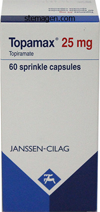
Topiramate
Margaret L. Godley, PhD, CBiol, MIBiol
- Clinical Scientist, Honorary Fellow in Pediatric Urology,
- Institute of Child Health, University College London and
- Great Ormond Street Hospital for Children, London, United
- Kingdom
Topiramate dosages: 200 mg, 100 mg
Topiramate packs: 30 pills, 60 pills, 90 pills, 120 pills, 180 pills, 270 pills

Cheap topiramate 200mg with amex
The lack of ability to use the leg and chronically putting the leg in a dependent place might trigger peripheral edema medicine qid buy 100mg topiramate, a finding often mistaken for venous disease in these sufferers treatment viral conjunctivitis generic 100 mg topiramate overnight delivery. With severe ischemia medicine glossary purchase topiramate 100mg amex, any pores and skin perturbation, including bedclothes or blankets, might trigger pain; in ischemic neuropathy, this causes a lancinating ache within the foot. The embolized material consists of fibroplatelet debris and ldl cholesterol crystals. A frequent reason for atheroembolism is iatrogenic disruption of the vessel, whether from catheterization or surgery. Patients typically have pulses palpable right down to the ankle, as a result of the emboli require a patent pathway to distal parts of the extremities. On examination, the patient will have areas of cyanosis or violaceous discoloration of the toes or portions of the toes and areas of livedo reticularis. Acute limb ischemia could occur from thrombosis in situ or from thromboemboli of huge fibroplatelet accumulations that originate in the coronary heart or large arteries and occlude conduit arteries (see Chapter 44). These sufferers have an accelerated course and will current with the "five Ps" of acute ischemia: ache, pallor, poikilothermia, paresthesia, and paralysis. Other causes of ulcers embrace neuropathy, venous illness, and trauma (see Chapter 61). Nonvascular causes of foot ache embrace neuropathy, arthritides such as gout, fasciitis, and trauma (see Box 18. A blood pressure distinction of 10 mm Hg or extra could additionally be indicative of innominate, subclavian, axillary, or brachial artery stenosis. A very distinguished or forceful pulse may happen in patients with aortic regurgitation or excessive cardiac output states. This most commonly occurs within the setting of iliac artery illness when there could additionally be adequate collateral vessels to maintain perfusion to distal arteries. Examiner ought to place his/her fingers within the curve under the malleolus with mild stress and reposition as needed. The abdominal aorta additionally ought to be palpated to elicit proof of an aortic aneurysm if permitted by physique habitus. Once palpated, the stomach and several other peripheral vessels also ought to be auscultated. Palpation of an expansile or pulsatile periumbilical mass is indicative of an abdominal aortic aneurysm. Bruits ought to be sought over the carotid and subclavian arteries, in the abdomen, within the decrease back, and over the femoral arteries. Presence of a bruit, indicative of turbulent blood flow, sometimes happens on account of arterial stenosis, but might point out extrinsic compression or arteriovenous malformation. A pores and skin examination must be carried out, in search of alterations in temperature, edema, indicators of energetic or healed lesions, or indicators of chronic ischemia-including thin shiny pores and skin, thickened yellow nails, and lack of hair. Inspection of the pores and skin may reveal trophic indicators of chronic ischemia, together with sympathetic denervation (impaired hair growth or impaired sweating) and sensorimotor neuropathy (lack of vibratory sense). After 1 minute, the affected person sits up and the leg is positioned in a dependent position. This affected person with extreme peripheral artery disease (note beforehand amputated second digit) develops a ruborous appearance of the forefoot with dependent positioning as a end result of arteriolar and venular dilation. Arterial fissures most commonly develop in the heel, toes, within the web house between the toes, or in segments subjected to strain (the ball of the foot). The ulcers, in contrast to venous ulcers, are dry; nonetheless, the devitalized tissue is susceptible to infection, which may generate a purulent exudate. The cuffs are placed on each arm above the antecubital fossa and above each ankle. The cuffs are sequentially inflated above systolic stress and then are slowly depressurized. As the cuff is slowly deflated, reappearance of a Doppler signal signifies the systolic strain on the stage of the cuff. Because atherosclerosis might happen in subclavian and axillary arteries, brachial artery pressures should be measured in each arms (A). In each ankle, stress must be measured at the dorsalis pedis pulse (B) and the posterior tibial pulse (C). Brachial artery pressures must be measured in each arms because atherosclerosis might happen in subclavian and axillary arteries. Arterial calcification happens more commonly in patients with diabetes or end-stage renal disease, and in the elderly. Exercise accentuates arterial gradients by growing the turbulence across the flow-limiting lesion and reducing muscular arteriolar resistance to considerably attenuate lower-extremity perfusion strain. In reality, arterial pressure at the ankle might reach zero in patients who develop claudication and get well greater than 10 minutes after exercise cessation. Physiological testing Physiological or functional testing most commonly happens in a noninvasive vascular laboratory (see Chapter 12). Sphygmomanometric cuffs are positioned on the proximal thigh, distal thigh, calf, and ankle. The cuffs are inflated sequentially to suprasystolic stress after which deflated to decide systolic pressure at each website. Arterial stress gradients of greater than 20 mm Hg between thigh cuffs and 10 mm Hg between cuffs under the knee indicate the presence of a stenosis. Pressure is measured within the toes and a ratio of toe stress to brachial artery strain is generated; a worth of 0. Sequential Doppler pressures are measured by inserting sphygmomanometric cuffs on the proximal thigh, distal thigh, calf, and ankle. Simultaneously, a Doppler probe positioned over the dorsalis pedis or posterior tibial artery screens stress. As the cuff is slowly deflated, the reappearance of Doppler sign indicates systolic pressure on the level of the cuff. In this example, arterial pressures (mm Hg) are famous within the location of each sphygmomanometric cuff. The affected person has proof of a systolic gradient between each upper thigh cuffs, which is suggestive of right iliac and/or widespread femoral arterial occlusive disease, and a gradient between proper calf and ankle, which is suggestive of arterial occlusive disease in the infrapopliteal arteries. A vital gradient between the left decrease thigh cuff and the calf cuff is indicative of distal superficial femoral artery and/or popliteal artery occlusive disease. The pulse-volume waveform represents the product of pulse stress and vascular wall compliance. As the stenosis worsens, the waveform upstroke (anacrotic slope) is delayed, the amplitude is much less, and the downstroke (catacrotic slope) is slower. In the vascular laboratory, a prognosis of intermittent claudication and a quantification of exercise tolerance may be obtained via treadmill train testing. Many treadmill protocols exist to take a look at walking capacity, however each falls into one of two varieties: constant or graded exercise. The fall in ankle strain is instantly related to severity of arterial occlusive disease.
Buy 200mg topiramate fast delivery
If craditional suture is used medicine you can order online discount 100 mg topiramate otc, one should keep rigidity to sufficiently close the cuff 9 treatment issues specific to prisons cheap 200 mg topiramate amex. It is advisable to reduce the suture flush with the tissue to decrcue bowel injury danger from the barbed finish medications and mothers milk 2016 purchase 100 mg topiramate with mastercard. Generally, the night of the surgical procedure, the Foley casheter is eliminated, food regimen is advanced, and the patient is allowed to ambu� late. Delay of scmal exercise mirrors that fur abdominal hystem:comy, which is typically 6 weeks. Last, the rou~ of aifF dosure is likdy much less Important than the ~-listed dink:al points. Indications are diversified and include abnormal uterine bleeding analysis, infutility evaluation. Many provide a working channel, into which small operative instru� mcnts are threaded. The patient is placed in standard lithotomy place and the vagina is swgically prepared. If a stan- dard, single-channel, rigid hystcroscope is selected, the endoscope is placed throughout the outer sheath and locked into place. Discomfort related to a vaginal speculum could additionally be avoided by introducing the hysteroscope direcdy into the vagina using a no-tuuth mn! The uterine sound additionally may ttawnatize the cndocervix or endometrium, which alters muc:osal. The forceps arc rcinttoclua:d via the dwind fur additional biopsia, if needed. If diagnostic curettage is planned, it follows bysteroscopy and cavity inspection. Most throughout the endomctrial cavity an: grasped by the saing or stem with bym:roscopic grasping fun:eps and are easily atra. In this case, the arm is grasped and managed p~re is direcud toward the fundus to dislodge it. For diagnostic purposes, a hysteroscope geared up with a 0-, 12�, or 30-degree forward-oblique-view lens is appropriate. Systr:madcally, the hys- teroscope is moved to the fundus after which to the left and proper to pennit irupeaion of the tubal. Medium move is begun, and the rcscct08copc is imcrtcd into the cndoccrvical canal with direct hysteroteopic steering. Distending medium management, panicularly with hypotonic fluid, warrants a degree of safety which could be best offered in an operating suite. Thw, bleeding, infu:don, uterine perforadon, fluid ovctload, and gas emboliu:n are mentioned with the padcnt. Instead, resected fragments are allowed to float throughout the cavity as resection continues. Thus, the cavity may must be emptied previous to full resection to allow an unobstructed view throughout resection. Moreover, the mass is stored between the morcellator opening and the optics ofthe digital camera. The morcellator also provides suction, which may clear blood, tissue debris, and clots throughout resection of large growths. Better visual acuity and the continual retrieval of tissue fragments are two advantages to this method. A critical step at this point, and throughout the procedure, is to note the quantity of distending medium used and retrieved to calculate the fluid deficit. The hysteroscope and the morcellation gadget housed be coagulated with the same resection loop utilizing a coagulating current. If heavy or persistent bleeding ensues, a Foley catheter with a 30-mL balloon can be launched into the uterine cavity and inflated to tamponade vessels. Resection of bigger leiomyomas may be undertaken, though this may require a number of surgical classes to full and require an extended recovery (Camanni, 2010). To allow easier cervical dilation and resectoscope insenion, misoprostol can aid cervical softening (Chap. This method works rapidly on the time of surgery if the need for preoperative preparation was not anticipated (Phillips, 1997). Although the chance of postoperative an infection is low, because pelvic infections can have devastating impact on future fertility, antibiotic prophylaxis previous to extensive hysteroscopic resections, as with myomectomy, is cheap. Candidates embrace chose with abnormal uterine bleeding, with dysmenorrhea, or with infertility during which leiomyomas are suspected co be contributory. Tumors selected for resection ought to be either submucous or intramural but with a predominantly submucous component. During surgical procedure, pcdunculated submucous leiomyomas may be excised similarly to polyps (p. However, tumors with an intramural part require excision with a resecroscope, morccllator, or laser. Hysteroscopic myomectomy is associated with a greater threat of uterine perforation. This complication might observe cervical dilation, but more frequently results during aggressive resection into the myometrium. Because of this danger, women also are consented for laparoscopy to assess and deal with uterine or abdominal organ harm if perforation occurs. Additionally, sufferers planning to search being pregnant are knowledgeable of attainable intrauterine adhesion formation following resection and of uncommon uterine rupture throughout subsequent pregnancies (Batra, 2004; Howe, 1993). During hysteroscopic myomectomy, distention medium can move into open vascularure channels, primarily venous channels, throughout the myometrium. Termed hitravasation, this doubtlessly can lead to intravascular volume overload in the patient. Consequently, sufferers are counseled that more than one surgical session could additionally be required to finish these resections. Fonunatdy, devdopment of newer bipolar resectoscopes and hysteroscopic morcellating tools has helped reduce operating times and thus fluid deficits. Last, myomectomy is efficient remedy, however 15 to 20 percent of sufferers will finally require: reoperation. This may be hysterectomy or repeat hysteroscopic resection at a later time for either persistent or recurrent signs (Derman, 1991; Hart, 1999). Concurrent Ablation In selected girls with heavy menstrual bleeding and with no want for future fertility, endometrial ablation may be concurrently carried out (p. In one retrospective cohon of ladies with submucosal leiomyomas, endomecrial ablation coupled with myomectomy improved symptom outcome and lower remedy failure rates in contrast with hysteroscopic myomectomy alone (Shazly, 2016). Specific leiomyoma traits help predict which leiomyomas are appropriate for hysteroscopic resection and which as a substitute increase the scientific failure fee (Di Spiezio Sardo, 2008). Thus, previous to resection, ladies typically endure transvaginal sonography, saline-infusion sonography, or diagnostic hysteroscopy to determine these characteristics. A totally different system by Lasmar and coworkers (2005, 2011) equally considers the degree of tumor penetration into the myomecrium. In addition, larger tumor measurement, wider rumor base, tumors in the upper portion of the cavity, or selected found along the lateral wall receive larger scores (Table 9-3, p.

Cheap topiramate 100 mg with amex
Laterally 4 medications buy generic topiramate 200mg online, each ovary is hooked up to the pdvic wall by the suspensory ligament of the ovary medications post mi order topiramate 100 mg with visa, clinically generally identified as the infondibulopelvic ligament medicine pouch purchase topiramate 100mg on line, which accommodates the ovarian vessels and nerves. The proper ovarian vein drains into the inferior vena cava, and the left ovarian vein drains into the left renal vein. Lymphatic drainage of the ovaries follows the ovarian vessels to the decrease abdominal aona, the place they drain into the paraaonic nodes. The i11fo11dibulum of the uterine tube is the distal continuation of the ampullary section and accommodates the ftmbriated portion at its distal end. This end has many frondlike projections that provide a large surface area for ovum pickup. The uterus is innervated by fibers of the uterovaginal plexus, also called the Frankenhauser ganglion. The recesses throughout the vaginal lumen Ovaries and Uterine (Fallopian) Tubes Ovaries the ovaries and fallopian tubes represent the uterine aelntxa. A nonkeratinized squamow epithdium and its adjacent lamina pmpria line the vaginal lumen. The vagina is separated from the bladder anteriorly and the rectum posteriorly by the vaginal advcntitia. The continuation of this adventitial layer laterally contributes and blends into the pa. Here, the dense fibromuscular tissue of the pcrincal body separates the vaginal from the anal partitions (OcLanc:ey, 1999). Anatomy 807 Posterior vaginal fornix Veslcocervlcal space c�supravaglnal septum� fibers in decrease part) Vesicouterine peritoneal reflection ~ Rectovaglnal area (filled with loose connective tissue) 1/J. Vesicocervical and Vesicovaginal "Potential" Spaces the vmcocm>ical space begin& below the vesicouterine peritoneal fold or reflection. The vcsicoccrvical house: continues down as the vuicovaginai space, which extends to the junction of the proximal and middle third& of the urethra. Clinically, throughout an stomach hysterectomy or cesarean delivery, surgeons simply raise and incise the vesicouterine peritoneal fold to create a "bladder flap" after which dcvclop the vesicocervical and vcsicowginal spaces. The posterior cul-d~ac is bordered by the vagina anteriorly, the rectosigmoid posteriorly, and the uterosacral ligaments latcrally. The inferior extensions of the utcrosacral ligament fibers, also called rectal pillars, are fibers of the posterior par. These 6bcrs join the vagina to the lateral partitions of the rectum and to the sacrum. They also separate the midllne rectovaginal area from the extra lateral pararcctal spaces. Vaginal Support Although the connective t:i&sue within the pdvis i& continuous and interdependent, Delancey (1992) has described thm: levels of vaginal connective tissue suppon. In the standing place, lcvc1 I help 6bcrs are each vertically oriented (cardinal ligaments) and posteriorly oriented (utcrusacral ligaments) (Chen, 2013). RectovaginalSpace 1his potential area: is straight away adjacent to the posterior surface of the midvagina. These lateral attachments of the vaginal partitions blend into the arcus tendineus fascia pdvis and to the medial aspect of tbe levator ani mwcles, and in doing so create the anterior and posterior latc. These grooves run along the vaginal sidewalls and provides the vagina an "H" form when viewed in cross part. Laterally it attaches to the pubovaginalis muscle and pctineal membrane, and posteriorly to the perincal physique. The distal part of the vagina drains with the vulvar lymphatics to the inguinal nodes. Last, vaginal innervation comes from inferior extensions of the uterovaginal plexus, a component of the inferior hypogastric pl. Anteroinferiorly and laterally, the bladder contacts the unfastened connective and fatty tissue that fills the rct. The reflection of the bladder onto the abdominal wall ia triangular, and the triangle apc:x. The mucosa of the bladder consists of transitional epithelium and underlying lamina propria. The bladder is divided into a hotly (dome) and a forulus (base) roughly at the level of the ureteric orifices. Additionally, the middle rectal attety from the interior iliac anery contributes to the posterior vaginal wall supply. The distal partitions of the vagina also receive contributions from the interior pudenda! Lymphatic drainage Anatomy Vesico-uterine pouch (anterior cul-<1e-ilac) Peritoneum Median umbilical ligament Transversalis fascia 809 Aortic bifurcation Retropublc house Clitoris Vaginal opening Perinealbody Sacral promontory Uterosacral ligament Veslcocervtcal area wilt! The pelvic ureter programs in the pelvic sidcwall rctroperitoncum and is discussed later (p. Together with the sphincter urethrae, they represent the ftriateJ urogmilal sphincter comp/a. This oomplc:x supplies constant tonus and supplies emergency reflex exercise mainly in the distal half of the urethra to sustain continence. Distal to the depth of the perineal membrane, the walls of the urethra encompass fibrous tissue, serving as the nozzle that directs the urine stream. Duct openings of the two most prominent glands, termed Skene glands, are seen on the inner surface of the external urethral orifice (p. Although nonetheless controversial, the pudendal nerve is believed to innervate 810 Aspects ofGynecologic Surgery the anterior and lateral portions of the proximal two thirds of the rectum arc lined by peritoneum. In girls, this culdc-sac is positioned approximately 5 to 6 cm from the anal orifice and can Sphincter ure1hrae m. But, near its termination, it dilates to form the rectal ampulla, which begins under the posterior cul-de-sac peritoneal fold. The rectum lumen contains several, normally three, transverse folds lschiopubic ramus (cut) that include a mucosa, submucosa, and circular layers of the bowel walls. An further di&cuasion of decrease urinary tract innercontinence by supporting fecal matter above the anal canal. It descends Below the anorcctal junction, the intestine extends because the anus, which on the anterior surface of the aaaum for approximately 12 cm has a collection ofanal valves to help its closure. At their decrease border and ends in the anus after passing through the lcvator hiatus. Here, the mucosa of the colon the posterior surface of the rectum is retroperitoneal. Anatomy 811 offers approach to a uansitional layer of non-hair-bearing squamous epithelium. They tie in opposition to the sacrum and levator plate posteriorly and in opposition to the vagina anteriorly. Next, the rectum and ampulla above the pdvic ground obtain a direct department from the middle.

Buy cheap topiramate 200 mg line
The decrease extremity strain analysis ought to start at the ankle degree and proceed proximally symptoms stomach ulcer topiramate 100 mg without prescription. If disease distal to the ankle is suspected treatment 1st 2nd degree burns purchase 200 mg topiramate mastercard, pedal or digital artery obstruction can be evaluated with cuffs sized appropriately for the toes medicine plies discount topiramate 200 mg on-line. Segmental Doppler Pressure Interpretation Segmental limb pressures are in contrast with the very best arm stress. This is accomplished by dividing each of the ankle pressures by the higher of the brachial artery pressures. A 20 mm Hg or higher reduction in pressures from one stage to the next is considered vital and indicates stenosis between these two ranges. In healthy topics, the excessive thigh stress determined by cuff usually exceeds the brachial artery stress by approximately 30 mm Hg. If only one excessive thigh strain is lower than the brachial stress, then an ipsilateral iliofemoral artery stenosis is inferred. The right leg has a strain drop between the low thigh and calf, according to superficial femoral/popliteal artery stenosis. The left leg has a pressure drop on the stage of the high thigh relative to the brachial artery and right high thigh, according to iliofemoral artery stenosis. This ought to be performed in a warm room, as a result of cold-induced vasoconstriction could decrease the digital strain. Pulse Volume Recording Interpretation the identical cuffs used to measure segmental pressures could also be attached to a plethysmographic instrument and used to report the change in volume of a limb section with each pulse, designated the heart beat quantity. Each cuff is inflated in sequence to a predetermined reference stress, as much as sixty five mm Hg. The change in quantity in the limb section causes a corresponding change in pressure in the cuff throughout the cardiac cycle. The configuration of the normal pulse volume waveform resembles the arterial stress waveform and is composed of a pointy systolic upstroke followed by a downstroke that accommodates a prominent dicrotic notch. The normal waveform has a pointy upstroke, dicrotic notch, and a interval of diastasis. The mildly irregular waveform has a delay within the upstroke and a straightened downslope (blue line). The moderately irregular waveform has a delay in upstroke (blue line), flat systolic peak, and diminished amplitude. The severely irregular waveform has a flat systolic peak and very diminished amplitude. Pulse waveforms may also be obtained using photoplethysmography, recording reflected infrared light. Waveform form is assessed similarly in pulse quantity and photoplethysmography recordings. Low photoplethysmographic waveforms in the toes establish elevated threat of amputation, in addition to the toe stress. It is helpful to assess functional capacity and determine the distance sufferers with claudication are able to walk. The fixed load treadmill test is performed at a velocity of 2 miles per hour and an incline of 12%. Graded exercise protocols increase the grade and/or speed in 2- to 3-minute phases. The Gardner protocol is probably the most commonly used graded protocol to evaluate walking exercise capacity. It begins at a velocity of two mph and an incline of 0% and the grade increases by 2% progressively every 2 minutes, permitting for a wider vary of responses to be measured. It is usually used to decide scientific trial end points, similar to change in walking time in response to therapy. The ankle pressures are obtained beginning with the symptomatic leg, adopted by the highest brachial stress. The pressures are repeated approximately each 1 to 2 minutes till they return to baseline. The time before ankle stress returns to normal is elevated in more extreme illness. Transcutaneous oximetry By exploiting the variations in color absorbance of oxygenated and deoxygenated hemoglobin, transcutaneous oximetry can decide the state of blood oxygenation. Deoxygenated blood absorbs more red mild, whereas oxygenated blood absorbs more infrared gentle. Red and infrared gentle is emitted and passes through a comparatively translucent structure, such because the finger or earlobe. A photodetector determines the ratio of purple and infrared light obtained to derive blood oxygenation. When measured repeatedly, oxygenation peaks with every heartbeat as fresh, oxygenated blood arrives in the zone of measurement. One probe is placed on the chest as a management to ensure that the oxygen pressure is from 50 to 75 mm Hg. Measurements are obtained from the probe, which is sequentially positioned from proximal to distal segments of the limb. Physical principles of ultrasonography Ultrasound Image Creation An ultrasound transducer, or probe, emits sound waves in discrete bundles or pulses into the tissue of interest. The fraction of returning waves is dependent upon the density and size of the tissue examined. The depth of tissue is decided by the point required for pulse emission and return. Thus, by integrating the variety of returning pulses and the time required for return, a B-mode, or gray-scale, image may be created. Transducer probes with higher frequencies picture superficial tissues better than probes with lower frequencies however lose depth imaging because of attenuation of the returning emitted pulses. Improvements in technology have permitted a widening of the bandwidth of vascular transducers and may enhance gray-scale imaging using harmonics of the fundamental frequency. Because the tissue compresses and expands in response to the appliance of ultrasound, the basic wave might turn into distorted, impairing image quality. However, the distortion also creates harmonics of the unique frequency that could be detected by the transducer. Detection of Blood Flow Normal blood flow is laminar in a straight phase of an artery. If thought of as a telescopic sequence of move rings, blood moves ahead most quickly within the middle ring and velocity decreases within the outer rings as blood comes nearer to the vessel wall. The cardiac cycle, outlined by its pulsatile nature of move, causes a continual variation in blood move velocity, highest with systole and lowest with diastole. The concentric or laminar flow of blood may be disturbed at a traditional branching point or with abnormal vessel contours, such as these attributable to atherosclerotic plaque. Disturbed or turbulent flow causes a a lot greater lack of pressure than does laminar move. Flow in a standard vessel is proportional to the difference of strain between the proximal and distal end of the vessel.

Order 200mg topiramate overnight delivery
The timing speculation: do coronary risks of menopausal hormone therapy vary by age or time since menopause onset treatment locator purchase topiramate 200 mg otc. Common genetic loci influencing plasma homocysteine concentrations and their impact on threat of coronary artery illness treatment plant generic 200mg topiramate free shipping. Effects of decreasing homocysteine ranges with B nutritional vitamins on heart problems treatment jock itch purchase 100mg topiramate visa, most cancers, and cause-specific mortality: meta-analysis of eight randomized trials involving 37 485 individuals. Lipoprotein(a) as a possible causal genetic danger issue of cardiovascular disease: a rationale for elevated efforts to perceive its pathophysiology and develop focused therapies. Plasma fibrinogen stage and the chance of main cardiovascular illnesses and nonvascular mortality: an individual participant metaanalysis. Additive worth of immunoassay-measured fibrinogen and high-sensitivity Creactive protein ranges for predicting incident cardiovascular events. Effects of antibiotic therapy on outcomes of sufferers with coronary artery disease: a metaanalysis of randomized controlled trials. Repeated replication and a prospective meta-analysis of the affiliation between chromosome 9p21. Six new loci associated with blood low-density lipoprotein ldl cholesterol, highdensity lipoprotein cholesterol or triglycerides in humans. From loci to biology: useful genomics of genome-wide association for coronary illness. Phenotypic penalties of a genetic predispostion to enhanced nitric oxide signaling. Evaluation of the pooled cohort equations for prediction of cardiovascular threat in a contemporary prospective cohort. Longitudinal and secular tendencies in whole cholesterol levels and influence of lipidlowering drug use among Norwegian men and women born in 1905-1977 in the population-based Tromso Study 1979-2016. Trends in weight problems prevalence among kids and adolescents within the United States, 1988-1994 by way of 2013-2014. Subendothelial lipoprotein retention because the initiating course of in atherosclerosis: update and therapeutic implications. Distinct features of chemokine receptor axes in the atherogenic mobilization and recruitment of classical monocytes. Leukocytes link native and systemic inflammation in ischemic cardiovascular disease. A rock and a tough place: chiseling away at the multiple mechanisms of aortic stenosis. Arterial and aortic valve calcification inversely correlates with osteoporotic bone remodelling: a job for inflammation. Macrophage-derived matrix vesicles: an alternative novel mechanism for microcalcification in atherosclerotic plaques. Vascular smooth muscle cell calcification is mediated by regulated exosome secretion. Macrophage apoptosis exerts divergent effects on atherogenesis as a perform of lesion stage. Plaque development in coronary arteries with minimal luminal obstruction in intravascular ultrasound atherosclerosis trials. The contribution of plaque and arterial transforming to de novo atherosclerotic luminal narrowing in the femoral artery. Arterial remodelling: an unbiased pathophysiological element of atherosclerotic disease progression and regression. Effects of coronary heart illness risk elements on atherosclerosis of selected regions of the aorta and proper coronary artery. Childhood cardiovascular threat factors and carotid vascular modifications in maturity: the Bogalusa Heart Study. Maternal-fetal cholesterol transport in the placenta: good, dangerous, and goal for modulation. High prevalence of coronary atherosclerosis in asymptomatic teenagers and younger adults: proof from intravascular ultrasound. Combined noninvasive evaluation of endothelial shear stress and molecular imaging of irritation for the prediction of infected plaque in hyperlipidaemic rabbit aortas. Extent and course of arterial remodeling in secure versus unstable coronary syndromes: an intravascular ultrasound study. Cytokines and growth components positively and negatively regulate intersitial collagen gene expression in human vascular smooth muscle cells. Plaque erosion: a brand new in vivo diagnosis and a possible main shift in the administration of patients with acute coronary syndromes. Flow perturbation mediates neutrophil recruitment and potentiates endothelial harm via tlr2 in mice - implications for superficial erosion. Hypochlorous acid, a macrophage product, induces endothelial apoptosis and tissue factor expression: involvement of myeloperoxidasemediated oxidant in plaque erosion and thrombogenesis. Impact of statins on serial coronary calcification during atheroma progression and regression. Mechanisms of progression in native coronary artery disease: position of healed plaque disruption. Nonculprit plaque traits in patients with acute coronary syndrome attributable to plaque erosion vs plaque rupture: a 3-vessel optical coherence tomography research. These have traditionally been thought-about as a manifestation of atherosclerosis but-as rising epidemiological and pathomechanistic evidence suggests-may, in reality, characterize a distinct disease. Structurally, this ends in considerably greater elastin content material of the thoracic aorta compared with the stomach phase, leading to greater mechanical distensibility and buffering cyclical mechanical stress from pulsatile cardiac action (aortic Windkessel function). Genetically, aneurysmal aortopathies (typically in thoracic aortic segments) could occur as a consequence of heritable, single-gene easy muscle cell protein mutations leading to irregular function or signaling, or premature breakdown. This phenomenon is especially important within the infrarenal belly aorta, which lacks the nutritive intramural vasa vasorum found in more proximal aortic segments. The comparatively hypoxic surroundings might induce subsequent neovascularization and irritation. Accordingly, aging leads to heterogeneous aortic stiffness and resultant stiffness gradients alongside the stomach aorta. However, these mechanical stimuli may also elicit subtler organic responses via quite so much of signaling mechanisms in varied vascular cell types. Epigenetics/micrornas in aortic aneurysms A latest appreciation has developed for the crucial position played by epigenetic elements in disease regulation. Unfortunately, our in depth knowledge of these processes has not but translated into efficient medical therapies. Localization of aortic disease is related to intrinsic variations in aortic structure. Matrix metalloproteinases and tissue inhibitors of metalloproteinases: construction, perform, and biochemistry.

Strophanthus Seeds (Strophanthus). Topiramate.
- Artery disease, heart problems, high blood pressure, and stomach problems.
- Dosing considerations for Strophanthus.
- Are there any interactions with medications?
- What is Strophanthus?
- Are there safety concerns?
- How does Strophanthus work?
Source: http://www.rxlist.com/script/main/art.asp?articlekey=96254
Quality topiramate 200mg
Patient Preparation Bleeding is a common problem with pelvic lymphadenecromy and could additionally be exacerbated with obese patients medicinebg purchase topiramate 200mg mastercard, grossly enlarged or densely adhered lymph nodes treatment upper respiratory infection buy topiramate 100 mg, and pelvic vessel anatomic variants symptoms ruptured spleen buy 200mg topiramate mastercard. This surgical procedure may be carried out beneath basic or regional anesthesia with a patient supine. A midline vertical or transverse stomach incision that allows access to the previously famous anatomic boundaries is appropriate for this procedure. A Pfannenstiel incision offers restricted exposure and is reserved for selected patients. Pelvic and paraaortic lymph nodes are routinely inspected during preliminary stomach exploration. Unc:x:pected grossly optimistic nodes may indicate that a proposed operative plan should be abandoned (for instance, radical hysterectomy for cervical cancer) or revised (Whitney, 2000). With an incision made parallel and superficial to the artery, distal nodal tmue is freed. Mobilization of this tissue exposes the deep circumftcz: iliac vein, which crosses laterally over the distal atcmal iliac artery. Once completed, this cxtemal iliac nodal group dissection later permits safe enuy into the obturator space, outlined in Step 7. Spatially, nodes that have been dWeacd off the exterior iliac vessels aiid the fatty nodal tissue bridging the cnemal iliac vein and the internal iliac artery lie in the same airplane. Beginning on the frequent iliac artery bifurcation, the free nodal bundle is clcwtcd and placed on rigidity. Initial sharp dissection of the interior iliac nodal group continues caudally alongside the internal iliac vwcls and thc. Lateral arterial or venous branche& may need vascular clip application and a:anscction. During this dissection, nodal tissue may be identified behind the external iliac vmds and added to the specimen. With the vein retractor in place, the obturator nodal tissue is grasped with forceps. With upward traction applied, blunt forceps or a suction tip moved gently sidoto-side disrupts nodal tissue attachments to the obturator nerve. Finn fibrotic attachments may be electtosurgically transectc:d beneath direct visualization. At the cephalad end of the bundle, nodes are fastidiously separated sharply from the inferior side of the exterior iliac vein whereas avoiding obturator nerve injwy. To take away this group, the upper retractor blade is readjurted to expose the dlrta. The colon may requite mobilization using dccttosurgial di"ec:tion along the white line of T oldt. Lateral fatty-lymphoid tissue could additionally be eliminated by first grasping and elevating with forceps and utilizing electroswgial dissection to establish a plane. Notably, uanscaion of the obturator nerve is ideally instantly noted inttaop-erativd. Surgical blunt dissection n:dmiques decrease the danger of inadvertent vessd or nerve lnjuiy, but these might improve the possibility of postop-erative lymph. Moreover, removal of enlarged paraaortic nodes may provide optimum debulking of ovarian cancer and may confer a survival benefit in chosen endomc:trial and cc:rvical cancer sufferers (Cosin, 1998; Havrilesky, 2005). Paraaortic lymphadenectomy implies the bilateral full: removing of all nodal tissue: from inside an space with well-defined anatomic boundaries: inferior mesenteric artery (cephalad), midlength of common iliac artery (caudad), ureter (lateral), and aorta (medial). The completeness of the procedure will range by clinical setting, but an adequate dissection requires that lymphatic tissue: a minimum of be: demonstrated pathologically from both the right and left sides (Whitney, 2010). Most usually, that is perfurtned throughout ovarian cancer staging or in high-risk endometrial most cancers circumstances to debulk rumor and more accuratdy stage these cancers (Mariani, 2008; Morice, 2003). Once bowel has been cleared, the peritoneum overlying the aorta and proper frequent iliac artery must be visible. Also, as described on web page 1162, the: urc:tc:r is isolated and hdd laterally on a Penrose drain to keep away from its damage. Lymphadenectomy could also be carried out underneath common or regional anesthesia with a patient supine. A midline vertical stomach incision that enables entry to the previously famous anatomic boundaries is suitable for this process. Low transverse incisions supply limited exposure and are reserved for sdecred patients. The paraaortic lymph nodes are routinely palpated during preliminary belly exploration. The index and middle fingers arc: then used to straddle the: aorta and palpate for lymphadenopathy. Suspicious or grossly constructive paraaortic nodes are typically eliminated as an preliminary step. Unexpected positive nodes might indicate that the proposed operative plan must be abandoned or revised (Whitney, 2000). For most cases, during which no adenopathy is present, the: dissection is normally performed final due to the potential for triggering catastrophic bleeding that may in any other case restrict further surgery. Beginning atop the midportion of the proper frequent iliac artery, a right-angle clamp is used to guide dectrosurgical blade: incision of the posterior parietal peritoneum. Continuing cephalad in the midline, sharp incision of the peritoneum is extended via the caudal and then left lateral facet of the duodenal peritoneal reflection to mobilize the duodenum cephalad. An upper midline sdf-retaining retractor blade is repositioned to retract this bowel. In overweight women, operative visibility is hindered, and thus process: complexity and operative times are significantly larger. Exposure and correct retractor positioning is perhaps the most important part of this procedure. The sigmoid colon and descending colon are gently retracted in a lower left course, whereas small bowd and transverse colon are packed into the upper stomach by laparotomy sponges. Modified Trendelenburg patient positioning additionally is helpful to shift bowd horn the operative fidd. Additional sharp dissection alongside the right paracolic gutter peritoneum (white line ofToldt) may be: necessary to sufficiently mobilize and move Right Paraaortlc Nodes. With the ureter still held laterally, the surgeon first establishes the medial border of the best paraaortic nodal group. Atop the midportion of the: right common iliac artery, the lymph node bundle is devated with forceps to reveal fibrous bands connecting it to the artery. A right-angle clamp is positioned beneath these: bands, that are then sharply divided co free the distal bundle from the artery. Using decttosurgical cutting atop the best widespread iliac artery, cephalad and slightly medial dissection continues following the vessd course. To establish the: lateral border of this nodal group, the ureter is once more hdd laterally.
Syndromes
- Diabetic neuropathy
- Shows affection
- Pertussis immunization (vaccine)
- Excess hair growth on the face, neck, chest, abdomen, and thighs
- Fever and chills that come and go
- Certain types of skull fractures can cause bruising around the eyes, even without direct injury to the eye.
Buy topiramate 100mg
A& a n:sult symptoms for pregnancy topiramate 100mg for sale, the tissue is excised in two pic:ccs medications given for uti 200mg topiramate with mastercard, and both arc despatched fur analysis treatment multiple sclerosis cheap topiramate 200 mg overnight delivery. With plac:ement of Sturmdorf sunues, a operating locked suture line closes the cone bed by circumferentially folding the minimize ectoce. Surgeries for Benign Gynecologic Disorders is directed to reduce and take away the cone-biopsy specimen. A higher power density is used to create a chopping effect, for instance, 25 W with a 1-mm spot measurement (power density = 2500 W/cm2). During excision of the cone specimen, nonreBective tissue hooks could additionally be wanted to retract the cctoccrvical edge away from the laser beam path and to create tissue rigidity alongside the aircraft of incision. However, due to the larger incision, postoperative bleeding can develop more generally and is treated with Mansel paste or other topical hemostat. Patients require postoperative surveillance for disease persistence or recurrence, and this is described intimately in Chapter 29 (p. Tuia mne spana from the ccnter of the cryoprobe to a perimeccr 2 mm indtJe the outer iceball edge. Although some use these iceball dimensions to direct therapy, the World Health Organization (2014) recommends a doublefreeze method for cryotherapy, described within the steps beneath. Importantly, a single-&eeze methodology is imuHiclent, and dyspluia recurs incessantly within the fi. The particular indicatiat11 and long-term charges of success for ayothenpy arc dia:ussed in Chapter 29 (p. Anaiomically, a shon celYb: that becoml:ll flush with the vaginal vault wider mod. Ciraunfi:rcntial grooves on the ayoprobe base permit it to be saewed securely to the tip of the cryogun. Selection of an acceptable cryoprobe is individualized however should cover the trans-formation zone and lesion. A water-based lubricant jelly is applied m the top of the cryoprobe to guarantee even ti&sue contact. The set off is held fur 3 minutes u the ic:eball c:ncnds put the outer margin of the ayoprobe. The 8 crytiprobe is gently teased away from the wall, after wbkh the process is continued. Attempts to remove it prior to full defrosting could cause patient discomfort and bleeding. Accordingly, sufferers abstain from intercourse through the four weeks following surgery. Depending on patient symptoms, work and train might resume shortly following therapy. Therefore, moistened doth toWds arc draped outside the vulva to absod> misdirected vitality. The colposcope-laser meeting is introduced into position and centered on the ectocei:vilt. These dots serve as landmarli:s and are connected in an arching pattern to create a circle. Laser ablation for many women is an outpa� tient process and carried out in both an operating suite or an workplace, depending on laser equipment location and affected person clwacteristics. As with any 1006 Atlas of Gynecologic Surgery counsding relating to expectations fur anatomic: consequence and fur sexual function. Most rca>mmend a 5- to 10-mm circumferential surgical margin surrounding the lesion Qoura, 2002; American College of O~tetricians and Gynecologists, 2019c). However, nonual tissue can also absorb the stain and deform true disease margins, and use of toluicline blue is thus not beneficial. Wide native excision oflesions, ablative methods, and medical therapies arc dfec:tivc and commonly seleaed (Chap. If malignancy is suspected, extensive local excision is performed to determine or exclude oc:cult invasion (American College of O~tetrlcians and Gynecologi5u, 2019c:). This mixed approach makes use of C02 luer vaporb:ation at sites the place o:clsion may lead to dysfunction or poor cosmesis (Cardosi, 2001). For extra widespread disease, easy vulvcctomy may be applicable and is described in Section 46-24 (p. Whereas smaller labial or pcri� ncal lesions could simply be exdsed utilizing native analgesia in an office setting, larger lesions or those involving the u. The Patient Evaluation Prior to excision, full analysis of the decrease reproductive tract for evidence of invasive disease is completed as outlined. Most prcinwsive lesiom fail to prolong deeper than I mm on non-hair-bear� ing areas suc:b. Adson forceps or pores and skin hooks can elevate and retract the pores and skin matgin away from the incision line. Diucction beneath the lesion begins at the incision periphery and progresses towards the centcr of the proposed aclsion space after which to the other incision margin. Disease re<:Urrencc is expounded to the presence or absence of di~c-frec sw:gic:al margins. However, disease regularly recurs, and even in those patients with tissue margins negative for illness, recurrence ranges from 15 to forty percent (Modc:aitt, 1998; SatmaJ:y. Las frequent problems are chronic vulvodynia, dyspareunia, and scarring or altered vulvar appearance. Reappro:dmation of the woWld edges with out pressure ciccrcases the danger of postopc. Accordingly, a swgcon may need to sharply Wldennine the wound margins with fantastic acissors to mobilize the akin and instant underlying subcutaneous tissue. The edges of the skin are then rcapproximat�l with inwruptcd stitches utilizing 3-0 or 4-0 gauge 9 Wound Closure. Although the proccdute permits for tissue cwluation, tissue disruption may preclude enough examination of all components of the specimen and th. Irrigation is used to management the appreciable warmth generated by the vibrating titanium tip (23 kHz) of the hand piece and to suspend the fragmented tl. The bigger of the 2 is located in the midportion of the proper labium minus, and the smaller is extra anterior and toward the clitoris. Irrigation and aspiration charges may be various relying on the necessity of the operator. Only close oont:act with the pores and skin of the vulva is required; no stress is nca:ssary�. One example ls if multlfucal illness iiivolves each hair-bearing and nonhair-bearing areas, such as the clitoris, where excision may not be best.

Safe 200 mg topiramate
These are thought of in cases in which anatomic distortion is anticipated or the uterus giant medications for high blood pressure buy discount topiramate 200 mg on-line. The bowel is gently displaced from the pelvis into the stomach to broaden avail� ready working house and viewing symptoms kidney disease buy topiramate 100mg lowest price. For bigger uteri medicine gabapentin 300mg capsules 100mg topiramate for sale, if the uterine funclus is dose to or above the level of the umbilicw, the optical port is placed roughly three to four cm above the fundus for optimum viewing. Irrigating fluids and C02 used fur insuftlation can, with time, crem peritoneal cdema. The opening within the peritoneum then is c:x:tendc:d caudally and cephalad along the apected axis of the ureter. After broad lipent incision bilaterally, the vesiccuterine fold is grasped with atraumatic forceps, elevated away from the underlying bladder, and inc. Loosely connected connections can be bluntly divided by gently pushing in opposition to the c:ervix and. As this tissue is dissected, the e Round Ug1ment Tr1nsectlon, the proximal spherical ligament is graspccl and divided. With this, the tube and ovaiy are fu:c:cl &om the uterus and cui be placed in the ov:arian fem. For elimination of one or both ovaries, the n> ligament is graspccl and pulled up and. This incision is directed caudally and tentrally to the midllne above the veslc:outerine fold. The posterior leaf requires incision caudally to the levd of the uterosacral ligament. Development of this area allows the bladder to be moved caudally and off the decrease uterus and upper vagina. Cystotomy happens most regularly to the dome throughout this sharp or blunt dissection (Harkki, 2001). Scarring within the vesicouterine space from prior cesarean supply or cndomctriosis can increase this danger. With laparoscopic hysterectomy, after the uterine arteries arc transected, the surgical strategy is convened co that for vaginal hysterectomy and is completed as outlined in Section 43-13 (p. In this transition, the patient is repositioned from low lithotomy to standard lithotomy place throughout the same booted support stirrups. During this inspection, inttaabdominal pressures are lowered to better establish sources of bleeding. A clear liquid diet may be initiated the day of surgical procedure and superior quickly as tolerated. Postoperative complications in general mirror these for belly hysterectomy with the exception that superficial surgical web site an infection rates are decrease. After the uterine arteries are identified, the areolar connective tissue surrounding them is grasped, placed on pressure, and incised. This skdctonizing of the vessels results in superior occlusion of the uterine artery and vein. After vaginal completion of the hysterectomy, consideration is redirected to laparoscopic inspection of the pelvis for signs of bleeding. Pwtopc:wM:ly, ettdomcaium throughout the lowu ua::rine segment may be retained with l. Ran:s quoted in early studies attain 24 percent however are lower in more reccot investigations and vary fi:om 5 to 10 percc. With laparoscopic gyneoologic surgical procedure, the choice to present VfE prophylaxis fu:ton patient- and proc:c>dure-related VfE dangers (Gould. Specifically, adhesions bctwccn the bladder and the lower uterine phase in the vesic:outerine area or these within the posterior cul-<le-sac may make elimination of the cemx tough. Certain conttaindications to preserving the cervix are sought prior to sdccting supracc. After rumor acision, elimination may be completed by a number of tcc:hniqucs described in steps 3, four, and 5. Patient Preparation A blood pattern is typed and crossmatched for potential transfusion. Ifconsidered, bowd preparation prior to laparoscopy may assist with colon manipulation and pdvlc anatomy visualization by cvawating the rcc:tosigmoid. Onc:e a patient is deemed eligible for a lapuoscopic approach, preoperative analysis mirrors that for lap~ scopic hysterectomy, described in the last section. The corpus is amputated &om the cervix at some extent just below the interior a:tvical os and superior to the uterosa. First, for smaller specimens, a minilaparotomy incision tanging from three to four cm may be made to atract the corpus. The um-ine manipulator is lifted anteriorly, and an Alllt damp is placed on the potterior vaginal wall 2 to 3 an &om the potterior c;erviconginal junaion. Alternatively, a c:olpotomy may be created laparoccopically by incising the posterior cul-de-Ge with a monopolar imuument, a Harmonic scalpd, or Endo Shean close to the mvkowginal junction. Por larger uteri, the addition of a tissue retrieval bag during tiAue extraction through a colpotomy iocifion can create a closed envirorunent for sO. Following cruaction, a colpotomy incision is dosed with interrupted stitches or a running suture line uaing. One= irWde the abdomen, the bag 11 unfolded to allow the specimen and gas to be contained. During energy morcellation, the corpus specimen is grasped securely with a toothed in. Improved slicing i4 usually restored with thil step and usually presents enough bJade lif-e to complete the proc:edure. Following mon:dlation, the g;as is rdeaxd, and the bag and its contents are eliminated. Limiarions of at present out there retrieval hap Involve pouch size, working aperture diameter, tcnsilc energy of the bag, and bag pcnneability (Cohen, 2016). The uterus is also sounded to determine cavity depth for correct manipulator placement. Once once more deflated, the tip is passed by way of the cervical os, into the endometrial cavity, and to the fundus. To securely anchor the cup and cervix, stitches enter the ectocccvix and exit just lateral to the endocervix. Once in position, the proximal rim of the cup will delineate the cervicovaginal junction. If the Koh Cup is used, a pneumo-occluding balloon is positioned behind the colpotomy cup.
Generic topiramate 200 mg line
Another is venous absorption of large volumes of sure distending media throughout lengthy operative hysteroscopy circumstances (Chap medicine zalim lotion effective topiramate 100 mg. In addition medicine cabinets with lights generic topiramate 200 mg mastercard, extrarenal sodium losses may follow profuse diarrhea mueller sports medicine buy generic topiramate 200 mg online, vomiting, or nasogastric suctioning. Similarly uncommon, extreme renal excretion of sodium might accompany diuretic overuse or adrenal insufficiency. Severe hyponatremia can result in metabolic encephalopathy and associated cerebral edema, seizures, elevated intracranial pressure, and even respiratory arrest. Overaggressive correction may end up in a specific demyelination dysfunction often identified as central pontint mytlinolysis. In those with out symptoms, cautious replacement with isotonic fluids and remedy of underlying situations will correct most circumstances. In those with acute neurologic signs, 3-percent saline may be given in a 100-mL infusion over half-hour and repeated an extra two instances if needed (Nagler, 2014; Verbalis, 2013). Hypokalemia is often caused by diarrhea or by abnormal renal losses secondary to metabolic alkalosis. Mild hypokalemia is usually asymptomatic, but nonspecific signs seen with development embrace generalized weak spot and constipation. Compared with N substitute, oral potassium is safer as a result of it enters the circulation extra slowly and reduces the chance of iatrogenic hyperkalemia. Magnesium depletion can cause hypokalemia refractory to alternative efforts, and magnesium could must be concomitantly replenished (Whang, 1985). Pseudohyperkalemia may end up from traumatic hemolysis, released from muscles distal to a tourniquet, or from cellular launch in a clotted specimen tube. If hypokalemia is an unsuspected discovering in an asymptomatic affected person, repeat measurement is sound. Instead, digitalis and [3-receptor antagonists may cause transcellular shifts in potassium. With administration, basic principles emphasize protecting myocardium, shifting potassium intracellularly, and enhancing potassium excretion. Intravenous calcium gluconate administered as 10 mL of a 10-percent answer over 2 to three minutes � Hypernatremia Hypematremia is outlined as a serum sodium concentration > one hundred forty five mEq/L. Common causes are loss ofhypotonic physique fluids such as diarrhea, gastric secretions, and sweat. The resulting plasma hypertonicity draws water out of cells to preserve intravascular fluid compartment quantity. To restore brain cell volume, the brain metabolically generates compensatory compounds, termed idiogenic osmoles, which Postoperative Considerations antagonizes the impact ofpotassium on myocardial repolarization and the conduction system. Alternatively, a ~2-agonist, similar to inhaled albuterol, can drive an analogous potassium shift. Atelectasis is widespread however is extra likely a coincidental quite than causal discovering (Mavros, 2011). Logically, catheterization length correlates positively with this infection risk. For example, ladies with ovr usually complain of unilateral lower-extremity edema and erythema. As for wounds, fever related to surgical site an infection usually develops 5 to 7 days after the procedure. Last, medicines commonly used postoperatively-such as heparin and numerous antibiotics-may trigger a rash, eosinophilia, or drug fever. The yearly incidence in hospitalized patients approximates 5 circumstances per a hundred,000 persons (Galindo, 2019). An acute occasion, similar to surgery, trauma, or parturition might precipitate thyroid storm. Hypotension, arrhythmias, and cardiovascular collapse leading to dying are others. A ~-blocking agent and a thionamide, similar to propylthiouracil or methimazole are provided. An iodine solution may be administered to block the release of thyroid hormone, and glucocorticoids are given to cut back the conversion of thyroxine (T4) to triiodothyronine T3) Ross, 2016). Thus, preliminary assessment of a girl with postoperative fever is individualized and begins with a targeted history and physical examination. The infiltration of leukocytes and launch of cytokines helps provoke the proliferative phase of wound restore. During this, two activities happen simultaneously-the development of granulation tissue to fill the wound and the formation of epithelium to cowl the wound. The final stage, remodeling, restores the structural integrity and practical aptitude of the new tissue. Postoperative hyperglycemia is a well-recognized threat issue for surgical site infection (Hopkins, 2017). Other undesired hyperglycemia effects are dehydration, electrolyte abnormalities, and possibly ketoacidosis in patients with sort 1 diabetes. Thus, maintaining random blood glucose levels <200 mg/dL is advocated Oacoher, 1999). Recommendations concerning perioperative management of oral hypoglycemic brokers and insulin could be present in Tables 39-6 and 39-7 (p. Care is taken to keep away from hypoglycemia, which can develop especially throughout oral consumption modifications. The reported incidence of superficial separations ranges from 3 to 15 % (Owen, 1994; Taylor, 1998). Fascial dehiscence occurs much less frequently and is associated with greater morbidity Carlson, 1997). Infection or sutures held beneath too much pressure are infamous causes and result in Fever is a response to inflammatory mediators, termed pyrogens. The inflammatory cascade additionally produces a number of cytokines in response to surgery, cancer, trauma, and infection Wortel, 1993). Thus, fever is common after surgical procedure and is self-limited typically (Garibaldi, 1985). However, for those with persistent signs, a scientific method to patient evaluation helps differentiate inflammatory from infectious etiologies. Fevers that develop more than 2 days after surgery are more likely to be infectious. More broadly, causes are categorized by 922 Aspects of Gynecologic Surgery Time since surgery t 0-24 hours I >24 hours t High-risk & low-risk No evaluation essential except symptomatic t High-risk�, symptomatic High-risk, asymptomatic w/low-grade feverb Low-risk, asymptomatic t Laboratory and radiologic analysis t Urine culture when catheterized t Follow for extra temperature increase or signs of an infection t Begin with history and bodily examination No laboratory or radiologic analysis needed �High risk: (1) Cancer, with extra advanced stage of disease; (2) lmmunosuppressed; (3) Bowel resection; (4) Temperature> 38. These layers then separate with solely minimal will increase in intraabdominal strain. Prevention Rates ofwound dehiscence are affected by basic affected person health, surgical method, and dangers related to wound infections. These embody age older than 65 years, pulmonary disease, malnutrition, weight problems, malignancy, immune compromise, diabetes mellitus, and hypertension (Hodges, 2014; Riou, 1992).
References
- Jeremic B, Shibamoto Y, Igrutinovic I. Single 4 Gy reirradiation for painful bone metastases following single fraction radiotherapy. Radiother Oncol 1999;52(2):123-127.
- Cooke GS, Campbell SJ, Sillah J, et al. Polymorphism within the interferon-c/receptor complex is associated with pulmonary tuberculosis. Am J Respir Crit Care Med 2006; 174: 339-343.
- Galie N, Barbera JA, Frost AE, et al. Initial Use of Ambrisentan plus Tadalafil in Pulmonary Arterial Hypertension. N Engl J Med. 2015;373:834-844.
- Hoffman JIE, Christianson R: Congenital heart disease in a cohort of 19,502 births with longterm follow-up. Am J Cardiol 1978; 42:640-647.
- Elkind MM, Sutton-Gilbert H, Moses WB, et al. Radiation response of mammalian cells grown in culture. V. Temperature dependence of the repair of x-ray damage in surviving cells (aerobic and hypoxic). Radiat Res 1965;25:359-376.
- Wilson FC, Manly T, Coyle D, et al. The effect of contralesional limb activation training and sustained attention training for self-care programmes in unilateral spatial neglect. Restor Neurol Neurosci 2000;16:1-4.

