Cleocin
Lorenzo Biassoni, MSc, FRCP, FEBNM
- Honorary Senior Lecturer, Institute of Child Health,
- University College London
- Consultant in Nuclear Medicine,
- Department of Radiology, Great Ormond Street Hospital for
- Children, London, United Kingdom
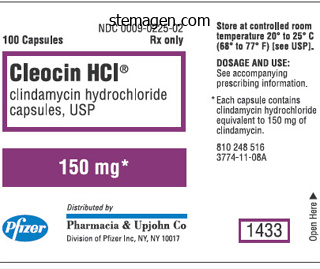
Cleocin dosages: 150 mg
Cleocin packs: 30 pills, 60 pills, 90 pills, 120 pills, 180 pills
Order cleocin 150mg visa
Partial fibulectomy: Weight-bearing acne 12 weeks pregnant cheap 150mg cleocin otc, intact fibula or early union of the fibula or early union of the fibula for two results skin care wholesale cheap cleocin 150 mg otc. Therefore acne jensen dupe discount 150 mg cleocin otc, partial fibulectomy by allowing nearer apposition and weightbearing on the tibia might promote union. However, it may have advantage over tibial intramedullary nailing in sufferers who had external fixators, since it avoids the pin tracks. The gap may be due to congenital pseudarthrosis of fibula or because of resection of fibula use as a bone graft in grice subtalar arthritis. To prevent valgus deformity of the ankle, it is recommended to fuse the distal fragment with tibia. When the fragments of a nonunion are in good place, fibrous union may be suitable with passable perform. The severity of later arthritic changes is about proportional to the irregularity of the articular floor of the patella. When the fragments are separated, partial or full excision of the patella, as for a fresh fracture, is indicated. Femur Supracondylar fracture: If the fractures are severely osteoporotic, screws used with the plate ought to be augmented with bone cement, longer plate must be used, autogenous bone graft is mandatory. When comminution is extreme or the distal fragment is small, fixation could be completed by an intramedullary nail traversing the femur, knee joint and proximal tibia. We use tricortical graft from the iliac rest in the medullary canal to enhance the soundness and fee of union. Intramedullary nail of humeral nonunion has not as profitable as in the nonunion of femur or tibia. The longer defect could be bridged with a tricortical hole or a fibular graft combined with shortening of 4�5 cm. One finish of the transplant must be inserted within the metaphyseal part, proximally or distally. The Ilizarov methodology of inside bone transplant also can be used for humeral nonunions with bone loss. Nonunion of lateral condyle of humerus: Nonunion of lateral condyle of humerus occurs in childhood with tuberous, valgus deformity, instability and science of strain of the ulnar nerve. Nonunion in kids hole treated as much as 4 months, by bone discount, bone grafting and secure fixation by lack screws. In adults, when the elbow is asymptomatic, no remedy is indicated aside from anterior transposition of the ulnar nerve for aid of ulnar neuritis. Technique: Freshen, appose the fracture surfaces, and fix the fragment to the humerus with two screws. Nonunion of Monteggia fracture, radial head and as a lot of the neck as needed are resected. The fragments of the ulna are fastened with an intramedullary nail or a compression plate, and iliac grafts are placed in regards to the nonunion. Defects within the radial head or neck or the distal 5 cm of the ulna are treated by excising the fragment. Very large defects in the radius can be handled by making a single-bone fora by an operation. When the fracture web site is opened, shingling (decortication) of the fragments 2 cm on either side of the nonunion aspect and bone graft are carried out. Bone grafting with a small incision or a cancellous bone through a tube could additionally be necessary. Large gaps also may be treated with the Ilizarov external fixator and inner bone transport. Pelvis and Acetabulum Nonunion of pelvis and acetabulum do agree often and required remedy. Patients with good bone inventory are treated with inflexible inside fixation and cancellous bone grafting. Proximal Humerus A tension band building that fixes the rotator cuff and proximal fragment to the remainder of the shaft, in addition to two or three cancellous screws within the proximal fragment is recommended. In addition a locking plate with screws and pressure band wire through the rotator cuff is used. Combination of pressure band and buttress plate technique for nonunion of proximal humerus can be utilized. NoNuNioN of fractures of LoNg BoNes gaps in each bones of the fora, when gaps are present in both bones, the sclerotic finish must be excised, plating and grafting is done. Equalizing the bones, if the gaps are longer, whole fibula or tricortical graft can be used. Amputations:three It is necessary to take a decision of amputation of severally injured limp with irreparable damage to the muscle, tendons, nerves or vessels; or unsatisfactory pores and skin protection. To reconstruct such a limp takes a very lengthy time, patient might undergo extreme financial hardships and if it is unsuccessful and amputation is then advised affected person becomes very unhappy. Indications for amputations are: � Senseless foot with broken muscles � Function is severely compromised � Reconstruction is impossible. Amputation in such a patient is kinder to the patient with proper fitting, affected person goes back to work at earlier date. Bacterial adherence and the glycocalyx and their role in musculoskeletal an infection. Fresh autogenous and osteochondral allografts for the remedy of segmental collapse in osteonecrosis of the hip. Vascularized autografts for reconstruction of skeletal defects following decrease extraordinarily trauma. The choices of therapy are: � Closed reduction � Open reduction � Traction in abduction � Subtrochanteric osteotomy � Resection of the hip and pelvic osteotomy � Arthrodesis and � Hip substitute. Chronic Infection and Infected Nonunion granulation tissue develops and is ultimately transformed right into a layer of dense fibrous tissue. This membrane isolates the host from the infected space and acts as a barrier around the sequestra and devitalized bone. The mechanical axis is all the time a straight line connecting two joint center points, whether or not within the frontal or sagittal airplane. The anatomic axis line could additionally be straight within the frontal airplane however curved in the sagittal plane, as in the femur. The mechanical axis is the line from the center of proximal joint to the middle of the distal joint. Therefore, the mechanical axis line is straight in both the frontal and sagittal planes of the femur and tibia. In straight bones (A and C), the anatomic axis follows the straight mid-diaphyseal path. Therefore, the mechanical axis of the tibia is definitely slightly lateral to the midline of the tibial shaft. The femoral anatomic axis intersects the knee joint line generally 1 cm medial to the knee joint heart, within the vicinity of the medial tibial spine. When prolonged proximally, it usually passes through the piriformis fossa simply medial to the higher trochanter medial cortex.
Buy cleocin 150mg on line
The block is carried out at some extent where the plexus is decreased to its few components and a small volume native anesthetic is required to achieve a high success rate skin care 4men wendy order cleocin 150 mg without a prescription. The interscalene groove is identified and traced decrease down towards the clavicle skin care forum buy cleocin 150mg cheap, right here the pulsations of subclavian artery is felt and a 24-guage needle is inserted superior to palpating finger and the needle directed caudad and barely medially acne no more book discount cleocin 150 mg on-line. A paresthesia elicited in the middle two fingers increases the success rate of block to a close to one hundred pc. The straightforward anatomical landmarks, a small short bevel needle, acceptable needle path, concentration and volume of native anesthetic increase the success and minimize the issues of block. Axillary Approach the axillary method is hottest method amongst the novice. In the supine place, the arm is kidnapped to 80�85�, externally rotated and flexed. The axillary artery is palpated and a 22�24 G needle is directed superior to the palpating finger. A click is felt because the needle enters the perivascular, perineural sheath paresthesia could or is most likely not elicited, 30�35 cc of native anesthesia is injected. During injection, distally a thumb compression is given to obliterate the sheath in order that the native anesthesia bathes the axillary plexus. After injection is full firm pressure and therapeutic massage is finished for 7�10 minutes, this will increase the success price. Haasio and Rosenburg have described the use of business set for steady brachial plexus (Contiplex set; B/Braun). The interscalene method with its indirect approach seems to be extra best for the shoulder procedures. This approach is more appropriate for extended analgesia in chronic pain aid and the catheters are more stable in this place. Urmey in 1993 described the Combined Axillary with Interscalene block to obtain more complete unfold of the native anesthetic above and beneath the clavicle. Lower Limb Block8 Lumbar Plexus Block Lumbar plexus block, being a real plexus block has a a lot greater success price in achieving anesthesia of the whole lumbar plexus. Additionally, the relatively recent introduction of equipment for steady blocks makes it potential not only to administer the block, but in addition to introduce a catheter for delay pain administration, which has triggered a further curiosity. Anatomy of Lumbar Plexus Lumbar plexus consists of paravertebral branches of the roots of L1 to L4. This area is limited superiorly by the insertion of the muscle psoas on the physique of the vertebra and behind by its insertion on the transverse strategy of the vertebrae. This compartment posteriorly is bordered by the lumbar rachis and the peridural area, limited anteriorly by the aponeurosal continuation of the fascia iliaca, thus producing a true sheath, which permits diffusion of local anesthetics within the sheath. Along with a block of the sciatic nerve, we are in a position to obtain full anesthesia of the lower limb. Similarly, insertion of the lumbar plexus catheter and steady infusion technique is indicated for postoperative analgesia after extensive hip and knee surgical procedure. Contraindications9 � � � � � Infection in the lumbosacral region Polytrauma contraindicating lateral decubitus position Disorders of coagulation Prior surgery of the retroperitoneum Anticoagulant or antiplatelet remedy. For the catheter approach, the creator uses a set with 100 mm lengthy needle, which is designed to enable introduction of the catheter by way of the needle. The patient is positioned in the lateral decubitus position with the facet to be blocked up, tilted 30� ahead and the leg to be anesthetized flexed at the knee at 90�. Anatomical Landmarks In sitting place, a line is drawn vertically up and one other line is drawn horizontally from L3 to intersect the primary line. The kind in spindle corresponds to the drawing of the muscle psoas with the pool displaying the space between the muscle psoas and the quadratus lumborum. Puncture the needle is launched at the level of puncture perpendicular to the pores and skin. Conclusion the lumbar plexus block is an efficient anesthetic technique in the elderly and those with cardiac and pulmonary lesions. The Single Injection Technique For a brief surgical procedures, single injection a quantity of 20�25 mL of anesthetic solution is necessary. It is necessary to rule out the epidural unfold, intraperitoneal, epidural or too cephalad placement of the catheter. Continuous Technique6 An epidural catheter may be inserted in the psoas compartment after the plexus has been recognized. The depth and the direction of the needle are noted and the epidural needle is inserted in the same path and at the same depth. Sciatic Nerve Block8 Technique the patient is positioned on the aspect with the facet to be blocked facing up. A line is drawn between the posterior superior iliac backbone and the higher trochanter. The midpoint of this line is recognized, and a line is drawn perpendicular (caudally) to the primary line. This mark should overlie a line drawn between the higher trochanter and the sacral hiatus. Though, the creator admits the issue in blocking the a quantity of nerves within the lower limb and inconsistent analgesia makes it a doubtful starter. Indications the sciatic nerve block is used to present anesthesia and analgesia for surgery on the lower extremity. It is often combined with lateral femoral cutaneous, femoral and obturator nerve blocks. The sciatic nerve block may be mixed with the saphenous nerve block at the knee to provide anesthesia and analgesia for lower leg and foot surgical procedure. Popliteal Fossa Block8 Anatomy the sciatic nerve travels by way of the posterior aspect of the higher leg till it reaches the upper facet of the popliteal fossa, which is located on the back of the upper leg behind the knee. Its higher lateral border is the medial aspect of the biceps femoris muscle, and its medial border is the lateral facet of the semitendinosus ligament. When the sciatic nerve reaches the higher aspect of the popliteal fossa, it divides into two branches: the tibial nerve and the widespread peroneal nerve. The tibial nerve is bigger and passes straight via the popliteal fossa and enters the lower leg between the heads of the gastrocnemius muscle. The widespread peroneal nerve passes extra laterally and travels under the biceps femoris muscle. It then wraps anteriorly across the head of the fibula and divides into the deep and superficial peroneal nerves. Anatomy of Fascia Iliaca Compartment the femoral vessels lie within the femoral sheath between the fascia lata and iliaca whereas the femoral nerve lied deep to fascia iliaca and separate from the vessels. Indications the block is effective particularly in pediatric group for the hip and the femur surgical procedures together with gentle general anesthesia and for postoperative pain aid for the hip and the femur surgical procedures. Technique the needle is inserted at the most cephalad level of the popliteal fossa at the junction of the biceps femoris and semitendinosus muscles. A 23-gauge stimulating needle is inserted via the skin and superior until a motor response is evoked-a plantar flexion (tibial nerve) is obtained. Occasionally, a twin response is obtained together with the dorsiflexion/eversion of the foot.
Discount cleocin 150mg with amex
The overlying pores and skin could turn into stretched skin care buy cleocin 150 mg free shipping, thin and glossy and should exhibit distended veins skin care network barnet ltd safe 150 mg cleocin. Motion of the adjoining joint is unimpaired till the muscular tissues acting on the joint turn out to be concerned by the infiltrating tumor skin care 50th and france purchase 150 mg cleocin. Rarely the tumor might breach the articular surface and enter the joint impairing movement. Osteosarcoma Osteogenic sarcoma (osteosarcoma) is outlined as a major malig nant tumor by which the malignant mesenchymal cells produce osteoid and/or immature bone. It is the most common main malignant tumor of bone, excluding these of hematopoietic origin with an incidence of zero. Osteosarcomas could be broadly categorised into intramedullary, surface and extraskeletal. High grade osteosarcomas embody typical osteosarcoma, telangiectatic osteosarcoma, small cell osteosarcoma and excessive grade surface osteosarcomas. Periosteal chondrogenic type of osteosarcoma is an intermediate grade osteosarcoma. Conventional intramedullary osteosarcoma accounts for 80�90% of all osteosarcomas. Radiology Plain radiographs of classic excessive grade osteogenic sarcoma reveal a exceptional osteoblastic lesion but the tumor could show a broad variety of radiographic look. The roentgeno graphic findings of osteosarcoma could include a variable combination of radioopacities of osteogenesis and the radiolucencies due to damaging changes and replacement with osteoid tissue. The radiographic changes are initially noted within the metaphysis of an extended bone located eccentrically and outgrowing from the medullary canal to the extraskeletal region. The tumor displays representative features of an aggressive lesion, similar to a permeative development sample, vague margins, and cortex erosion. Age: Peak incidence during second decade in the adolescent years, but a second peak is seen in advanced age within the fifth decade. Fine lines of elevated density, representing newly shaped spicules of bone radiate laterally from and at right angles to the floor of the shaft, giving the everyday "sunburst" appearance. Nonneoplastic bone can also be deposited in layers by the periosteum, producing a lamellated appearance. The radiograph may present presence of a pathological fracture or skip metastasis. Magnetic resonance imaging is excellent for describing lesions particularly in the marrow, which is useful to determine the extent of resection, to display screen for skip lesions and to determine whether juxtacortical tumors invade the medullary canal. During the active progress interval while the epiphyseal plate continues to be intact, it often acts as a barrier to extension of the tumor into the epiphysis. After epiphyseal closure, the tumor could extend into the epiphysis however the articular cartilage bars further extension into the joint. Osteosarcoma is composed of spindle, epithelioid, oval, round, polygonal, multinucleate or pleomorphic cells, most with a combination of cell sorts. The characteristic feature of osteosarcoma is osteoid which is dense, pink and amorphous materials. Historical knowledge exhibits that survival fee is less than 20% with ablative surgery alone however fashionable chemotherapy has helped increase survival to 60�70%. Doxorubicin, cisplatin, highdose methotrexate, etoposide and ifosfamide have demonstrated antitumor activity in osteosarcoma. Most present protocols incorporate these brokers in three or four drug mixtures. Chemotherapy for osteogenic sarcoma usually features a preoperative, socalled neoadjuvant phase (for 3�4 cycles), adopted by surgical procedure and subsequent postoperative or adjuvant chemotherapy. A whole body bone scan screens for bony metastases, which are the second most typical web site of metastasis, in addition to skip lesions. Approximately 15�20% of patients current with radiographic metastases, mostly to the lung, however metastases also can develop in bone and infrequently in lymph nodes Pathology Conventional osteogenic sarcoma assumes a wide variety of histologic patterns which may be typed in accordance with the predomi common malignanT bone Tumors custom prostheses and procurement of allografts. Importantly, preoperative chemotherapy permits us to analyze the histologic response to chemotherapy within the surgical specimen. Though theoretically the thought appears attractive to administer totally different agents postoperatively when a affected person exhibits poor response to the preoperative brokers, altering postoperative chemotherapy in poor responders has not been shown to enhance outcomes. Radiation remedy though as soon as commonly used before the emergence of modern chemotherapy now not plays part of the usual therapy for main tumors. Postoperative radiotherapy may be indicated in sufferers with optimistic or shut surgical margins especially for the websites like pelvis, thorax, head and neck, etc. Palliative radiotherapy may be useful in incurable or metastatic patients for alleviation of native signs like ache, bleeding, fungation or metastatic symptoms like dyspnea, spinal cord compression, brain metastases, and so on. The chief complaint is a localized painless swelling and typically mechanical interference with the motion of the neighboring joint. On examination, a circumscribed, bony exhausting swelling is discovered which is fastened to the underlying bone. If a high grade part is identified in the excised specimen then the affected person ought to receive postoperative chemotherapy, much like a traditional high grade osteosarcoma. Low grade intramedullary osteosarcoma: the remedy is essen tially similar to parosteal osteosarcoma, requiring only surgery with out systemic chemotherapy. Periosteal osteogenic sarcoma arises from the diaphyseal cortex or periosteum, frequently positioned in the diaphysis of long bones mainly the femur and tibia. The tumor has a specific function that it incorporates a remarkable cartilaginous part which often makes it troublesome to distinguish from chondrosarcoma. Secondary osteogenic sarcoma is rare in younger sufferers however accounts for more than half of the patients over 60 years of age. Ewing Sarcoma Ewing sarcoma is the third most typical main tumor of bone general, however the second most typical malignant bone tumor of late childhood and early adulthood accounting for approximately 1% of childhood cancers. Although the exact cell of origin is unclear, this small round blue cell tumor is thought to come up from primitive mesenchymal cells. In axial locations such because the sacrum and pelvis, radiographic adjustments could be refined and infrequently missed on preliminary examination. A rare form of periostealbased Ewing sarcoma has additionally been reported that arises on the periosteum of lengthy bones with saucerization of the cortex however with out underlying medullary extension. Magnetic resonance imaging is superb for describing lesions, especially in the marrow, as usually the marrow extent of disease is larger than that evident on plain radiographs. Etiology Ewing sarcoma is much commoner in the white inhabitants as compared to the African and Asian population. Age: the height incidence of Ewing sarcoma is within the first 2 a long time of life Sex: Slight preponderance in males, with a ratio of 1. Site: the tumor happens throughout the skeleton but the most frequent websites of involvement are the pelvis, lengthy bones, ribs and the vertebral column. In Ewing sarcoma a bone marrow biopsy is necessary to look for disseminated disease. Approximately 25% of patients present with metastasis, most commonly to the lung, but metastasis can even develop in bone and rarely in lymph nodes. About 10% of patients may current with a pathologic fracture because the preliminary symptom. Occasionally, the affected person could present with indicators and constitutional symptoms of systemic an infection, therefore the tumor is often confused with an infection.
Cheap cleocin 150mg free shipping
Moth eaten destruction is just like acne 6dpo purchase 150 mg cleocin visa moth eaten garments with holes of destroyed bone acne 5 dpo buy cheap cleocin 150mg. Permeative destruction is an illdefined acne popping cleocin 150mg fast delivery, diffuse, considerably subtle harmful means of bone. Radiographs of each knees (A) and the proper wrist (B) present multiple osteochondromas (arrows) involving the metadiaphyses of the visualized lengthy bones with dysplastic changes. Multiple myeloma is differentiated from metastases by a generalized lower in bone density and chilly spots on the bone scan. Carcinoma breast is answerable for 70% of skeletal metastases in girls whereas nearly all of skeletal metastases in men are from carcinoma prostate and lung. Osteolytic, expansile damaging lesions with a large zone of transition within the metadiaphyseal region are the usual feature. Matrix calcification; stippled, popcorn like or irregular is seen in additional than twothird of the circumstances. Because of the high water content of the chondroid matrix, cartilaginous tumors are bright on T2W pictures. They are seen as osteolytic, expansile lesions with a lobulated contour and endosteal scalloping. Malignant degeneration into chondrosarcoma is more frequent with a quantity of osteochondromas (hereditary a number of exostosis, diaphyseal aclasis). The cartilage cap is seen brilliant on T2W pictures and is the site of malignant degeneration. It affects sufferers between 10 years and 25 years of age, arises from the metaphysis of lengthy bones and nearly half of all osteosarcomas occur around the knee joint. The lesion is often dark on each T1W and T2W images due to the osseous matrix. The plain radiograph of the knee and distal femur exhibits a well outlined, eccentric, mixed osteolytic and sclerotic lesion (arrow) with a pointy lobulated margin involving the distal femoral metaphysis medially. The plain radiograph of the leg reveals a long-segment lesion (arrows) with cortical thickening with a narrow zone of transition alongside the anterior aspect of the tibial shaft with radiolucent lacunae inside the lesion, giving a "soap bubble" appearance. Associated bowing of the tibial shaft is seen as properly as a spherical, osteolytic, welldefined lesion in the epiphysis. Fibrous Neoplasms Fibrosarcoma Fibrosarcomas are uncommon major malignant bone tumors of fibrous origin and usually have an effect on people within the second to fifth decade. This benign lesion is seen in the immature skeleton in patients lower than 20 years of age. The majority of them occur within the decrease limbs, notably within the tibia and femur. Distinction between fibrous dysplasia, adamantinoma, and osteofibrous dysplasia may be difficult on imaging alone and histopathology is required for affirmation. The plain radiograph of the thumb reveals an expansile, osteolytic lesion (arrow) with a slender zone of transition and internal trabeculae, however without an obvious matrix involving the whole proximal phalanx from proximal to distal epiphysis years of age. It arises in long tubular bones such because the femur, tibia, fibula and flat bones such because the pelvic bones. Radiological appearances embrace a diaphyseal permeative lesion with a delicate onionskin periosteal response. It normally presents as multiple osteolytic lesions that are darkish on T1W and brilliant on T2W images as different round cell tumors. Plain radiograph (A) of the humerus shows a well-defined, expansile diaphyseal lesion with a slender zone of transition and fracture (white arrow). This is different from aneurysmal bone cyst, which shows multiple fluid-fluid ranges inside the lesion Plasma Cell Tumors Solitary plasma cell tumors are referred to as plasmacytomas and polyostotic, multisystem disease is called multiple myeloma. Multiple myeloma is the commonest malignant bone tumor and affects aged patients. Multiple osteolytic, punchedout lesions are seen in the skull, vertebrae and pelvis. They affect children and adolescents and originate within the metaphyses of long bones and posterior elements of the vertebrae. The classical appearance includes an osteolytic, eccentric lesion in the epiphysis with no sclerotic margin, normally inside a centimeter of the articular margin. They arise in the Radiology of Bone TumoRs metaphyses and are central or medullary. Its classical appearance includes a groundglass medullary lesion sometimes affecting the entire bone. Eosinophilic Granuloma this multisystem illness often happens in the second and third decades. Skeletal affection contains geographical lesions within the skull and backbone with vertebra plana usually seen, associated with Chapter 92 Role of Nuclear Imaging Venkatesh Rangarajan, Nilendu Purandare, Sneha Shah, Archi Agrawal Introduction Nuclear drugs is distinct from anatomical imaging techno logies by the reality that it makes use of data of the physiology of the organ in query to acquire its image. Eight hundred to one thousand one hundred megabecquerels are injected and entire body scan is typically carried out after 3�4 hours. The localization of the radiopharmaceutical is due to perfusion, vascular causes, hemadsorption and uptake in hydroxyapatite and at supplementary binding websites in bone. Radiotherapy, bisphosphonate remedy, trade reactions and drug reactions cause altered biodistribution. Focal muscle uptake can be seen in rhabdomyolysis and nontraumatic causes like poliomyelitis, muscular dystrophy and dermatomyositis. The position of bone scanning as a diagnostic modality is proscribed in main tumors of bone as a end result of they produce nonspecific findings (intense uptake of tracer corresponding to the scientific mass) at the primary tumor website. On a bone scan, this osteoblastic activity reveals up as soon because the injury occurs, as a spotlight of elevated tracer uptake. Osteomyelitis, osteoid osteoma, a latest fracture or a granuloma may be as "scorching" as a metastatic focus. Graft viability may be examined utilizing the threephase bone scan wherein the vitalization shows uptake of tracer as a result of vascularization and osteoblastic repopulation of the grafted bone. Super scan is a term to describe in depth uptake of radiotracer in each axial and appendicular skeleton with absence of visualization of kidneys. This is seen in hypertrophic pulmonary osteoarthropathy and metabolic bone diseases. It is an imaging approach that gives details about the metabolic changes related to most cancers. Cancer cells show elevated anaerobic glycolysis (Warburg effect) which is believed to be as a outcome of upregulation of glucose transporters and hexokinase and reduced levels of glucose6phosphatase thus limiting further metabolism of the tracer in cancer cells. This can permit focused biopsy from the most metabolically viable portion of the tumor, which can help in ascertaining the correct histological grade. This can have potential implications on management in the form of whether or not to change or intensify chemotherapy or to decide whether or not to salvage or amputee the limb in chemo nonresponsive tumors. Distortion of normal anatomy, symmetry and tissue planes following surgery or radiation therapy makes detection of tumor recurrence difficult. Degradation of image quality as a result of artifacts produced by metallic prosthesis limits analysis of local tumor recurrence. Its potential benefits and limitations in comparability with conventional imaging modalities might want to be additional studied. It is useful in differentiating solitary plasmacytomas from disseminated multiple myelomas.
Purchase cleocin 150 mg fast delivery
All the peg holes on the proximal peg row are at all times crammed because these provide the stability crucial to prevent dorsal redisplacement of the fractures acne home treatments discount cleocin 150 mg free shipping. The proximal row pegs follow anatomical contour and assist dorsal side of subchondral plate skin care lines order cleocin 150mg amex. Smooth pegs provide the strongest help to subchondral bone and are routinely used but a threaded peg is required to seize dorsal comminuted fragments acne neutrogena purchase 150 mg cleocin. The distal row of peg holes supplies additional assist to the central and volar facet of the subchondral plate. Both, the transverse and vertical limbs have small holes to place in Kirschner wires for temporary fixation of proximal and distal fracture fragments and for plate alignment. The implant is of value in "advanced articular fracture of the distal radius" (subgroup C3. In newer development, implants with locking screw facility are actually available that mix the advantages of compression as well as of locking plate method as a result of the new system is ready to use either locking or compression screws. Locking screws in the head of the plate kind a stable angular screwplate construct. The locking femoral buttress plate was biomechanically superior in its capacity to resist utilized loads and had less irreversible deformation. It is inserted via 3�4 cm lengthy surgical publicity, which is fascinating in aged and polytraumatized patients. Proximal Femur Sliding hip screw and plate, Medoff plate, Gottfried plate and screws are well-known examples of precontoured plate. Its bullet tip allows simpler application of a minimally invasive surgical method. The thinned plate profile, particularly designed for the distal finish offers easy contouring of the plate and takes the peculiarities of the metaphyseal space into consideration. The long gap helps to optimize finetuning of the reduction within the longitudinal axis. The dense web of integrated holes in the thinned plate space of the distal end overlaying the malleolar region allows a closer insertion of the screws and therefore, provides the next buy Distal Femur Plates A dynamic compression screw or a condylar plate is the system of choice because it maintains reduction of the segments which are often intraarticular. The integrated holes present a choice of dynamic compression and angular stability in a single implant. The angulation (11 degrees) of the two outermost hole items toward the middle of thinned plate space permits a more in-depth juxtaarticular plate placement. The undercuts on the floor abutting bone face preserve good visualization of the periosteum. Plates with comparable design are available for fractures within the metaphyseal areas that reach into proximal tibia, the proximal and distal shaft of the humerus, fibula, and proximal and distal radius in addition to ulna. Drill bit failure with implant involvement: an intraoperative complication in orthopaedic surgery. Biomechanical evaluation of the less invasive stabilization for the inner fixation of distal femur fractures. Biomecha nical evaluation of the less invasive stabilization system, angled blade plate, and retrograde intramedullary nail for the fixation of distal femur fractures: An osteoporotic cadaveric mannequin in Orthopedic Trauma Association 18th Annual assembly last program. Treatment of proximal tibia fractures utilizing the much less invasive stabilization system: surgical expertise and early clinical results in seventy seven fractures. Treatment of complex proximal tibia fractures with the much less invasive skeletal stabilization system: J Orthop Trauma. Treatment of distal femur fractures utilizing the less invasive stabilization system: surgical experience and early scientific leads to 103 fractures. Treatment of distal femoral fractures in the elderly using a lessinvasive plating approach. In some situations, these pins pierce the limb utterly, while in others penetration is only from one facet. In all situations, the protruding pins are joined exterior the limb by a rigid scaffolding, of which many different designs exist. In external fixation, the fracture components may be realigned, compressed or distracted at will. The wound area is well exposed, local lavage, flushing, dressing and surgical procedures are simply carried out with minimal patient discomfort. The stabilization of the phase, thus, achieved facilitates limb elevation as early movements of adjacent joints. A good understanding of the ideas involved can be useful for any scholar of orthopedics. A History the exterior fixation is mostly attributed to Malgaigne, the Parsian surgeon, but the essential precept was recognized by Hippocrates, who did the most effective he may with the means out there to him (Le Vay). The mechanism, if nicely organized, will make the extension both correct and even according to the conventional alignment, and will cause no pain within the wound since the external stress, if any, shall be diverted partly to the foot and partly to the thigh, and the wound is both simple to study and handle. Henri Judet, a Frenchman and father of extra well-known brothers-Robert and Jean-was the first to transfix both the cortices of a bone in 1934. He wrote in particulars about instrumenta tion for osteosynthesis by external fixation. The concept of external fixation was taken up and developed in several ways by a number of surgeons (Anderson, 1934; Lewis, Breidenbach and Stader, 1942). The biomechanical rules have been poorly understood, the time was not ripe, and there were technical and technological problems which had been unsolvable at the time. Osteomyelitis, pin tract infections, delayed and nonunion were widespread occurrences (Johnson and Stovall, 1950). This development was largely unknown outside that nation till the start of the Eighties. In late 1960s, exterior fixation loved a revival as a outcome of persistent efforts of Burny and Vidal. In Eighties unilateral configurations were examined, as search for environment friendly fashions continued. Increased medical experience, modifications within the pins, improved designs of clamps and rods, higher metals and superior technical data renewed interest in exterior fixation. Wound entry is great and management of sentimental tissue injuries approaches best situations. The presence of a set bar-remote from the axis of the bone-reduces the flexibility to regulate for angulatory and rotatory deformity. However, presence of a joint between the pin clusters does overcome this problem to some extent. These frames enable a progressive buildup of parts and permit the treatment of complicated fractures. Comprehensive pin frames can deal with most situations but are likely to evolve in cumbersome configurations which can impede cosmetic surgery and make rehabilitation tough. The frames present enough stability even for probably the most complicated diaphyseal fracture complex. They replicate the structure of an extended tubular bone, therefore, are one thing like an exoskeleton. They are an elastic fixator as the tensioned wires, and weight bearing produces micromovement, which favors fast healing.
Paeonia mascula (Peony). Cleocin.
- Are there any interactions with medications?
- What is Peony?
- Muscle cramps, gout, osteoarthritis, breathing problems, cough, skin diseases, hemorrhoids, heart trouble, stomach upset, spasms, nerve problems, migraine headache, chronic fatigue syndrome (CFS), and other conditions.
- How does Peony work?
- Are there safety concerns?
- Dosing considerations for Peony.
Source: http://www.rxlist.com/script/main/art.asp?articlekey=96082
Cheap cleocin 150 mg free shipping
Reconstructive nerve surgical procedure acne neck 150mg cleocin free shipping, when possible and successful acne free severe cleocin 150mg otc, results both in motor restoration and delicate reafferentation of the central nervous system acne 2015 heels purchase cleocin 150 mg amex. Transcutaneous or medullary electrical neurostimulation is possible only in partial accidents of the brachial plexus (distal lesions of two nervous trunks or proximal lesions involving one or two roots). Stimulation of huge fibers peripherally or of the medullary dorsal column produces an inhibitory management within the dorsal horn of the spinal twine. Tricyclic antidepressants in additions to their antidepressor impact appear to have an analgesic impact, in all probability mediated by their affect on monoaminergic descending inhibitory techniques in the spinal wire. Some psychological strategies such as rest or hypnosis, in all probability acting by way of central inhibitory pathways, might help to control permanent pain and aggravation of pain by emotional stress. This destroys the realm of the spinal twine the place the spontaneous ongoing firing of neurons released from afferent inhibition is going down. A further series of fine results (20 out of 24 sufferers having >75% relief of pain) was reported by Bruxelle, Travers and Thiebaut in 1988. After performing an in depth cervical laminectomy and opening the dura, the arachnoid is dissected beneath magnification and the dorsolateral sulcus is identified. The degree is decided by recognizing the hooked up roots under and above the avulsed area. Pain relief is famous in the immediate postoperative period and is maintained at longterm followup. Complications � Cerebrospinal fluid fistula � Slight postoperative sensory or motor deficit of the homolateral lower limb which may persist in some instances without really impairing normal gait managemenT of adulT brachial plexus injuries � Sensory disturbances could prolong to the thoracic area with gentle intermittent constrictive sensations. They have had partial success however they seem to work (that too partially) provided that accomplished very early, like in the first few weeks after injury. Workers in fundamental biology are reporting something more fascinating in nonmammalian animals; two teams engaged on the ocean cucumber (an echinoderm)90 and the zebrafish91 have proven superb regeneration of the nervous system. The main cell accountable is the equal of the mammalian radial glial cell which manages to assist the organism in regeneration and bridging the gap. In mammals too the glia are obtainable in on the web site of an injury but currently appear to stay static there and in fact hinder regeneration to some extent. Conclusion All sufferers with brachial plexus damage want early referral to a person specializing in treating it. All sufferers can be offered some modality of treatment irrespective of time of referral. Acknowledgments Parts of the article printed within the Annals of the Indian Academy of Neurology have been reproduced here with the sort permission of the Editor Dr Satish Khadilkar and Medknow publications. Traumatic paralysis of the brachial plexus: preoperative problems and therapeutic indications. Paralysis in root avulsion of the brachial plexus neurotization by the spinal accessory nerve. Use of anterior nerves of cervical plexus to partially neurotize the avulsed brachial plexus. Restoration of prehension with the double free muscle technique following full avulsion of the brachial plexus: indications and longterm results. Preliminary experiences with brachial plexus exploration in children: start damage and vehicular trauma. Evoked potentials in the investigation of traumatic lesions of the peripheral nerve and the brachial plexus. Bases anatomo chirurgicales des neurotisations ppour avulsion radiculaires du plexus brachial. Intercostal nerve transfer in the remedy of brachial plexus injury of root avulsion type. Intercostal nerve transfer of the musculocutaneous nerve in avulsed brachial plexus injuries- evaluation of sixty six patients. La neurotizzazione degli ultimi nervi intercostali, mediante trapiante nervoso peduculato, nelle avulsioni radicolari del plesso brachiale. Neurotization of avulsed roots of the brachial plexus via anterior nerves of the cervical plexus. Nerve transfer to biceps muscle using part of ulnar nerve for C5/ C6 avulsion of the brachial plexus. Malungpaishrope K, Leechavengvongs S, Uerpairojkit C, Witoonchart K, Jitprapaikulsarn S, Chongthammakun S. Nerve switch to deltoid muscle using the intercostal nerves via the posterior strategy: an anatomic research and two case reports. Seventh cervical nerve root transfer from the contralateral wholesome facet for treatment of brachial plexus root avulsion. Transfer of brachialis branch of musculocutaneous nerve for finger flexion: anatomic study and case report. Selective neurotization of the median nerve within the arm to treat brachial plexus palsy. Clinical use of supinator motor department transfer to the posterior interosseous nerve in C7T1 brachial plexus palsies. Transfer of the supinator muscle to the extensor pollicis brevis for thumb extension 33. Cervical nerve root avulsion in brachial plexus accidents: magnetic resonance imaging classification and comparison with myelography and computerized tomography myelography. Comme aide diagnostique et pronostique dans les lesions traumatiques du plexus brachial. Trial surgical procedures of nerve transfers to avulsion injuries of the plexus brachialis. Les paralysies supraclaviculaires totalespossibilites chirurgicales et les resultats. Paralysis in root avulsions of the brachial plexus: Neurotization by the spinal accent nerve. Presentation on the assembly of the European Federation of Societies for Microsurgery held at Genova in Italy in May, 2010. An method to the supraclavicular and infraclavicular elements of the brachial plexus. Doublemuscle approach for reconstruction of prehension after full avulsion of brachial plexus. Spinal nerve root repair and reimplantation of avulsed ventral roots into the spinal twine after brachial plexus damage. Brachial plexus restore by peripheral nerve grafts immediately into the spinal cords in rats. Median nerve neurotization by peripheral nerve grafts instantly implanted into the spinal cord: 577 seventy nine. Brachial plexus repair by peripheral nerve grafts immediately implanted into the contralateral spinal cord.
Cheap cleocin 150 mg
The older patients have more rapid progression of damage to joint skin care food buy cheap cleocin 150 mg online, indicates that osteoarthritis was liable for a good portion of the damage famous at the onset of illness acne quitting smoking generic cleocin 150 mg visa. The signs of joint inflammation are pain acne zones generic cleocin 150mg amex, swelling, warmth and painful limitation of joint movement. Clinical proof for ankle involvement is cystic swelling anterior and posterior to the malleoli. Of those affected with foot and ankle illness, 90% have forefoot illness, 66% have subtalar joint involvement and solely about 9% have ankle disease. This erosive illness, along with making a gift of of ligaments, result in metatarsal unfold and thus, produce laterally deviated forefoot known as as fibular deviation. Hallux valgus is outlined when the first metatarsal and the base of first phalanx are at an angle greater than 20�. As illness progress, first toe tends to lie underneath the second and third toe (usually bunion is fashioned medially over metatarsal head). As disease progresses, there happens involvement of muscle, ligament supporting the arches of foot with additional gradual involvement of talonavicular joint resulting in pronation and eversion of the foot (pes planovalgus deformity). It includes pleuritis, pulmonary nodule, effusion, and interstitial lung illness (also referred to as as rheumatoid lung). When tendo-Achilles insertion or plantar aponeurosis insertion at calcanum is affected, then sufferers might complain of ill-defined heel pain (actually insertion of tendo-Achilles become inflamed and thickened). Spontaneous rupture of tendon has been reported when diffuse granulomatous irritation is current within the tendon. Extra-Articular Manifestations Rheumatoid arthritis is a systemic disorder which manifests as extra-articular involvement in seropositive sufferers usually. Sometimes it causes vasculitis of nerves resulting in painful neuropathy and even resulting in paralysis as foot drop. Rheumatoid factor is an antibody that binds to the Fc portion of an immunoglobulin G (IgG) molecule. Synovial fluid analysis: Analysis of synovial fluid may help to exclude different form of arthritis as gout infections. Radiological Features Early: Soft tissue swelling, juxta-articular osteoporosis and erosions. Investigations Laboratory Tests Complete automated blood counts, liver perform checks, renal perform tests, urine evaluation and viral markers should be carried out. Not only this, Ultrasonography of the Joint this is an efficient method of showing joint inflammation before the X-ray exhibits the damage. Regular monitoring of blood counts, liver enzymes and renal operate ought to be accomplished. This molecule is linked to an antibiotic generally recognized as sulfapyridine with an antiinflammatory agent known as as 5-aminosalicylic acid. Common antagonistic reactions, which may be there, are dyspepsia, pores and skin rashes, bone marrow suppression and oligospermia. Disease-modifying antirheumatic medication should be started as early as possible to minimize the joint damage. Prior to onset of therapy baseline investigations of liver perform check, renal operate test and full blood depend should be carried out. Various drug regimens which are adopted are: � Monotherapy, or � Combination remedy. The parenteral route of administration is most well-liked as a result of better bioavailability and tolerability. The anti-inflammatory impact of drug often seems after a minimum duration of 4�6 weeks. Adverse effects within the form of headache, dyspepsia and long-term usage results in retinal modifications, so, regular retinal examine must be accomplished. The lively metabolite of drug is long lasting (15�18 days), so elimination of molecule may be elevated by cholestyramine, every time required. Adverse reactions which can be seen are diarrhea, hypertension, pores and skin rashes, even alopecia and liver toxicity. Later, in combination therapy, one can opt for step-down remedy (from three medication, later can swap to two medication, or even one). Definitions vary widely and can imply either absence of clinical and radiological signs of disease whereas the treatment is on, or a state with minimal or no disease activity after the therapy is withdrawn. Limited remission may occur in being pregnant, which can have a flare up (in 90% cases) after childbirth. Recent Advances Immunosuppressive capacities of mesenchymal stem cells have been evaluated in humans. The American Rheumatism Association 1987 revised standards for the classification of rheumatoid arthritis. A crucial evaluation of the diagnostic features of the toes in rheumatoid arthritis. Increased radiographic harm scores on the onset of seropositive rheumatoid arthritis 20. Magnetic resonance imaging evidence of tendinopathy in early rheumatoid arthritis predicts tendon rupture at six years. Blood transfusion, smoking, and weight problems as risk components for the event of rheumatoid arthritis: outcomes from a primary care-based incident case-control examine in Norfolk, England. In distinction to rheumatoid arthritis, seronegative spondyloarthropathies are extra frequent in male with the lone exception of psoriatic arthritis the place both sexes are equally affected. Enthesopathy results in new bone formation and subsequent calcification and ossification at and round enthuses. In the paravertebral delicate tissues, enthesitis causes formation of recent bone inside the outer layers of the annulus fibrosis of the intervertebral disc. The margins of the disc are invaded by hyperemic granulation tissue arising from subchondral bone. Formation of bony bridges between adjacent vertebrae (syndesmophytes) and progressive ossification of extraspinal joint capsules and ligaments are characteristic of the disease. In the synovial joints, a proliferative persistent synovitis indistinguishable from rheumatoid arthritis might happen; nevertheless, subchondral bone and cartilage are invaded by reactive tissue originating from the bone. Inflammatory back pain is the commonest and the first manifestation in approximately 75% of patients. Limitation of chest expansion relative to age and sex-matched people help prognosis of SpA. Most sufferers have gentle chronic illness or intermittent flares with durations of remission. Progression occurs from the lumbosacral area proximally, with ossification of the annulus fibrosus that ends in fusion of the spine when illness has advanced (Traditionally known as the bamboo spine). Peripheral enthesitis entails irritation on the website of insertion of ligaments and tendons.
Buy 150mg cleocin mastercard
Fracture deformities (malunions skin care 4 less discount 150mg cleocin with mastercard, nonunions and fractures) with angulation and translation combined might look similar to acne free buy cheap cleocin 150mg on-line one another skin care house philippines 150 mg cleocin with visa. The graphic technique is used to illustrate the relationship between the planes of angulation and translation. Angulation-translation deformities are divided into angulation and translation in the same plane and in several planes. The variants of those differ in accordance with whether or not angulation and/or translation are in the anatomic or indirect planes. An oblique radiograph obtained perpendicular to the aircraft of maximum angulation would present angulation with no translation. Similarly, the orthogonal indirect radiograph of the airplane of most angulation can be on the fracture degree. In this case, it corresponds to the x-axis; (B) this tibial malunion has the angulation and translation deformities each in the sagittal airplane. The graph depicts the airplane of angulation and translation using the graphic technique. The angulation is in a single anatomic airplane, and the interpretation is in the other anatomic aircraft. The angle between the two strains is 90�; (B) In this instance of a unique left tibial malunion, the translation is in the frontal airplane, and the angulation is within the sagittal aircraft. Because just one is in an anatomic airplane, angulation and translation must be less than 90� aside. One of the airplane strains is on the x- or y-axis, and the other is in an indirect airplane. The other deformity part (angulation or translation) is kin an anatomic airplane. The aircraft of every part, when plotted graphically, exhibits the planes of angulation and translation to be different but lower than 90� apart. An oblique radiograph obtained in the aircraft of the indirect plane element will show only the anatomic aircraft part. An oblique aircraft radiograph obtained perpendicular to the indirect plane deformity will show the maximum deformity for that part of lesser magnitude to that measured within the anatomic plane. Because both dreformities are in an indirect airplane and since the indirect planes are less than 90� aside, four completely different indirect radiographs could be necessary to present the utmost and minimal angulation and translation components, respectively. The indirect radiograph that shows the utmost translation would additionally show some angular deformity of lesser magnitude to the actual indirect airplane angulation. The indirect aircraft radiograph obtained orthogonal to the previous one reveals the absence of translation deformity however the presence of angulation. The similar is true for translation deformity on the radiographs obtained in and perpendicular to the indirect plane of angular deformation. Osteotomy Correction of Angulation-translational Deformities Correction of angulation and translation after they happen concurrently is decided by the magnitude and airplane of each deformity and its significance in that aircraft. For instance, translation and angulation within the sagittal airplane are significantly better tolerated than in the frontal plane and due to this fact could not have to be corrected. The graph depicts the plane of angulation and translation, each are in the identical anatomic plane. Closing or opening wedge correction at this level will concurrently correct each the angulation and the interpretation by means of a single bone reduce. Osteotomy by way of the purpose of maximum translation entails sequential correction of the angulation after which translation or translation after which angulation. The bone at the previous fracture degree is commonly sclerotic, hypovascular, beforehand contaminated from an open fracture, and/or beneath poor delicate tissue protection. The a-t level is often a safer level for osteotomy by way of a previously unhurt degree with good soft tissue coverage and an open medullary canal. Opening, closing, or impartial wedge angular corrections could be performed at this stage. Straight or focal dome osteotomies at totally different levels require angulation with translation of the osteotomy site. When angulation and translation are in the identical oblique aircraft, the correction may be performed via the a-t level in the same manner as for anatomic airplane deformities. The primary disadvantage of corrections by way of the a-t level is the residual bump at the malunited earlier fracture web site. In the tibia, if the bump is on the subcutaneous medial border, it may be bothersome and esthetically displeasing. When the bump is on the medial subcutaneous border of the tibia, it is extremely obvious. With gradual correction, the reverse is most well-liked; (B) Closing wedge osteotomy-angulation first, then translation; (C) Opening wedge osteotomy-angulation first, then translation; (d) Opening wedge osteotomy-translation first, then angulation CorreCtions of Deformity of Limbs Both angulation and translation are corrected at this stage. This sort of correction lends itself to intramedullary fixation because the medullary canal could be realigned. If this translation is significant, it should be corrected by translation of the osteotomy. In the sagittal aircraft, the limiting factor for this sort of correction is the bone-to-bone contact on the osteotomy website. The angulation is corrected in its airplane, and the interpretation is corrected in its aircraft. Because the correction is through the original fracture area, realignment is related to good bone to bone apposition. An understanding of the relationship between angulation and translation is essential to the reduction of these deformities. Insignificant could refer to the magnitude of angulation and/or translation relative to the plane during which they occur. The osteotomy is carried out to right essentially the most vital component(s) of the deformity whereas accepting the much less significant component(s). One aircraft of angulation correction is the frontal aircraft, and the opposite is the sagittal plane. In considering all bypass options (strategies 1, 2, three and 5), one should take into consideration that a bump might remain regardless of correct realignment. Strategies 2 and three the a-t point in the frontal (strategy 2) or sagittal (strategy 3) aircraft is chosen as the primary deformity apex for the correction of angulation. For most bowing deformities, only a Multilevel fracture deformities observe the same planning steps as with multiapical or uniapical options. This leaves residual translation is corrected by translating the osteotomy in its airplane. This leaves two bumps on the bone, one on the original fracture level and one on the osteotomy website. This is probably the most sensible resolution; (C) Angulation is corrected first, then translation; (d) Fracture reduction follows the same technique as that offered in (B), translation first, then angulation. The correction will be performed utilizing two osteotomies as a end result of the two apices are far apart from each other. Angulation on the former osteotomy is carried out for frontal plane correction solely and at the latter osteotomy for sagittal airplane correction solely. There is also a very mild distal femoral valgus and a leg-length discrepancy; (B) the osteotomies have been carried out in the proximal metaphysis and within the mid diaphysis.
References
- Borg B, Kamperis K, Olsen LH, et al: Evidence of reduced bladder capacity during nighttime in children with monosymptomatic nocturnal enuresis, J Pediatr Urol 14(2):160.e1n160.e6, 2018.
- Chung L, Genovese MC, Fiorentino DF. A pilot trial of rituximab in the treatment of patients with dermatomyositis. Arch Dermatol 2007;143:763-7.
- Gowing L, Farrell M, Ali R, et al. Alpha2-adrenergic agonists for the management of opioid withdrawal. Cochrane Database Syst Rev. 2009;(2):CD002024.
- Fitch D. The small bowel see-through: an improved method of radiographic small-bowel visualization. Can J Med Radiat Tech 1995; 26:167-171.
- Christ T, Wettwer E, Wuest M, et al: Electrophysiological profile of propiverineo relationship to cardiac risk, Naunyn Schmiedebergs Arch Pharmacol 376(6):431n440, 2008.
- Iori, F., Franco, G., Leonardo, C. et al. Bipolar transurethral resection of prostate: clinical and urodynamic evaluation. Urology 2008;71:252-255.

