

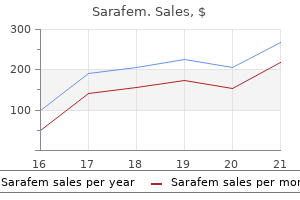
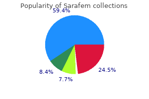
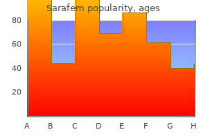
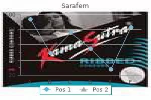
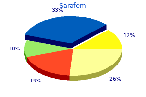
Sarafem
Michelle Leech MBBS(Hons), FRACP, PhD
- Consultant Rheumatologist, Monash Medical Centre
- Associate Professor and Director of Clinical Teaching Programs, Southern Clinical
- School, Monash University, Melbourne, Vic
Sarafem dosages: 20 mg, 10 mg
Sarafem packs: 30 pills, 60 pills, 90 pills, 120 pills, 180 pills, 270 pills, 360 pills

Discount 20mg sarafem with mastercard
Balloon dilation can additionally be the popular main mode of therapy in the infant and baby with aortic valve stenosis menstruation ovulation cycle purchase 20 mg sarafem free shipping. Care should be taken to keep away from oversizing the balloon which might lead to women's health center yorkton discount sarafem 20mg overnight delivery an unacceptable degree of valvar regurgitation breast cancer 9gag generic 10mg sarafem otc. It is important, nonetheless, to understand that within the neonatal interval when the ductus is patent, evaluation of a gradient across the aortic valve either by catheter or Doppler-derived strategies will underestimate the severity of the stenosis due to the low circulate across the valve. Depressed contractility, high grade obstruction to transaortic flow, and ductal blood flow into the aorta all contribute to low flow throughout the aortic valve. It is especially essential for the echocardiographer to measure all left coronary heart constructions in two planes. Assessment of the mitral valve size and mobility is just as important as for the aortic valve. The long axis size of the left ventricle as a proportion of the entire lengthy axis length of the center (atrioventricular valve annulus to apex) can also be a useful measurement. The decision whether to pursue a two ventricle or a one-ventricle approach might be guided by these calculations. Closure of the ductus should be documented by bodily examination and echocardiography. Prostaglandin-Dependent Critical Neonatal Aortic Valve Stenosis If the child is prostaglandin dependent a call should be made early in the neonatal interval whether to proceed to a one-ventricle (Norwood) or two ventricle pathway. There are currently multivariable scoring techniques (discussed below) to aid on this decision-making course of. The traditional state of affairs that results is that despite the very fact that a passable and profitable balloon valvotomy procedure is performed with minimal residual gradient, the kid stays ventilator dependent because of very excessive left atrial pressures. When the child has been ventilator dependent for two or 3 weeks and the surgical and intensive care staff realizes that a single-ventricle strategy shall be essential, by this time the pulmonary resistance is very high. This increases the danger of a Norwood process and may eliminate the choice of cardiac transplantation as properly. Fortunately, knowledge analyses by a quantity of teams are actually obtainable to assist guide this choice though they clearly have their limitations. The Rhodes Score the first try to develop a scoring system was developed by Rhodes et al. The presence of more than one of the "Rhodes elements" noted above suggests a high chance of demise if a two ventricle method is pursued. While very useful in neonates with isolated aortic stenosis, the Rhodes rating has proven to have decrease accuracy in neonates with multiple left heart obstructive lesions. A subsequent analysis included patients three months of age or much less with two or extra areas of left heart obstruction or hypoplasia. A complete of 116 sufferers have been directed toward a biventricular restore by having both a balloon valvotomy (n = 83) or surgical procedure (n = 33), mostly an open valvotomy. Risk elements for dying have been a higher grade of endocardial fibroelastosis estimated by echocardiography, a lower Z score of the aortic valve diameter on the stage of the sinuses of Valsalva, and younger age at entry. An initial Norwood procedure was performed in 179 sufferers with survival at 5 years of 60%. The danger components for the Norwood method are mentioned in Chapter 23, Hypoplastic Left Heart Syndrome. Because of the big variety of patients in this research it was possible to use multiple logistical regression to develop a calculator which allows prediction for any particular person patient as to whether or not a biventricular repair is extra more doubtless to result in survival than a Norwood process. Early versions of the calculator had some software faults that required revision several occasions. Frequent reinterventions are usually needed including mitral valve replacement, modified Konno procedure, and aortic valve substitute with annular enlargement. If an infant Ross procedure is performed the homograft used to reconstruct the pulmonary outflow is careworn by the pulmonary hypertension secondary to elevated left atrial pressure and should require early and frequent replacement. If the recent end result of a study of patients with pulmonary atresia/intact septum. Intervention for Aortic Valve Stenosis past the Neonatal Period Balloon dilation of the aortic valve is a low-risk procedure which serves both to scale back the transvalvular gradient and to promote development of the aortic annulus. The latter is essential because aortic stenosis in youngsters is commonly associated with hypoplasia of the aortic valve. Balloon dilation of a stenotic valve early in life, notably if it leads to a light degree of aortic regurgitation, provides an essential stimulus for development of the valve. If echocardiographic assessment signifies leaflet commissural fusion which is more probably to be improved by balloon dilation, then intervention is indicated for a peak Doppler-derived gradient of greater than 30�40 mmHg and a peak to peak catheter-derived gradient of greater than 20�30 mmHg. These gradients are significantly decrease than those beforehand used as the threshold for surgical intervention in kids. Early aggressive intervention by balloon dilation permits the child to grow and exercise and promotes annular growth. At current, there are basically no indications for major surgical intervention for a stenotic aortic valve with adequate annular dimensions. Ross later launched the pulmonary autograft procedure41 which has subsequently been combined with the Konno process for patients with annular hypoplasia and notably those with related tunnel subaortic stenosis. However, aortic valve restore for predominant regurgitation is described in Chapter 21, Valve Repair and Replacement. Repair often contains techniques to get rid of a rigid raphe or commissural fusion which each contribute to stenosis when regurgitation coexists with stenosis. Aortic Valve Replacement with Aortic Annular Enlargement Aortic valve replacement for pure aortic valve stenosis with a traditional aortic annular diameter is nearly by no means indicated in the pediatric age group. Posterior Enlargement of the Aortic Annulus: Manougian and Nicks Procedures these procedures are used alone when a modest diploma of enlargement of the aortic annulus is required. They could be utilized along with an anterior Konno-type annular procedure when extra aggressive enlargement is required (see below). Posterior 426 Comprehensive Surgical Management of Congenital Heart Disease, Second Edition root replacement using an aortic homograft within the small aortic root. They are generally not performed along side the Ross process (see below). Nicks Procedure A commonplace reverse hockey-stick incision is made extending the incision inferiorly towards the area between the left/noncommissure and the bottom of the noncoronary commissure. A collagen impregnated woven Dacron patch is used to supplement the annulus although in the smaller youngster autologous pericardium may be most popular. Inverting sutures placed beneath the prosthesis allow a bigger prosthesis to be positioned relative to everting sutures positioned above the prosthesis. Another choice is to place all sutures from outside the aorta with care to avoid compromise of the coronaries. The coronary heart is de-aired within the ordinary fashion and when rewarming is completed discontinuation of bypass ought to be routine. Manougian Procedure the incision is as for the Nicks procedure however is extended across the intervalvular fibrosa into the anterior leaflet of the mitral valve on the commissure between the left and noncoronary leaflets. However, it can be simply picked up within the supplementing patch suture line and, in fact, serves a helpful function in pledgetting the suture line. The Manougian process may be performed in conjunction with homograft replacement of the aortic root. In these circumstances, the annulus is supplemented by the mitral valve part of the homograft.
Generic 10mg sarafem with amex
Extensions o the inerior hypogastric plexus send autonomic bers alongside the blood vessels breast cancer statistics buy discount sarafem 20mg on-line, which orm visceral plexuses on the partitions o the pelvic viscera menstrual blood art safe 10mg sarafem. The celiac womens health hershey pa cheap 20 mg sarafem mastercard, superior mesenteric, and inerior mesenteric plexuses are interconnected. The celiac plexus, surrounding the foundation o the celiac (arterial) trunk, accommodates irregular proper and let celiac ganglia (approximately 2 cm long) that unite superior and inerior to the celiac trunk. The parasympathetic root o the celiac plexus is a branch o the posterior vagal trunk, which accommodates bers rom the right and let vagus nerves. Vasomotion (control o blood fow) at this degree infuences water and electrolyte movement. Corresponding plexuses with smaller, sparser ganglia lengthen to the pancreas, gallbladder, and cystic and major biliary ducts. Abdominal Viscera 531 the motor neurons o these plexuses are intrinsic or enteric ganglia that serve nominally as postsynaptic neurons or the parasympathetic system. In addition to unctioning as relay neurons, receiving and passing on eerent impulses despatched by presynaptic parasympathetic neurons, they also receive enter rom postsynaptic sympathetic bers (making them a third-order neuron in that system). They have huge interconnectivity with surrounding eerent neurons, each directly and via interneurons, as properly as axons terminating on clean muscle and glands. In addition, there are intrinsic aerent neurons with cell our bodies within the plexuses that monitor mechanical and chemical conditions within the intestine and talk with the eerent neurons offering local (short) refex circuitry, as nicely as sending inormation centrally. Schematic illustration o the group o the enteric nervous system within the intestinal wall. Flow chart demonstrating lengthy (extrinsic) and short (intrisic) reexes involving the enteric nervous system. With regard to the smooth muscle sphincters, the roles o the sympathetic and parasympathetic methods reverse, with the sympathetic system maintaining tonus and the parasympathetic system inhibiting it. Relatively nonpermeable capillaries associated with the ganglia present a diusion barrier resembling the blood�brain barrier o cerebral blood vessels. Approximate spinal wire segments and spinal sensory ganglia involved in sympathetic and visceral aerent (pain) innervation o belly viscera are proven. Visceral aerent bers conveying ache sensations accompany the sympathetic (visceral motor) bers. The ache impulses move retrogradely to those o the motor bers along the splanchnic nerves to the sympathetic trunk, by way of white speaking branches to the anterior rami o the spinal nerves. Then they move into the posterior root to the spinal sensory ganglia and spinal cord. The abdomen (oregut) receives innervation rom the T6 to T9 levels, small intestine through transverse colon (midgut) rom the T8 to T12 levels, and descending colon (hindgut) rom the T12 to L2 levels. Starting rom the midpoint o the sigmoid colon, visceral pain bers run with parasympathetic bers, the sensory impulses being conducted to S2�S4 sensory ganglia and spinal wire ranges. These are the same spinal wire segments concerned in the sympathetic innervation o those portions o alimentary tract. The fbers traverse the paravertebral ganglia o the trunks with out synapsing, persevering with as elements o abdominopelvic splanchnic nerves. The sympathetic fbers move to prevertebral ganglia, most o that are clustered around the main branches o the stomach aorta. Ater synapsing within the ganglia, the postsynaptic sympathetic fbers be a part of the presynaptic parasympathetic fbers, traveling via peri-arterial plexuses around the branches o the belly aorta to attain the viscera. A continuation o the abdominal aortic plexus inerior to the aortic biurcation (the superior and inerior hypogastric plexuses) conveys sympathetic innervation to most o the pelvic viscera. The sympathetic fbers mainly innervate the blood vessels o stomach viscera and are inhibitory to the parasympathetic stimulation. The parasympathetic fbers synapse on or in the partitions o the viscera with intrinsic postsynaptic parasympathetic neurons, which terminate on the sleek muscle or glands o the viscera. Parasympathetic innervation: the vagus nerves supply parasympathetic fbers to the digestive tract rom the esophagus via the transverse colon. Parasympathetic stimulation promotes peristalsis and secretion (although a lot o the latter is usually hormonally regulated). Sensory innervation: Visceral aerent fbers ollow the autonomic fbers retrograde to sensory ganglia. Aerent fbers conveying ache sensation rom belly viscera orad (proximal) to the center o the sigmoid colon run with the sympathetic fbers to the thoracolumbar spinal sensory ganglia; all other visceral aerent fbers run with the parasympathetic fbers. Thus, visceral aerent fbers conveying reex inormation rom the gut orad to the middle o the sigmoid colon cross to vagal sensory ganglia; fbers conveying each pain and reex inormation rom the gut aborad (distal) to the center o the sigmoid colon move to spinal sensory ganglia S2�S4. Its mainly convex superior surace aces the thoracic cavity, and its concave inerior surace aces the belly cavity. The diaphragm is the chie muscle o inspiration (actually, o respiration altogether, because expiration is largely passive). It descends during inspiration; however, solely its central half moves as a outcome of its periphery, because the xed origin o the muscle, attaches to the inerior margin o the thoracic cage and the superior lumbar vertebrae. The pericardium, containing the center, lies on the central half o the diaphragm, miserable it barely. The diaphragm curves superiorly into right and let domes; normally, the right dome is greater than the let dome owing to the presence o the liver. During expiration, the proper dome reaches as high as the fifth rib and the let dome ascends to the fifth intercostal house. The level o the domes o the diaphragm varies based on the phase o respiration (inspiration or expiration). The muscular half o the diaphragm is located peripherally with bers that converge radially on the trioliate central aponeurotic half, the central tendon. The central tendon has no bony attachments and is incompletely divided into three leaves, resembling a wide cloverlea. Although it lies near the center o the diaphragm, the central tendon is nearer to the anterior part o the thorax. Costal half: consisting o broad muscular slips that attach to the interior suraces o the inerior six costal cartilages and their adjoining ribs on both sides; the costal parts orm the proper and let domes. Lumbar part: arising rom two aponeurotic arches, the medial and lateral arcuate ligaments, and the three superior lumbar vertebrae; the lumbar part orms right and let muscular crura that ascend to the central tendon. The right crus, larger and longer than the let crus, arises rom the rst three or our lumbar vertebrae. The thoracic wall and cage have been eliminated to show the attachments and convexity o the right dome o the diaphragm. The eshy sternal, costal, and lumbar elements o the diaphragm (outlined with broken lines) attach centrally to the treoil-shaped central tendon, the aponeurotic insertion o the diaphragmatic muscle fbers. The right and let crura and the brous median arcuate ligament, which unites them as it arches over the anterior aspect o the aorta, orm the aortic hiatus. The diaphragm can be hooked up on both sides to the medial and lateral arcuate ligaments. The medial arcuate ligament is a thickening o the ascia overlaying the psoas major, spanning between the lumbar vertebral bodies and the tip o the transverse process o L1. The lateral arcuate ligament covers the quadratus lumborum muscular tissues, continuing rom the L12 transverse course of to the tip o the twelfth rib. The superior side o the central tendon o the diaphragm is used with the inerior surace o the brous pericardium, the strong, external half o the broserous pericardial sac that encloses the guts.
Buy 20mg sarafem with amex
The superior urinary organs (kidneys and ureters) menstrual migraine treatment generic 10mg sarafem visa, their vessels women's health center of clarksville tn order 20 mg sarafem with visa, and the suprarenal glands are main retroperitoneal structures on the posterior belly wall women's health center yonkers ny sarafem 20mg generic. Perinephric at (perirenal at capsule) surrounds the kidneys and their vessels because it extends into their hollow facilities, the renal sinuses. The kidneys, suprarenal glands, and the at surrounding them are enclosed (except ineriorly) by a condensed, membranous layer o renal ascia, which continues medially to ensheathe the renal vessels, mixing with the vascular sheaths o the latter. Ineromedially, a fragile extension o the renal ascia is prolonged along the ureter as the periureteric ascia. The ureter crosses the external iliac artery simply beyond the widespread iliac biurcation. The gonadal arteries (testicular arteries, as in this male, or ovarian arteries, in emales) cross anterior to the ureters and supply ureteric branches to them. The superior mesenteric artery arises superior to and runs anteriorly throughout the let renal vein, compressing the vein against the abdominal aorta posteriorly. The posterolateral abdominal wall has been incised between the muscles o the anterolateral belly wall and the muscular tissues o the back. This transverse part o the kidney shows the relationships o the muscles and ascia. Because the renal ascia surrounds the kidney as a separate sheath, it have to be incised in any surgical procedure on the kidney, whether rom an anterior or a posterior strategy. However, motion o the kidneys occurs throughout respiration and when altering rom the supine to the erect position, and vice versa. Normal renal mobility is roughly 3 cm, approximately the peak o one vertebral physique. Superiorly, the renal ascia is continuous with the ascia on the inerior surace o the diaphragm (diaphragmatic ascia); thus, the primary attachment o the suprarenal glands is to the diaphragm. Ineriorly, the anterior and posterior layers o renal ascia are only loosely united, i hooked up at all. They lie retroperitoneally on the posterior belly wall, one on both sides o the vertebral column at the level o the T12�L3 vertebrae. Abdominal Viscera 517 At the concave medial margin o every kidney is a vertical clet, the renal hilum. Structures that serve the kidneys (vessels, nerves, and structures that drain urine rom the kidney) enter and exit the renal sinus by way of the renal hilum. The hilum o the let kidney lies near the transpyloric aircraft, roughly 5 cm rom the median airplane. The transpyloric plane passes via the superior pole o the proper kidney, which is approximately 5. Each kidney strikes 2�3 cm in a vertical direction in the course of the movement o the diaphragm that happens with deep respiration. During lie, the kidneys are reddish brown and measure roughly 10 cm in size, 5 cm in width, and 5. Superiorly, the posterior aspects o the kidneys are related to the diaphragm, which separates them rom the pleural cavities and the twelfth pair o ribs. More ineriorly, the posterior suraces o the kidney are related to the psoas main muscular tissues medially and the quadratus lumborum muscle. The subcostal nerve and vessels and the iliohypogastric and ilio-inguinal nerves Median plane Scapular line Diaphragm Liver Spleen T10 T11 Transpyloric aircraft Left kidney twelfth rib Right kidney Ureter Iliac crest Ilium Dimple indicating posterior superior iliac backbone T12 L1 5cm descend diagonally throughout the posterior suraces o the kidneys. The let kidney is expounded to the abdomen, spleen, pancreas, jejunum, and descending colon. At the hilum, the renal vein is anterior to the renal artery, which is anterior to the renal pelvis. Within the kidney, the renal sinus is occupied by the renal pelvis, calices, vessels, and nerves and a variable amount o at. Each kidney has anterior and posterior suraces, medial and lateral margins, and superior and inerior poles. However, because o the protrusion o the lumbar vertebral column into the stomach cavity, the kidneys are obliquely positioned, lying at an angle to one another. Consequently, the transverse diameter o the kidneys is oreshortened in anterior views. The lateral margin o each kidney is convex, and the medial margin is concave the place the renal sinus and renal pelvis are located. The renal pelvis is the fattened, unnel-shaped enlargement o the superior finish o the ureter. The renal pelvis receives two or three major calices (calyces), each o which divides into two or three minor calices. Each minor calyx is indented by a renal papilla, the apex o the renal pyramid, rom which the urine is excreted. The lobes are seen on the external suraces o the kidneys in etuses, and proof o the lobes could persist or a while ater delivery. The ureters are muscular ducts (25�30 cm long) with narrow lumina that carry urine rom the kidneys to the urinary bladder. They run ineriorly rom the apices o the renal pelves at the hila o the kidneys, passing over the pelvic brim at the biurcation o the frequent iliac arteries. The belly elements o the ureters adhere closely to the parietal peritoneum and are retroperitoneal all through their course. From the again, the surace marking o the ureter is a line becoming a member of some extent 5 cm lateral to the L1 spinous process and the posterior superior iliac spine. The ureters occupy a sagittal plane that intersects the information o the transverse processes o the lumbar vertebrae. The anterior lip o the renal hilum has been reduce away to expose the renal pelvis and calices throughout the renal sinus. The renal pyramids comprise the amassing tubules and orm the medulla o the kidney. These constricted areas are potential websites o obstruction by ureteric stones (calculi). Although the name "suprarenal" implies that the kidneys are their major relationship, their main attachment is to the diaphragmatic crura. [newline]They are separated rom the kidneys by a skinny septum (part o the renal ascia-see the Clinical Box "Renal Transplantation," p. The crescent-shaped let gland is medial to the superior hal o the let kidney and is expounded to the spleen, abdomen, pancreas, and the let crus o the diaphragm. Each gland has a hilum, where the veins and lymphatic vessels exit the gland, whereas the arteries and nerves enter the glands at multiple sites. Each suprarenal gland has two parts: the suprarenal cortex and suprarenal medulla. The suprarenal cortex derives rom mesoderm and secretes corticosteroids and androgens. These hormones cause the kidneys to retain sodium and water in response to stress, increasing the blood quantity and blood strain. The suprarenal medulla is a mass o nervous tissue permeated with capillaries and sinusoids that derives rom neural crest cells related to the sympathetic nervous system. The chroman cells o the medulla are related to sympathetic ganglion (postsynaptic) neurons in both derivation (neural crest cells) and unction.
Buy discount sarafem 20 mg on line
Dermatome maps o the body are primarily based on an accumulation o scientific fndings ollowing spinal nerve injuries women's health center abington purchase 20mg sarafem fast delivery. This map relies on the research o Foerster (1933) and reects each anatomical (actual) distribution or segmental innervation and clinical expertise menstruation cycle calendar 10mg sarafem. Another in style however more schematic map is that o Keegan and Garrett (1948) menopause breast changes generic sarafem 20 mg without prescription, which is appealing or its regular, more simply extrapolated pattern. Note that in the Foerster map, C5�T1 and L3�S1 are distributed virtually entirely within the limbs. As they emerge rom the intervertebral oramina, spinal nerves are divided into two rami. Posterior (primary) rami o spinal nerves provide nerve bers to the synovial joints o the vertebral column, deep (epaxial) muscle tissue o the back, and the overlying skin in a segmental sample. As a common rule, the posterior rami remain separate rom one another (do not merge to orm main somatic nerve plexuses). Anterior (primary) rami o spinal nerves provide nerve bers to the much bigger remaining space, consisting o the skin and hypaxial muscle tissue o the anterior and lateral areas o the trunk and the upper and decrease limbs. The anterior rami that are distributed solely to the trunk generally stay separate rom one another, additionally innervating muscular tissues and pores and skin in a segmental pattern. However, primarily in relationship to the innervation o the limbs, the majority o anterior rami merge with one or more adjoining anterior rami, orming the most important somatic nerve plexuses (networks) in which their bers intermingle and rom which a new set o multisegmental peripheral nerves emerges. Posterior rami are distributed to the synovial joints o the vertebral column, deep muscles o the back, and the overlying pores and skin. Maps o the cutaneous distribution o peripheral nerves are primarily based on dissection and supported by clinical fndings. Although the posterior rami (not shown) usually stay separate rom each other and ollow a distinctly segmental sample o distribution, most anterior rami (20 o 31 pairs) participate within the ormation o plexuses, that are primarily involved within the innervation o the limbs. The anterior rami distributed only to the trunk usually stay separate, however, and ollow a segmental distribution similar to that o the posterior rami. It is thereore essential to distinguish between the distribution o the bers carried by spinal nerves (segmental innervation or distribution-i. The overlapping within the cutaneous distribution o nerve bers conveyed by adjoining spinal nerves additionally occurs within the cutaneous distribution o nerve bers conveyed by adjacent peripheral nerves. Communication happens between cranial nerves, and between cranial nerves and upper cervical (spinal) nerves; thus, a nerve that originally conveys solely motor bers may obtain sensory bers distally in its course, and vice versa. Except or the rst two (those involved within the senses o scent and sight), cranial nerves that convey sensory bers into the brain bear sensory ganglia (similar to spinal or posterior root ganglia), the place the cell bodies o the pseudounipolar bers are situated. Although, by denition, the term dermatome applies only to spinal nerves, comparable areas o skin supplied by single cranial nerves could be identied and mapped. Proprioceptive sensations are normally subconscious, providing inormation concerning joint place and the strain o tendons and muscles. This inormation is mixed with input rom the vestibular equipment o the (continued on p. Adjacent anterior rami merge to orm plexuses by which their fbers are exchanged and redistributed, orming a new set o multisegmental peripheral (named) nerves. The peripheral nerves derived rom the plexus comprise fbers rom multiple spinal nerves. Although segmental nerves merge and lose their identification when plexus ormation ends in multisegmental peripheral nerves, the segmental (dermatomal) pattern o nerve fber distribution remains. The somatic motor system permits voluntary and reexive motion caused by contraction o skeletal muscles, similar to occurs when one touches a sizzling iron. The cell bodies o somatic motor and presynaptic visceral motor neurons are situated in the grey matter o the spinal wire. These bers are sometimes designated as branchial motor, reerring to muscle tissue derived rom the pharyngeal arches in the embryo. Their proximal stumps start to regenerate, sending sprouts into the realm o the lesion; however, this progress is blocked by astrocyte prolieration at the injury website, and the axonal sprouts are soon retracted. Thereore, only at these places can the surgeon selectively part either unctional element or the relie o intractable ache or spastic paralysis (rhizotomy). When nerves are stretched, crushed, or severed, their axons degenerate mainly distal to the lesion because they rely upon their nerve cell our bodies or survival. I the axons are broken but the cell bodies are intact, regeneration and return o unction may happen. Pressure on a nerve commonly causes paresthesia, the pins-and-needles sensation that occurs when one sits too lengthy with the legs crossed, or example. No surgical repair is needed or this type o nerve injury as a outcome of the intact connective tissue coverings information the growing axons to their destinations. Sprouting occurs at the proximal ends o the axons, but the growing axons might not attain their distal targets. A chopping nerve injury requires surgical intervention as a outcome of regeneration o the axon requires apposition o the reduce ends by sutures through the epineurium. Anterograde (wallerian) degeneration is the degeneration o axons detached rom their cell our bodies. Prolonged ischemia (inadequate blood supply) o a nerve might result in damage no much less extreme than that produced by crushing and even chopping the nerve. Transient paresthesias are amiliar to anyone who has had an injection o anesthetic or dental repairs. The somatic sensory system transmits sensations o touch, pain, temperature, and position rom sensory receptors. The somatic motor system innervates only skeletal muscle, stimulating voluntary and refexive movement by inflicting the muscle to contract, as happens in response to touching a scorching iron. A unctional distinction o pharmacological significance or medical apply is that the postsynaptic neurons o the two divisions typically liberate dierent neurotransmitter substances: norepinephrine by the sympathetic division (except within the case o sweat glands) and acetylcholine by the parasympathetic division. As the aerent component o autonomic refexes and in conducting visceral ache impulses, these visceral aerent bers also play a task within the regulation o visceral unction. The cell our bodies o postsynaptic neurons o the sympathetic nervous system occur in two places, the paravertebral and prevertebral ganglia. The superior paravertebral ganglion (the superior cervical ganglion o each sympathetic trunk) lies on the base o the cranium. The ganglion impar orms ineriorly where the two trunks unite at the level o the coccyx. Almost immediately ater coming into, all the presynaptic sympathetic bers depart the anterior rami o these spinal nerves and move to the sympathetic trunks through white rami communicantes (communicating branches). Within the sympathetic trunks, presynaptic bers ollow one o our attainable programs: 1. Enter and synapse instantly with a postsynaptic neuron o the paravertebral ganglion at that level. Pass through the sympathetic trunk with out synapsing, continuing through an abdominopelvic splanchnic nerve (a department o the trunk concerned in innervating abdominopelvic viscera) to reach the prevertebral ganglia.
Cheap 10mg sarafem visa
The accidents normally outcome rom a minor all on the hand women's health tone zone workout buy sarafem 10mg mastercard, with the orce being transmitted up the orearm bones o the prolonged limb breast cancer yoga generic 10mg sarafem with mastercard. Because o impaction o the ragments women's health york pa generic sarafem 20mg without a prescription, the racture website is usually stable and the person is able to transfer the arm passively with little ache. An avulsion racture o the higher tubercle o the humerus is seen mostly in middle-aged and elderly individuals. In youthful people, a racture o the larger tubercle may result rom impaction with extreme abduction or fexion o the arm. Muscles (especially the subscapularis) that stay connected to the humerus pull the limb into medial rotation. Fractures o the shat o the humerus end result rom a direct blow to or torsion o the arm, producing numerous sorts o ractures. In a transverse racture o the humeral shat, the pull o the deltoid muscle carries the proximal ragment laterally. Indirect injury resulting rom a all on the outstretched hand could produce a spiral or indirect racture o the humeral shat. Overriding o the indirect ends o an obliquely ractured bone may lead to shortening o the limb. Because the humerus is surrounded by muscular tissues and has a well-developed periosteum, properly aligned bone ragments often unite properly. An intercondylar racture o the humerus results rom a extreme all on the fexed elbow or with high-impact accidents such as in a motorized vehicle accident. The olecranon o the ulna is driven like a wedge between the medial and lateral components o the condyle o the humerus, separating one or each parts rom the humeral shat. The ollowing components o the humerus are in direct contact with the indicated nerves: Surgical neck: axillary nerve. Fractures o each the radius and the ulna in older people and athletic adults are normally the outcome o extreme injury. A direct harm normally produces transverse ractures on the same stage, normally in the center third o the bones. Because the shats o these bones are rmly bound together by the interosseous membrane, a racture o one bone is likely to be related to dislocation o the closest joint. A full transverse racture o the distal 2 cm o the radius, referred to as a Colles racture, is the most common racture o the orearm. The distal ragment is displaced dorsally and is oten comminuted (broken in pieces). The racture outcomes rom orced extension o the hand, usually because the end result o attempting to ease a all by outstretching the upper limb. This racture is oten reerred to as a dinner ork deormity as a end result of a posterior angulation (bending) happens within the orearm simply proximal to the wrist and the traditional anterior curvature o the relaxed hand. The posterior bending is produced by the posterior displacement and tilt o the distal ragment o the radius. Because o the wealthy blood supply to the distal finish o the radius, bony union is normally good. When the distal end o the radius ractures in kids, the racture line might lengthen by way of the distal epiphysial plate. Epiphysial plate injuries are frequent in older youngsters as a outcome of o their requent alls by which the orces are transmitted rom the hand to the radius and ulna. The therapeutic course of may end in malalignment (displacement) o the epiphysial plate and disturbance o radial progress. Avascular necrosis o the proximal ragment o the scaphoid (pathological demise o bone, resulting rom insufficient blood supply) might occur and produce degenerative joint illness o the wrist. Radius Ulna Palmar carpal branch Scaphoid (fractured, with necrotic proximal fragment) Trapezoid Radial artery Triquetrum Lunate Pisiform Hook of hamate Capitate Trapezium (A) Fracture o Scaphoid the scaphoid is essentially the most requently ractured carpal bone. It oten results rom a all on the palm when the hand is abducted, the racture occurring across the narrow half o the scaphoid. On palpation, pain is produced in the anatomical snu field on the lateral side o the wrist, particularly during dorsifexion and abduction o the hand. Initial radiographs o the wrist may not reveal a racture; oten, this injury is (mis)diagnosed as a severely sprained wrist. Radiographs taken 10�14 days later reveal a racture as a outcome of bone resorption has occurred there. Bones o Upper Limb 157 Fracture o Hamate Fracture o the hamate might end in nonunion o the ractured bony elements as a outcome of o the traction produced by the hooked up hypothenar muscular tissues. Because the ulnar nerve is close to the hook o the hamate, the nerve may be injured, inflicting decreased grip power o the hand. The head o the bone rotates over the distal end o the shat, producing a fexion deormity. Because o the extremely developed sensation within the e ngers, these accidents are extremely painul. Fractures o the proximal and middle phalanges are normally the end result o crushing or hyperextension accidents. Because o the close e relationship o phalangeal ractures to the fexor tendons, the bone ragments should be careully realigned to restore regular unction o the ngers. Fracture o Metacarpals the metacarpals (except the 1st) are carefully sure collectively; therefore, isolated ractures are probably to be secure. The higher limb consists o our increasingly mobile segments: the proximal three (shoulder, arm, and orearm) serve primarily to place the ourth phase (hand), which is used or greedy, manipulation, and touch. The clavicle serves as a movable crane-like strut (extended support) rom which the scapula and ree limb are suspended at a distance rom the trunk that permits reedom o motion. Shocks acquired by the higher limb (especially the shoulder) are transmitted via the clavicle, leading to a racture that most commonly happens between its center and lateral thirds. This triangular at bone is curved to conorm to the thoracic wall and supplies massive surace areas and edges or attachment o muscles. These muscles (1) transfer the scapula on the thoracic wall at the physiological scapulothoracic joint and (2) prolong to the proximal humerus maintaining the integrity o-and producing motion at-the glenohumeral joint. The coracoid course of o the scapula is the location o attachment or the coracoclavicular ligament, which passively supports the upper limb, and a web site or muscular (tendon) attachment. Humerus: the lengthy, strong humerus is a cell strut- the frst in a collection o two-used to position the hand at a height (level) and distance rom the trunk to maximize its efciency. The spherical head o the humerus permits an excellent vary o movement on the mobile scapular base; the trochlea and capitulum at its distal end acilitate the hinge actions o the elbow and, at the identical time, the pivoting o the radius. The lengthy shat o the humerus enables reaching and makes it an eective lever or power in liting, as properly as offering surace space or attachment o muscles that act primarily at the elbow. Added surace area or attachment o exors and extensors o the wrist is supplied continued on subsequent page 158 Chapter 3 Upper Limb the Bottom Line (continued) by the epicondyles, the medial and lateral extensions o the distal end o the humerus.
Discount 20 mg sarafem with visa
Mobile gallbladders are topic to vascular torsion and inarction (sudden insuiciency o arterial or venous blood supply) women's health center rome ga sarafem 20mg amex. Gallstones A gallstone is a concretion in the gallbladder menopause keene nh cheap sarafem 10mg amex, cystic duct menstrual odor treatment buy 20mg sarafem with visa, or bile duct composed chiefy o ldl cholesterol crystals. Over a 20-year period, two thirds o asymptomatic folks with gallstones remain symptom ree. For gallstones to trigger clinical symptoms, they want to acquire a measurement sucient to produce mechanical damage to the gallbladder or obstruction o the biliary tract (Townsend et al. The distal end o the frequent bile duct is a slender part o the biliary passages and is a typical site or impaction o gallstones. Note the concave contour o the distal finish o the visible duct at the superior margin o the calculus. Another frequent website or impaction o gallstones is a sacculation (Hartmann pouch) that will seem at the junction 5 Gallbladder wall Gallstones Liver Diaphragm (A) Longitudinal ultrasonic scan of gallbladder with gallstones. When this pouch is large, the cystic duct arises rom its upper let side, not rom what seems to be the apex o the gallbladder. I a peptic duodenal ulcer ruptures, a alse passage might orm between the pouch and the superior part o the duodenum, allowing gallstones to enter the duodenum. Infammation o the gallbladder may cause pain within the posterior thoracic wall, or right shoulder owing to irritation o the diaphragm. Cholecystectomy People with severe biliary colic often have their gallbladders removed. The cystic artery mostly arises rom the proper hepatic artery within the cystohepatic triangle (Calot triangle). In current medical use, the cystohepatic triangle is dened ineriorly by the cystic duct, medially by the widespread hepatic duct, and superiorly by the inerior surace o the liver. Careul dissection o the cystohepatic triangle early during cholecystectomy saeguards these essential structures ought to there be anatomical variations. Errors during gallbladder surgery generally end result rom ailure to respect the frequent variations in the anatomy o the biliary system, particularly its blood provide. Beore dividing any structure and removing the gallbladder, surgeons identiy all three biliary ducts, as well as the cystic and hepatic arteries. Continued infammation could break down (ulcerate) the tissue boundaries between the gallbladder and an element o the gastrointestinal tract adherent to it, leading to a cholecysto-enteric stula. Because o their proximity to the gallbladder, the superior half o the duodenum and the transverse colon are more than likely to develop a stula o this sort. The stula would enable a big gallstone, incapable o passing though the cystic duct, to enter the gastrointestinal tract. A cholecysto-enteric stula also permits gasoline rom the gastrointestinal tract to enter the gallbladder, providing a diagnostic radiographic signal. Portal Hypertension When scarring and brosis rom cirrhosis hinder the hepatic portal vein within the liver, strain rises in the vein and its tributaries, producing portal hypertension. The giant volume o blood fowing rom the portal system to the systemic system at the websites o portal�systemic anastomoses may produce varicose veins, particularly within the lower esophagus. The veins could become so dilated that their walls rupture, leading to hemorrhage. Bleeding rom esophageal varices (abnormally dilated veins) on the distal finish o the esophagus is oten severe and may be atal. In extreme instances o portal obstruction, the veins o the anterior stomach wall (normally caval tributaries) that anastomose with the para-umbilical veins (normally portal tributaries) could turn into varicose and look considerably like small snakes radiating beneath the skin around the umbilicus. This condition is reerred to as caput medusae as a outcome of o its resemblance to the serpents on the head o Medusa, a personality in Greek mythology. Initially, portocaval anastomoses or portosystemic shunts had been laparotomy procedures by which the two veins were surgically connected, often where they lie near one another posterior to the liver. Another means o reducing portal stress was to be part of the splenic vein to the let renal vein, ater splenectomy (splenorenal anastomosis or shunt). Once in the hepatic vein, the unopened stent is pushed by way of the parenchyma o the liver into the portal vein. The spleen is totally covered by peritoneum, besides on the splenic hilum, where the splenorenal ligament (conveying splenic vessels to the spleen) and gastrosplenic ligament (conveying the short gastric and let gastro-omental vessels to the stomach) connect. Although it receives protection rom the overlying let 9th�11th ribs, the relatively delicate spleen is the abdominal organ most vulnerable to oblique trauma. Strong blows to the stomach may cause a sudden increase in intra-abdominal strain that will rupture the spleen, leading to prouse intraperitoneal hemorrhage. The splenic vein runs parallel and posterior to the tail and physique o the pancreas because it runs rom the spleen to the hepatic portal vein. The primary pancreatic duct runs a similar course within the pancreas, continuing transversely via the head to merge with the bile duct to orm the hepatopancreatic ampulla, which enters the descending half o the duodenum. As an endocrine gland, the pancreas receives an plentiful blood supply rom the pancreaticoduodenal and splenic arteries. Although it receives vasomotor sympathetic and secretomotor parasympathetic nerve fbers, regulation o pancreatic secretion is primarily hormonal. The exocrine pancreas is seldom the trigger o scientific problems, although diabetes, involving the endocrine pancreas, has become increasingly frequent. It is our main metabolic organ, initially receiving all absorbed oodstus, except ats. It can be our largest gland, unctioning as an extrinsic intestinal gland in producing bile. The liver occupies essentially all o the proper dome o the diaphragm and extends to the apex o the let dome. Consequently, it enjoys safety rom the decrease thoracic cage and moves with respiratory excursions. The liver is divided superfcially by the alciorm ligament and a groove or the ligamentum venosum into a big anatomical proper lobe and a much smaller let one; ormations on its visceral surace demarcate caudate and quadrate lobes. The liver is roofed with peritoneum except or the bare space, demarcated by peritoneal reections comprising the coronary ligaments. Based on interdigitated branchings o the portal triad (hepatic portal vein, hepatic artery, and intrahepatic bile ducts) and hepatic veins, the continuous parenchyma o the liver may be divided into right and let livers (plus the caudate lobe). The liver can be urther subdivided into our divisions and then eight surgically resectable hepatic segments. The liver, just like the lungs, receives a twin blood supply, with 75�80% o the blood arriving through the hepatic portal vein, meeting the nutritional needs o the hepatic parenchyma; 20�25% arrives through the hepatic artery, delivered primarily to the nonparenchymal elements. Biliary ducts and gallbladder: Right and let hepatic ducts drain the bile produced by the right and let livers into the widespread hepatic duct, which thus carries all bile rom the liver. The frequent hepatic duct merges with the cystic duct to orm the bile duct, which conveys the bile to the descending part o the duodenum.
Purchase 10 mg sarafem
This junction occurs approximately on the degree o the L2 vertebra menstrual kits buy sarafem 10mg with mastercard, 2�3 cm to the let o the midline women's health clinic rockingham order sarafem 20 mg online. Most o the duodenum is xed by peritoneum to buildings on the posterior belly wall and is taken into account partially retroperitoneal pregnancy jokes cartoons sarafem 20mg for sale. Note the convolutions o the small intestine in situ, encircled on three sides by the large gut and revealed by elevating the larger omentum. The convolutions o the small gut have been retracted superiorly to demonstrate the mesentery. This orientation drawing o the alimentary system indicates the general position and relationships o the intestines. The duodenum, pancreas, and spleen and their blood supply are revealed by removal o the abdomen, transverse colon, and peritoneum. The stomach aorta and inerior vena cava occupy the vertical concavity posterior to the pinnacle o the pancreas and third half o the duodenum. The uncinate process is the extension o the head o the pancreas that passes posterior to the superior mesenteric vessels. The bile duct is descending in a fssure (opened up) within the posterior part o the head o the pancreas. Descending (second) part: longer (7�10 cm) and descends along the right sides o the L1�L3 vertebrae. Ascending (ourth) half: brief (5 cm) and begins on the let o the L3 vertebra and rises superiorly as ar as the superior border o the L2 vertebra. The rst 2 cm o the superior half o the duodenum, instantly distal to the pylorus, has a mesentery and is mobile. This ree part, known as the ampulla (duodenal cap), has an appearance distinct rom the remainder o the duodenum when noticed radiographically using distinction medium. The superior part o the duodenum ascends rom the pylorus and is overlapped by the liver and gallbladder. The proximal half has the hepatoduodenal ligament (part o the lesser omentum) connected superiorly and the higher omentum connected ineriorly. The descending part o the duodenum runs ineriorly, curving across the head o the pancreas. These ducts often unite to orm the hepatopancreatic ampulla, which opens on an eminence, called the main duodenal papilla, positioned posteromedially within the descending duodenum. The anterior surace o its proximal and distal thirds is roofed with peritoneum; however, the peritoneum refects rom its center third to orm the double-layered mesentery o the transverse colon, the transverse mesocolon. It is crossed by the superior mesenteric artery and vein and the basis o the mesentery o the jejunum and ileum. The ascending part o the duodenum runs superiorly and alongside the let side o the aorta to attain the inerior border o the body o the pancreas. Contraction o this muscle widens the angle o the duodenojejunal fexure, acilitating motion o the intestinal contents. The suspensory muscle passes posterior to the pancreas and splenic vein and anterior to the let renal vein. The arteries o the duodenum arise rom the celiac trunk and the superior mesenteric artery. The celiac trunk, via the gastroduodenal artery and its branch, the superior pancreaticoduodenal artery, supplies the duodenum proximal to the entry o the bile duct into the descending part o the duodenum. The superior mesenteric artery, by way of its branch, the inerior pancreaticoduodenal artery, supplies the duodenum distal to the entry o the bile duct. The pancreaticoduodenal arteries lie in the curve between the duodenum and the head o the pancreas and provide each buildings. The foundation o this transition in blood provide is embryological; this is the junction o the oregut and midgut. The veins o the duodenum ollow the arteries and drain into the hepatic portal vein, some immediately and others indirectly, via the superior mesenteric and splenic veins. The anterior lymphatic vessels drain into the pancreaticoduodenal lymph nodes, positioned along the superior and inerior pancreaticoduodenal arteries, and into the pyloric lymph nodes, which lie along the gastroduodenal artery. The shut positional relationship o these organs ends in sharing o blood vessels, lymphatic vessels, and nerve pathways, in entire or partially. Abdominal Viscera 465 o the pancreas and drain into the superior mesenteric lymph nodes. Eerent lymphatic vessels rom the duodenal lymph nodes drain into the celiac lymph nodes. The nerves o the duodenum derive rom the vagus and larger and lesser (abdominopelvic) splanchnic nerves by means o the celiac and superior mesenteric plexuses. The nerves are next conveyed to the duodenum by way of peri-arterial plexuses extending to the pancreaticoduodenal arteries (see also "Summary o the Innervation o Abdominal Viscera," p. The third part o the small intestine, the ileum, ends on the ileocecal junction, the union o the terminal ileum and the cecum. Together, the jejunum and ileum are 6�7 m lengthy, the jejunum constituting approximately two ths and the ileum roughly three ths o the intraperitoneal section o the small intestine. The terminal ileum often lies within the pelvis rom which it ascends, ending within the medial side o the cecum. The mesentery is a an-shaped old o peritoneum that attaches the jejunum and ileum to the posterior abdominal wall. The origin or root o the mesentery (approximately 15 cm long) is directed obliquely, ineriorly, and to the best. It extends rom the duodenojejunal junction on the let aspect o vertebra L2 to the ileocolic junction and the best sacro-iliac joint. The average size o the mesentery rom its root to the intestinal border is 20 cm. Between the 2 layers o the mesentery are the superior mesenteric vessels, lymph nodes, a variable quantity o at, and autonomic nerves. The arteries unite to orm loops or arches, referred to as arterial arcades, which give rise to straight arteries, called vasa recta. Specialized lymphatic vessels in the intestinal villi (tiny projections o the mucous membrane) that absorb at are called lacteals. They empty their milk-like fuid into the lymphatic plexuses within the walls o the jejunum and ileum. The lacteals drain in flip into lymphatic vessels between the layers o the mesentery. Within the mesentery, the lymph passes sequentially by way of three teams o lymph nodes. Eerent lymphatic vessels rom the mesenteric lymph nodes drain to the superior mesenteric lymph nodes. Lymphatic vessels rom the terminal ileum ollow the ileal branch o the ileocolic artery to the ileocolic lymph nodes. The sympathetic bers within the nerves to the jejunum and ileum originate within the T8�T10 segments o (continued on p.
References
- Brunzell JD, Schrott HG. The interaction of familial and secondary causes of hypertriglyceridemia: Role in pancreatitis. Trans Assoc Am Physicians 1973;86:245.
- Boue Y, Lavolaine J, Bouzat P, et al: Neuologic recovery from profound accidental hypothermia after 5 hours of cardiopulmonary resuscitation. Crit Care Med 42(2):e167-e170, 2014.
- Schijf LJ, Becx MCJM, de Bruin PC, Van der Vegt SGL. Whipple's disease: easily diagnosed, if considered. Neth J Med 2008; 66:392.
- Drachenberg CB, Hirsch HH, Papadimitriou JC, et al. Polyomavirus BK versus JC replication and nephropathy in renal transplant recipients: a prospective evaluation. Transplantation. 2007; 84(3):323-330.