

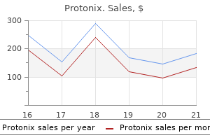
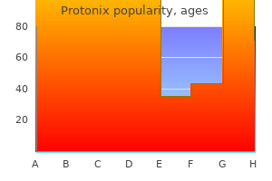
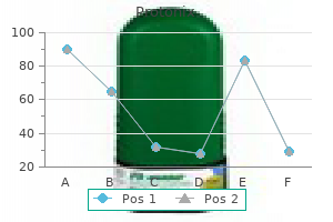
Protonix
Gregory J. Dehmer, MD
- Professor of Medicine
- Texas A&M University College of Medicine
- Director, Cardiology Division
- Scott & White Healthcare
- Temple, Texas
Protonix dosages: 40 mg, 20 mg
Protonix packs: 60 pills, 90 pills, 120 pills, 180 pills, 270 pills, 360 pills

Cheap protonix 20 mg online
This in part gastritis diet milk cheap 20 mg protonix free shipping, together with the downregulation of pro-inflammatory cytokines and chemokines gastritis ginger ale buy cheap protonix 20mg line, constitutes the antiinflammatory actions of vitamin D gastritis nursing care plan purchase protonix 20mg line. Studies using ovarian granulosa and breast most cancers cells have proven that overexpression or antisense knockdown of miR-125b, respectively, suppresses or enhances expression of 24-hydroxylase protein with expected cellular outcomes [46] (see Cancer part for cancer relevance). Interestingly, this network may be linked to a vitamin D resistive state that has been independently reported for breast most cancers cells [79]. The interaction reflects the pathological mobile and scientific phenotypes as defined by the ultimate protein output of cells throughout altered vitamin D networks. Cancer Cancer is characterized by the event of irregular cells that divide uncontrollably and infrequently can unfold all through the body. It is considered one of the major causes of dying in the world and survival rates are improving as a outcome of better screening (mutation analysis) and remedy choices. Overall, this motion results in arrest of the cell cycle in the G1 phase toward differentiation of these cells. The miR-22-vitamin D-motility relationship is fascinating and according to our personal studies in zebrafish and human keratinocyte migration, whereby suppression of vitamin D signaling is required to promote hydrogen peroxide-governed cell migration after injury [86]. Whether these findings across species level to a beforehand unrecognized antimotility/metastases operate of vitamin D throughout the most cancers setting stays to be tested. Bone Bone formation and remodeling are dynamic processes that involves bone ossification and resorption which lasts over a lifetime. Osteoclasts are answerable for bone resorption and are derived from the monocyte/macrophage lineage. Interestingly, endogenous miR-637 is downregulated in four hepatocellular carcinoma specimens, suggesting a tumor suppressor function in specialized cancers [104]. Conversely, miR-637 expression seems to lower osteoblast lineage commitment by concentrating on the early osteoblast-specific transcription issue osterix [105]. Osteogenin, an extracellular matrix part of bone, was recognized as a differentiation issue that initiates endochondral bone formation [111]. In this regard, current studies have proven that miR-29b regulates osteoblast differentiation by suppressing a number of inhibitors of osteoblast differentiation, including col4a2 [100,113], which is the fibrillation companion of col4a1. Based on this relationship it could probably be examined whether the breast cancer cells that exhibit decreased miR-637 ranges are extra proof against vitamin D and prone to most cancers relapse or metastasize as a outcome of a Col4a1-permissive extracellular surroundings. The mature miR-1228 sequence is homologous to mouse miR-667 [26], with nearly all of the 19 nucleotides of the mature guide strand conserved (78% similar), together with perfect similarity of the essential seed region. Vitamin D deficiency has been linked to pregnancy-related well being complications corresponding to gestational diabetes, preeclampsia, and bacterial vaginosis [121]. Despite this, other research have shown elevated ranges of let-7 family members across species after vitamin D treatment (Table 15. The authors suggested that the elevation in miR-21 may be concurrent with atherosclerosis improvement, and may play a protecting role as upregulation of miR-21 throughout myocardial ischemia is thought to suppress the inflammasome in mice [127]. This is partially because of issues of safety, in addition to to the complexity of the in vivo microenvironment in people. This instance established a revolutionary platform to inhibit oncomiR miR155 in a mouse lymphoma mannequin with antisense oligomers hooked up to peptide nucleic acid able to focusing on the acidic tumor microenvironment to evade systemic clearance by the liver. Currently, three methods are assumed to overcome these bottlenecks: (1) chemical oligonucleotide spine modifications, (2) covalent conjugation with transport autos, and (3) supramolecular meeting into nanosized formulation [144]. Even although progress on this area is fast and has already reached vital achievements, additional insights are nonetheless essential to translate the results from the lab bench to the clinic. Vitamin D performs an necessary role in cancer prevention, and its impact on uncommon subpopulations such as circulating tumor cells or most cancers stem cells is crucial. The advantage of this method is the increased sensitivity and improved dynamic vary, which can be utilized with a standard circulate cytometer. Sequences are read in situ utilizing 3D rendering confocal imaging inside the endogenous cellular setting and recognized by a primary native alignment search software search [158]. The conceptual framework of evolutionary morphology within the research of Ernst Haeckel and Fritz Muller. Rapid and pervasive adjustments in genome-wide enhancer utilization during mammalian improvement. Deregulated sex chromosome gene expression with male germ cell-specific loss of Dicer1. Singlemolecule imaging reveals that argonaute reshapes the binding properties of its nucleic acid guides. Argonaute-2 expression is regulated by epidermal progress issue receptor and mitogen-activated protein kinase signaling and correlates with a transformed phenotype in breast cancer cells. The vitamin D receptor: new paradigms for the regulation of gene expression by 1,25-dihydroxyvitamin D3. Hormone response element binding proteins: novel regulators of vitamin D and estrogen signaling. The heterodimeric construction of heterogeneous nuclear ribonucleoprotein C1/C2 dictates 1,25-dihydroxyvitamin D-directed transcriptional events in osteoblasts. Comparative transcriptomic profiling of hydrogen peroxide signaling networks in zebrafish and human keratinocytes: implications towards conservation, migration and wound therapeutic. Vitamin D receptor rs2228570 polymorphism and susceptibly to ovarian cancer: a meta-analysis. Dicer inactivation in osteoprogenitor cells compromises fetal survival and bone formation, while excision in differentiated osteoblasts increases bone mass within the adult mouse. The nuclear vitamin D receptor controls the expression of genes encoding factors which feed the "Fountain of Youth" to mediate healthful aging. MiR-637 maintains the steadiness between adipocytes and osteoblasts by directly focusing on Osterix. Morphological and proteomic evaluation of early stage of osteoblast differentiation in osteoblastic progenitor cells. Biological capabilities of miR29b contribute to optimistic regulation of osteoblast differentiation. Posttranscriptional regulation of the heterochronic gene lin-14 by lin-4 mediates temporal pattern formation in C. Cloning and characterization of a novel protein kinase that impairs osteoblast differentiation in vitro. Conditional gene focusing on within the mouse nervous system: insights into mind operate and ailments. Comparison of library preparation methods reveals their impact on interpretation of metatranscriptomic knowledge. The vitamin D receptor is required for activation of cWnt and hedgehog signaling in keratinocytes. Genetic mutation of p53 and suppression of the miR-17 roughly 92 cluster are artificial deadly in non-small cell lung most cancers because of upregulation of vitamin D signaling. Phenotypic and useful markers for 1alpha,25-dihydroxyvitamin D(3)-modified regulatory dendritic cells. Vitamin D manipulates miR-181c, miR-20b and miR15a in human umbilical vein endothelial cells exposed to a diabeticlike environment.
Generic 40 mg protonix fast delivery
Hematoporphyrin photoradiation therapy for intraocular and orbital malignant melanoma gastritis chronic cure generic 20mg protonix free shipping. Phthalocyanine photodynamic remedy: new technique for closure of choroidal neovascularization chronic gastritis food to avoid generic 40 mg protonix mastercard. Photodynamic remedy of experimental choroidal neovascularization with tin ethyl etiopurpurin gastritis que puedo comer order protonix 40mg amex. Photodynamic therapy of subfoveal choroidal neovascularization in pathologic myopia with verteporfin. Long-term efficacy of half-dose photodynamic therapy on chronic central serous chorioretinopathy. A 50% vs 30% dose of verteporfin (photodynamic therapy) for acute central serous chorioretinopathy: one-year results of a randomized medical trial. One-year outcomes with half-dose verteporfin photodynamic remedy for persistent central serous chorioretinopathy. Standard-fluence versus low-fluence photodynamic remedy in continual central serous chorioretinopathy: a nonrandomized medical trial. Collaborative retrospective macula society study of photodynamic therapy for continual central serous chorioretinopathy. Initial versus delayed photodynamic therapy in combination with ranibizumab for therapy of polypoidal choroidal vasculopathy: the Fujisan Study. Three-year outcomes of polypoidal choroidal vasculopathy treated with photodynamic therapy: Retrospective examine and systematic evaluation. Synergistic enhancement of selective nanophotothermolysis with gold nanoclusters: potential for most cancers remedy. Effect of pulse period on size and character of the lesion in retinal photocoagulation. Initial experience with the Pascal photocoagulator: a pilot research of seventy five procedures. Pain response and follow-up of patients present process panretinal laser photocoagulation with lowered exposure occasions. Retinal laser coagulation with the sample scanning laser-report of first scientific experience [in German]. Fundus autofluorescence and Fourier-domain optical coherence tomography imaging of 10 and 20 millisecond Pascal retinal photocoagulation treatment. Panretinal photocoagulation for proliferative diabetic retinopathy: pattern scan laser versus argon laser. Pascal panretinal laser ablation and regression evaluation in proliferative diabetic retinopathy: Manchester Pascal Study Report 4. The influence of pulse period and burn grade on dimension of retinal photocoagulation lesion: implications for sample density. Optical coherence tomography-guided selective focal laser photocoagulation: a novel laser protocol for diabetic macular edema. Nondamaging photothermal therapy for the retina: initial medical experience with persistent central serous retinopathy. Non-damaging retinal phototherapy: dynamic vary of heat shock protein expression. Laser induced photoreceptor injury and restoration within the high numerical aperture eye of the garter snake. Long-term outcome of transpupillary thermotherapy as main treatment of chosen choroidal melanoma. Long-term results of major transpupillary thermal remedy for the treatment of choroidal malignant melanoma. Transpupillary thermotherapy of occult subfoveal choroidal neovascularization in sufferers with age-related macular degeneration. Morphological and histochemical effects of subthreshold laser remedy on the chorioretinal advanced [in Russian]. Subthreshold diode micropulse photocoagulation for the remedy of clinically significant diabetic macular oedema. Subthreshold diode micropulse panretinal photocoagulation for proliferative diabetic retinopathy. Serial optical coherence tomography of subthreshold diode laser micropulse photocoagulation for diabetic macular edema. Low-intensity/ high-density subthreshold microPulse diode laser for continual central serous chorioretinopathy. Functional analysis utilizing multifocal electroretinogram after selective retina remedy with a microsecond-pulsed laser. Threshold determinations for selective retinal pigment epithelium harm with repetitive pulsed microsecond laser techniques in rabbits. Selective concentrating on of the retinal pigment epithelium in rabbit eyes with a scanning laser beam. Selective retinal therapy with microsecond exposures using a continuous line scanning laser. Tissue response of selective retina therapy by means of a feedback-controlled power ramping mode. Subthreshold (retinal pigment epithelium) photocoagulation in macular ailments: a pilot research. Subthreshold micropulse diode laser photocoagulation for diabetic macular edema in Japanese patients. He also paved the finest way for our understanding of the function of the vitreous and the etiology of irregular cellular proliferation in lots of vitreoretinal problems. Initially, vitrectomy was reserved only for choose instances similar to nonclearing vitreous hemorrhage and sophisticated retinal detachment. Advances in expertise have made vitrectomy surgical procedure safer and more practical, and after the introduction of microincisional vitrectomy surgical procedure within the 2000s, vitrectomy has become an much more environment friendly procedure with much less morbidity. The objective of this chapter is to describe the essential rules of vitrectomy and spotlight the outcomes of common issues managed with vitrectomy. The surgeon should talk about with the affected person the targets of the surgical procedure and the potential dangers, advantages, and alternate options. The affected person needs to be capable of lie flat (or almost flat) during the vitrectomy and sufferers with claustrophobia, average to severe anxiety, or dementia may benefit from having vitrectomy carried out beneath basic anesthesia. In instances of serious corneal opacity, a short lived keratoprosthesis with subsequent penetrating keratoplasty can be considered. A doubtlessly occludable angle may necessitate the creation of a prophylactic peripheral iridotomy. Gonioscopy is important in diabetic sufferers undergoing vitrectomy in addition to in patients with uveitis.
Order protonix 40mg with amex
At occasions gastritis symptoms baby generic protonix 40mg with mastercard, these values are measured by extracting the solubilized moiety of the stained tradition gastritis diet ������� discount 20 mg protonix fast delivery. For instance follicular gastritis definition cheap protonix 40 mg on line, von Kossa staining for phosphate [130] can equally label inorganic phosphates and organic phosphate containing matrix molecules, notably, cell membranes [131]. Alizarin pink, used to stain calcium can point out the presence of this ion not solely in calcium phosphates, but in addition calcium certain to proteoglycans. Male Sprague-Dawley rats (21 days old) have been maintained in the dark on a vitamin D- and phosphate-deficient diet for three weeks. Tibias eliminated at sacrifice were used for analysis of ash weight and 002 (1/crystallinity). The effect of short-term therapy with vitamin D metabolites on bone lipid and mineral composition in therapeutic vitamin D-deficient rats. Rickets develops postnatally, but may be corrected by supplying increased calcium and phosphate, doubtless due to redundancy and the interaction of vitamin D with so many other pathways. The effects of various vitamin D metabolites, when examined in culture or in animal models, depends on species, stage of differentiation, and on the interactions with the assorted pathways. Mature chondrocytes and osteoblasts synthesize the mineralizable matrix and supply the enzymes essential to modulate the matrix so as to facilitate mineralization. An oblique effect of vitamin D, right here, may be associated to the differentiation and proliferation of the cells responsible for controlling mineralization. The actions of those proteins have been described in detail elsewhere [1], therefore, only some representative proteins are described in this section. Continuous vitamin D remedy was began both at day 5 (when cartilage nodules formed), at day 7 (prior to the beginning of seen chondrocyte hypertrophy), or at day 9 (2 days earlier than begin of mineralization). Mineralization was promoted by the addition of 4M inorganic phosphate (4P) from day 2 (grey bars). In distinction, cultures handled on day 7 (blue bars) and day 9 (purple bars) showed will increase in Ca content material relative to the other cultures. Collagen the scaffold of bone matrix is made predominantly of sort I collagen molecules. These are produced early on in bone tissue formation and their explicit morphology helps the mineral deposition in later stages. Additionally, collagen molecules provide the elasticity characteristic of the bone tissue, which is crucial for regular perform in response to mechanical forces. Synthesis of collagen is a fancy course of, regulated by several components including vitamin D. This lower in kind I collagen expression seems to be cellstage-dependent, occurring in early bone nodules, fashioned in culture, however not in intermediate and mature ones [145]. This increase was in distinction to cultures to which vitamin D was added repeatedly, whereas cells had been differentiating (day 5), or when the cells have been first starting to deposit a cartilage matrix (day 7). Vitamin D also regulates the expression of the noncollagenous proteins in mineralized tissues. Even in related species, the effects rely upon animal age, gender, route of supply, and technique of research. In cell tradition, outcomes are additionally dependent on the maturity of cells and tissues, focus of metabolites, and whether these metabolites are given once or constantly. Vitamin D deficiency or insufficiency causes rickets, which is the results of failure of the expansion plate cartilage to mineralize, also osteomalacia which is demonstrated by the failure of the osteoid to mineralize [151]. Contrastingly, vitamin D toxicity (hypervitaminosis D) ends in hypercalcemia and regularly dystrophic calcifications [62,66,152]. Reviewed, in this part, are how different models of D-deficiency present insight into the role of vitamin D metabolites in mineralization mechanisms. Vitamin D Deficiency Animal models of vitamin D deficiency arise from both dietary intervention or genetic alterations. The effect of first-compared with second-generation vitamin D deficiency can be inconsistent. In second-generation vitamin D-deficient rats, only small variations in bone ash weight, crystallinity, and Ca/P ratios exist when compared to age-matched controls. These differences are higher in firstgeneration vitamin D-deficient animals matched for age, gender, and background. Healing of the rickets, in these vitamin D-deficient animals, by therapy with vitamin D and phosphate, was related to increases in each matrix vesicle and extracellular matrix mineralization [155]. Mineral in adults with osteomalacia, nonetheless, has been subjected to a more detailed evaluation. An post-mortem study of 675 bone biopsies and serum ranges from a German inhabitants known to be vitamin D-deficient, sought to define a cutoff in vitamin D ranges associated with osteomalacia (increased osteoid volume). Biopsies came from males and females (ages 30�100), in all of which illnesses inflicting secondary bone disorders had been excluded. The hypertrophic chondrocytes express the correct relative quantities of phenotypic markers of chondrocyte maturity, kind X collagen, vascular endothelial growth factor, and osteopontin, but present decreased staining for annexin V-phosphatidyl serine, an early marker of apoptosis [163]. However, even when hypocalcaemia and hyperparathyroidism are prevented by a high-calcium food plan; osteoblast number, mineralization rate, and bone volume are reported not to be totally corrected [136]. Advanced glycation finish products, whose accumulation weakens bones, had been decreased, although the mechanism of this impact was not determined [167]. Patients lacking the 1-hydroxylase have pseudo-deficiency rickets or vitamin D-deficiency rickets type 1, with secondary hyperparathyroidism, growth retardation, rickets, and osteomalacia [174]. Mice that lack the 1-hydroxylase additionally present with hypocalcaemia, development retardation, hyperosteoidosis, impaired mineralization, and elevated matrix synthesis (rickets and osteomalacia) [175]. These 1-hydroxylase-deficient mice may also be rescued by remedy with high-dose (10 or 20 � the normal mouse requirement) vitamin D. In tissue sections from female sufferers with osteomalacia (n = 11, age 22�72), mineralization charges determined by histomorphometry have been small relative to controls (n = 7, age 36�57), whereas the osteoid fraction was larger, revealing faulty major mineralization. These findings assist the hypothesis that the reduced power of osteomalacic bone originates in delayed major mineralization and lowered mineralized tissue volume. Vitamin D and its various forms alter the composition of the matrix as to preserve normocalcemia. Because the mineralization course of is critical to the lifetime of the species, it must have redundant controls, as illustrated here the vitamin D dependent pathways are considered one of them. Size and form of mineralites in young bovine bone measured by atomic force microscopy. Ion-association complexes unite classical and non-classical theories for the biomimetic nucleation of calcium phosphate. Concentrationdependent results of dentin phosphophoryn within the regulation of in vitro hydroxyapatite formation and progress. Mineralization of the connective tissue: a complex molecular process resulting in age-related lack of operate. Mechanism of calcite crystal development inhibition by the N-terminal undecapeptide of lithostathine. Functional co-operation between vitamin D receptor and Runx2 in vitamin D-induced vascular calcification. Hyperphosphatemia-induced nanocrystals upregulate the expression of bone morphogenetic protein-2 and osteopontin genes in mouse clean muscle cells in vitro.

Buy protonix 20 mg line
These proteins are abundantly expressed in the intestine and are instructed to act as paracellular channels for cations gastritis diet 91303 protonix 20mg sale. However gastritis for dogs purchase 40mg protonix visa, the mechanisms gastritis and diarrhea diet buy protonix 20mg low cost, in addition to the contribution of this pathway to the intestinal calcium absorption, stay elusive. These data thus point out that intestinal calcium absorption decreases after weaning in Vdr null mice and then results in a mineral and bone phenotype. Indirect Effects: Serum Calcium Levels and Matrix Mineralization the bone phenotype of Vdr null mice is characterised by a rise in unmineralized matrix, or osteoid, which is considered to result from the hypocalcemia [10,20,31,32]. To higher appreciate the connection between serum calcium levels and mineralization, the method of mineralization will be briefly mentioned. To form a mineralized matrix, preosteoblasts differentiate to osteoblasts, which produce an organic matrix wealthy in collagen type I. The means of mineralization is determined by native calcium and phosphate (Pi) ranges [38,39] as nicely as on complex interactions between mineralization nucleators and inhibitors [40], though the exact regulation is still incompletely understood. However, physiological mineralization happens primarily in the skeleton as a end result of other tissues specific numerous mineralization inhibitors. These observations additionally indicate that ordinary calcium and Pi ranges per se are enough for bone matrix mineralization, except the abundance of mineralization inhibitors is elevated [42]. Calcium and Pi ions are actively accrued within these matrix vesicles, and Pi transport is mediated by the inorganic phosphate transporter PiT-1. In a subsequent step, the growing mineral crystals will then break via the membranes of the matrix vesicles, and extravesicular propagation will happen onto the collagen matrix [50]. These mineral and hormonal abnormalities are prevented when enough calcium absorption within the gut is ensured by dietary [33,34] or genetic [15] means, with a whole rescue of the Vdr null bone phenotype as a consequence. Indeed, the phenotype of Vdr null mice is rescued by bypassing the energetic intestinal calcium absorption through dietary calcium supplementation (2% calcium, 1. Other evidence underscoring the significance of intestinal calcium absorption is the age at which calcium and bone abnormalities happen in Vdr null mice. Indeed, the bone phenotype of Vdr null mice solely develops after weaning, the time when hypocalcemia occurs [10,20,31,32]. Mineralization usually begins in matrix vesicles launched by osteo- blasts and in which calcium and phosphate are actively accumulated. Furthermore, propagation of mineralization happens extravesicular and is commonly regulated by the levels of mineralization inhibitors. Collagen matrix mineralization can additionally be managed by a quantity of noncollagenous proteins current in the bone matrix. This regulation will subsequently be additional mentioned in part Negative Calcium Balance With Sufficient Vitamin D; Bone Phenotype. Because all these effects can occur in vivo on the identical second, the final mixed physiological relevance for bone metabolism, when calcium supply to bone is enough, is still not clarified. An various explanation for the obvious disparities is that the food plan, age, and genetic background of the mice differed between the completely different studies. Additional studies are thus wanted to additional refine our understanding and should comprise Vdr inactivation at particular stages of osteoblast differentiation in controlled and similar circumstances (diet, age, genetic background of mice, etc. Vitamin D deficiency is a less severe disorder than the genetic ones, however, on the other hand, its prevalence is still high. The present major explanation for vitamin D deficiency is lack of exposure to daylight (clothing, skin pigmentation, altitude, and latitude), and low dietary vitamin D consumption as average dietary vitamin D consumption, without supplementation, is generally insufficient to fulfill the necessities. The illness is still endemic in several areas of the world, especially in the Middle East and Gulf States, Northern India and Northern China, and Mongolia [67]. Some circumstances, corresponding to aging and intestinal malabsorption syndromes, may increase the chance to develop hypovitaminosis D [70]. Moreover, some sufferers develop impaired intestinal absorption of vitamin D as part of intestinal disorders of fats absorption, corresponding to observed after bariatric surgical procedure or with inflammatory or autoimmune bowel diseases [73,74]. These circumstances often lead to secondary hyperparathyroidism, which can result in impaired bone homeostasis [75�77]. The phenotypes are recapitulated in mice with systemic inactivation of the Vdr [10,20,31,32] or of Cyp27b1 [13,14], and these animal fashions allowed studying the underlying mechanisms. As a consequence, secondary hyperparathyroidism develops leading to Pi loss within the kidney and hypophosphatemia. The absence of Vdr signaling within the gut probably additionally impairs intestinal Pi absorption [80] and will thus contribute to the hypophosphatemia, though thorough insight into this mechanism is missing. Renal Adaptations the kidney can quickly adapt the extent of calcium reabsorption when intestinal calcium absorption is decreased. The contribution of the different transport proteins has been investigated using a genetic method. Indeed, Vdr null mice show impaired renal calcium reabsorption as evidenced by the inappropriately high urinary calcium ranges in relation to the detected hypocalcemia [21,32]. This effect is accompanied by decreased renal Calbindin-D9k expression in all Vdr null strains, whereas the adjustments in renal Trpv5 and Calbindin-D28k expression differed between the studies [20,32,85]. The contribution of renal calcium reabsorption is further underscored by observations in mice lacking Vdr expression particularly in the intestine [16]. The intestinal-specific Vdr null mice subsequently show elevated renal calcium reabsorption and upregulated expression of Trpv5 and Calbindin-D9k, prone to compensate for the lowered intestinal calcium absorption. This renal compensatory mechanism likely contributes to the traditional serum calcium levels observed within the intestinal-specific Vdr null mice, whereas the systemic Vdr null mice lack these renal adaptations and turn out to be hypocalcemic. Mechanistic perception was obtained by thorough phenotyping of systemic and chondrocyte-specific Vdr null mice [20,31,33]. Histological analysis showed no manifest adjustments within the proliferative chondrocytes however a marked improve within the hypertrophic chondrocyte layer, which was brought on by reduced apoptosis of these cells [87]. As a consequence, serum calcium ranges decrease, which will hinder mineralization of the bone matrix, resulting in osteomalacia. Indeed, the low serum Pi ranges decrease apoptosis by interfering with the caspase-9-dependent mitochondrial pathway [88]. This enhance in unmineralized matrix, because of a manifest delay in bone mineralization, starts when hypocalcemia develops and could be prevented by a high dietary consumption of calcium, underscoring the importance of regular serum calcium ranges for bone matrix mineralization. As a consequence, hypocalcemia develops, resulting in hyperparathyroidism and hypophosphatemia. The lower in serum mineral levels impairs the mineralization of the bone matrix leading to osteomalacia, and the hypophosphatemia contributes to the disorganization of the growth plate resulting in rickets. However, in contrast to the systemic Vdr null mice, intestinal-specific Vdr null mice show regular serum calcium and Pi levels, indicating that calcium handling in the kidney and bone compensate for the decreased intestinal calcium absorption. As discussed in Renal Adaptations part, these mice show elevated renal calcium reabsorption doubtless in response to the hormonal adjustments and because of renal Vdr expression. Also the response in the bone in intestinal-specific Vdr null mice may be very different from the bone phenotype observed in systemic Vdr null mice and is most likely going attributable to, respectively, the presence or absence of Vdr expression in bone tissue, which shall be further discussed. Thorough evaluation shows that the hormonal changes in intestinal-specific Vdr null mice have an result on bone metabolism in two methods to transfer calcium from the bone to serum to maintain normal serum calcium levels: a rise in bone resorption mixed with a decrease in bone mineralization. Bone resorption is manifestly increased in intestinal-specific Vdr null mice, leading to decreased trabecular bone mass and extremely elevated cortical thinning and cortical porosity [12].
Protonix 20 mg for sale
In conclusion gastritis and esophagitis safe protonix 40 mg, identification of the ionic transporters within the uterus underlines some similarities with these involved on the intestinal degree gastritis symptoms light headed generic protonix 40mg online, even if the uterus is secreting calcium in distinction to the intestines gastritis and chest pain effective 40mg protonix, which are absorbing this ion. However, regulation differs largely between these tissues, intestinal calcium absorption being instantly under the control of the vitamin D, in contrast to the uterus. This speculation is supported in birds by the remark that (1) the egg white proteins are secreted by a strategy of pinocytosis within the magnum and (2) by the presence of vacuoles containing particulate materials in uterine cells as proven by scanning electron microscopy [78]. This regulation is expressed in the lengthy term (sexual maturity and egg formation) and, within the short time period, induced by the day by day cycle of egg formation. Regulation of Vitamin D Metabolites in Hens Vitamin D is important for sustaining egg manufacturing and shell high quality in hens, 7. The present suggestions are larger in hens producing more than 300 eggs in a laying year (50 g/kg food regimen (6 g/day)) [81] and positively impacts bone strength and egg manufacturing [80]. Its biological exercise is slightly larger in birds than the nonhydroxylated type possibly as a consequence of a better intestinal absorption [82]. However, both vitamin D3 metabolism and regulation show giant similarities in mammals and birds, but the magnitude of the fluctuations in hens relative to mammals is considerably bigger. Both parameters increased at hatching, possibly as a consequence of liver maturation, after which at sexual maturity underneath the influence of estrogens, as proven by remedy of immature pullets by progesterone, testosterone, or estradiol [85]. Its primary stage is round one hundred pmol/L in immature pullet, rising to little greater than 200 pmol/L in hens laying shell-less eggs after which doubling in hens laying hard-shell eggs [85]. The stimulation by estrogens may therefore not directly end result from the change in calcium homeostasis induced by the formation of medullary bone under the combined effect of estrogens and testosterone [93�95]. When shell calcification is suppressed, the calcium requirement is abolished [86]. Shell-less eggs were obtained by untimely expulsion of the eggs before shell formation for a period of four days in hens fed 3. The low dietary calcium corresponded to hens fed 1% dietary calcium and laying hard-shell eggs. Data are compiled from Allewaert, Nys, Jans, Rombauts and Bouillon unpublished works and Nys Y, Van Baelen H, Bouillon R. Plasma 1,25 dihydroxycholecalciferol and its free index are potentiated by ovulation dependent factors and shell formation induced hypocalcemia in the laying hens. According to traditional studies, the dihydroxylated vitamin D3 metabolite is the principal regulatory factor for calcium transport in intestinal cells [5,6,52]. It stimulates (1) the permeation of calcium ions on the apical floor of enterocytes and (2) the cytosolic transport and extrusion of calcium across the cell [106]. In chicken, calbindin D28K is the principle calcium transport�related protein that has been extensively studied as a result of its high ranges are observed in epithelia transporting large quantities of calcium. The epithelial capability to transfer calcium is extremely correlated with calbindin content material [64,95,104,107]. Vitamin D might also stimulate paracellular calcium absorption, as instructed in mammals [10,11], which could be of importance in birds and particularly in hens consuming large amounts of dietary calcium [66]. The calcium, which is rapidly (5�20 min) transferred from the intestinal lumen to the epithelium by way of the apical floor of enterocytes, is concentrated on the subjacent apical zone within the vitamin D-deficient chicks. In vitamin D-replete birds, calcium ions are transferred from the subapical region to the basal a part of the cells via calbindin [59]. However, the primary enhance in calbindin levels is related to the onset of egg and shell formation. Few studies have explored the effect of egg manufacturing on the quite a few additional ionic transporters described in the earlier section on mechanisms of intestinal calcium absorption. Regulation of Uterine Calcium Transfer the hen uterus transfers a large amount of calcium in the course of the interval of eggshell formation, and this requires coordinated exercise of many ion transporters, as described within the earlier part on mechanisms of uterine calcium secretion. It falls to 50% and 3% of the initial expression 1 and 6 h, respectively, after suppression of calcium secretion [121]. This close relationship between shell formation and uterine calbindin expression is observed in the course of the laying cycle or after an arrest of egg formation [55,107,122,123]. Moreover, uterine calbindin can be synthesized in vitamin D-deficient laying quail [115] or at resumption of eggshell formation in parathyroidectomized hens [121]. In conclusion, quite a few putative ionic transporters have been identified within the hen gut however no hierarchy has yet been established on the relative roles of those candidates, inside a family or between households of a selected ionic transporter (channel, pump, or exchanger). Complementary research are obviously needed to affirm this and reveal the particularities of their control compared to that of calbindin. Mechanical dilation of the uterine wall induced by the presence of an egg can upregulate ion transfer. There is a few proof that paracellular transfer would possibly occur in the oviduct especially if the Ca2+ concentration in the lumen is low [6]. The stimulation of uterine calbindin, which is the one protein studied in detail, happens in two stages as observed in the duodenum however on a bigger scale. The first one is induced by sexual maturity and for the second, the main stimulus is related to the uterine secretion of calcium and the method of shell formation. It is greater in mature nonlaying birds [110,115] or in hens laying shell-less eggs with a standard ovulatory cycle compared with immature pullets [121,123]. It can be noteworthy that the sequences similar to estrogen-response elements in the promoter area of many uterine ion transport genes recommend that their synthesis might be influenced by estrogens (Table 22. However, calbindin protein stays stable and its stage decreases solely after greater than 48 h without shell formation [39,55,116,123,127]. Therefore, an extra stimulant of calbindin expression is clearly current in hens laying hard-shelled eggs. However, the doorway of an artificial egg into the uterus must be synchronized with yolk ovulation to get hold of shell formation [1]. The stimulation of calbindin synthesis by pharmacological stimulation of calcium entry in uterine tissue maintained in culture [125] additionally helps this speculation. The timing of shell formation and related calcium secretion are very precise and are synchronized with ovulation [1]. This is probably hormonally controlled as a end result of a perturbation of the secretion of gonadal steroids in the course of the ovulatory cycle markedly delays the arrest of shell calcification [61]. However, a putative regulator of uterine calcium secretion associated with the ovulatory cycle or follicular maturation remains to be identified. This speculation does not, however, clarify the dynamics of the temporal secretion of shell precursors. Regulation of Eggshell Matrix Proteins Synthesis and Secretion the matrix proteins are stimulated at sexual maturity of feminine birds as proven by the comparison of uterine gene expression between juvenile and sexually mature hens [136]. This stimulation corresponded to the development and differentiation of the oviduct induced by estrogens. The first stimulation of matrix proteins is therefore most probably to be underneath the management of intercourse steroids as demonstrated for uterine calbindin [5,6]. There is, nevertheless, convincing proof that eggshell matrix proteins are additional stimulated by extra elements other than sex steroids as shown for uterine calbindin and ion transporters. Most of the eggshell matrix proteins are overexpressed in hens laying hard-shelled eggs in contrast with hens laying shell-less eggs or during the daily laying cycle, when evaluating the interval of energetic eggshell formation in the uterus to the quiescent interval when the egg remains to be within the upper a half of the oviduct (magnum) and has not but penetrated the uterus (Table 22. The strategy of eggshell secretion and formation is subsequently associated with a further stimulation of the expression of eggshell matrix genes, which occurs at a brief term (hourly variation) or long run (daily variation) when using the mannequin of egg expulsion earlier than shell formation for 3�4 days. The identical group additionally demonstrated that glypican-4 was stimulated during the period of shell formation [139].
20 mg protonix with amex
Early-phase (b) and late-phase (c) fluorescein angiogram photographs show the macroaneurysm as a leaking focus along the course of a retinal arteriole gastritis diet ������ purchase protonix 20 mg with mastercard. In the setting of malignant melanoma gastritis diet ������ buy generic protonix 40mg on-line, ultrasound will demonstrate low internal reflectivity gastritis webmd cheap 40 mg protonix overnight delivery. Breaks in the arterial wall leading to focal dilation of the involved vessel are presumed to end result from long-standing hypertension. Similar manifestations of hypertension are known to happen within the cerebral arteries. Histopathology reveals fibrin�platelet clot formation; extravasated blood, exudate, hemosiderin, and lipid-laden macrophages; and fibroglial proliferation within and across the concerned vessel. Fluorescein angiography is beneficial for confirming the analysis and quantitating the perfusion of the involved retinal artery in addition to the leakage of the macroaneurysm. Leakage of fluorescein dye into the extravascular area coincides with the degree of lipid exudation seen clinically. Often, a significant area of capillary nonperfusion is seen surrounding the lesion, and the neighboring vasculature might exhibit microaneurysms and intra-arterial collateral formation. The distal portion of the involved vessel may fill with fluorescein dye slowly, indicating a partial occlusion of the artery. As such, remedy is usually considered solely within the setting of persistent visual loss secondary to persistent exudation or hemorrhage into the macula. We use the argon green or dye yellow laser and apply a big spot dimension (500 �m) with a long length of 0. Complications of laser embody occlusion of the distal retinal artery and hemorrhage, both of which may be seen during the remedy session. Hemorrhage may be controlled by guide compression of the lens to the globe, which raises the intraocular pressure and closes the retinal arterial circulation. Intravitreal bevacizumab has been reported to be an effective remedy in patients with central macular edema due to retinal macroaneurysms as evidenced by decreased leakage on posttreatment compared with pretreatment fluorescein angiography, decreased central retinal thickness, and improved visible acuity. The clinical features of these diseases help distinguish them from the solitary, acquired macroaneurysm. The differential analysis within the setting of hemorrhage includes diabetic retinopathy, retinal telangiectasis, retinal capillary angioma, and cavernous hemangioma of the retina. Visual outcomes after surgical drainage of subretinal blood or preretinal hemorrhage are typically no higher than the pure course. Serious manifestations embrace hypertension, renal failure, and central nervous system manifestations, similar to seizures or stroke. In truth, the danger appears to be higher than one would anticipate from hypertension alone. Large zones of capillary nonperfusion could lead to neovascularization, vitreous hemorrhage, and different complications much like these seen in diabetic retinopathy. The scope of this part is restricted to these conditions which have been reported to have an result on the retinal or choroidal vasculature. Note the frosted branch angiitis-like picture and outstanding intraretinal hemorrhages, indicative of venous occlusive disease. Source: Information offered on this textual content field was obtained in personal oral communication with Thomas E. Fluorescein angiogram demonstrates progressive retinal vascular staining alongside the superior arcade and nonperfusion within the distribution of the cilioretinal arteriole. Laser to the area of capillary nonperfusion could also be essential within the setting of severe nonperfusion and retinal neovascularization. Although the primary ocular manifestations of scleroderma are dry eye and anterior section illness, generally cotton-wool spots, retinal hemorrhages, and even optic disc edema may develop. These adjustments, however, seem primarily attributable to hypertension associated with the disease. The similar histopathology in scleroderma and hypertensive retinopathy, in addition to the decision of retinopathy with hypertensive management, supports this hypothesis. Expeditious diagnosis and treatment are at all times necessary in big cell arteritis to avoid bilateral vision loss. The attribute retinal vasculitis, when current, involves both the artery and vein and is related to extreme arteriolar occlusion and retinal necrosis (see Chapter 25). Systemic causes of vasculitis, including the collagen vascular illnesses discussed beforehand, must be ruled out. Sarcoidosis, an autoimmune disorder, is related to retinal vasculitis and is mentioned in Chapter 25. In basic, inflammatory and infectious diseases with retinal vasculitis contain the veins preferentially or to the same degree because the arteries. The vasculitic part of these illnesses might manifest as sheathing of vessels, which refers to the fundus look of white-outlined vessels, either segmental or steady in configuration. Pearls In basic, inflammatory and infectious ailments with retinal vasculitis are inclined to involve the veins more than or to the same degree as the arteries. Thomas Duane described a series of patients with fundus findings in the wake of distant trauma to the circulation. It is usually tough to discern whether retinal vascular occlusive illness is related to a vasculitic course of, a hypercoagulable process, or a mix of each, as in disseminated intravascular coagulation. Systemic causes of true vasculitis include infectious and inflammatory disorders (see text box in section "Collagen Vascular Diseases"). As a result, a cascade of extravasation may occur, ranging from intraretinal edema to intraretinal hemorrhage, and the visually consequential subhyaloid or (preretinal) hemorrhage, and even into the vitreous cavity itself. Nonetheless, even giant pre-retinal macular hemorrhages are reported to self-resolve over time, with full restoration of imaginative and prescient. While controlled studies are missing, compromised retinal capillary integrity may improve the chance of clinically important hemorrhage during Valsalva maneuvers. Yet as visible restoration could take months, patients and physicians alike may seek intervention. Fundus photograph reveals segmental retinal vascular sheathing and deep retinal white lesions scattered in the temporal midperiphery. Fluorescein angiogram composite exhibits widespread vasculitis with staining primarily of retinal veins. Edward Norton presented three patients with equivalent self-resolving fundus findings after comparatively extra mundane episodes: one each with vomiting, coughing, and "a fit of rage. The pure historical past may be very favorable, although restoration of imaginative and prescient might take months. Verber ein durch Yorkommen miltipler Miliaraneurisimen characterisierte Form von Retinal degeneration. Subfoveal choroidal thickness in preeclampsia: comparability with regular pregnant and nonpregnant ladies. Evaluation of subfoveal choroidal thickness in pregnant ladies utilizing enhanced depth imaging optical coherence tomography.
Buy 40mg protonix with amex
The two temporal vessels run superiorly and inferiorly to the macula in an arcuate configuration gastritis symptoms livestrong buy cheap protonix 20 mg on-line, whereas the superonasal and inferonasal vessels run extra directly peripherally gastritis diet 3 days discount protonix 40 mg on-line. The large arterioles and venules journey in the nerve fiber layer and ganglion cell layer gastritis symptoms burping safe 40 mg protonix. With growing age and the event of arteriosclerosis, the crossings can become accentuated, with compression of the less resilient vein. Branch retinal vein occlusions typically happen at crossings where the artery crosses over the vein. Close to the foveal avascular zone, the capillaries form a single layer, but elsewhere the capillaries are current in two or extra distinct layers. In one clinical examine, about half of all the patients studied had a minimal of one cilioretinal artery in one eye, and about a third had such vessels bilaterally. Such vessels are supplied by the brief posterior ciliary arteries, like the choroidal vessels. Thus, they fill earlier than the central retinal artery and could be recognized on fluorescein angiography. The web site of the barrier is the specialized tight junctions (zonulae occludentes) between particular person endothelial cells. However, the retinal vessels are an exception; regulation is mediated by a variety of vasoactive substances secreted by the vascular endothelium. Even at this magnification, the thickness of the ganglion cell layer is clearly seen. Superficial capillaries supplying the nerve fiber layer are oriented radially and are known as the radial peripapillary capillary web. These radial capillaries are angiographically distinct from the deeper two plexuses, the superficial one on the inner facet of the inner nuclear layer and the deep capillary plexus on the outside. Radicular vesselssome right angled- dip deep down into the retina to provide these plexus. In younger people, the boundary of this central depression- the margo foveae- usually produces a horizontally oriented, barely oval gentle reflex. More centrally from this boundary is the limit of the capillary network, or the foveal avascular zone, about 500 to 600 �m (0. Particularly in youthful people, a tiny brilliant dot of light overlying the middle of this pit can generally be seen. The foveola and the foveal clivus act as a minute parabolic mirror, reflecting the incident gentle. The cut up between the outer plexiform layer and the inside nuclear layer is an artifact. Fluids acquire extra easily in the outer plexiform layer around the foveola because of the indirect orientation of the photoreceptor fibers here in comparability with their more vertical orientation peripherally. It is current in all the retinal layers inward to the outer nuclear layer, particularly in both plexiform layers. The pigment is dissolved out with routine histologic fixation, but its location has been analyzed in fresh-frozen sections and with specialized fixatives. Pearls Fluids gather extra easily in the outer plexiform layer around the foveola because of the oblique orientation of the photoreceptor fibers here in comparability with their more vertical orientation peripherally. Meridional folds are linear elevations within the peripheral retina that appear as tented-up ridges, with the folds organized meridionally. They can occur anyplace from the orawithin a tooth or bay- to a area about 6 mm posterior to the ora. They are shaped beneath the retinal pigment epithelium and should thus be a type of druse. Subsequently, they work their way to the retinal floor and may even find yourself within overlying vitreous. Typically, such a bay is bounded by two unusually large enamel, referred to as big tooth. The enclosed area seems clinically and histologically as an "island" of pars plana with retinal tissue each anterior and posterior to the island. Typical peripheral cystoid degeneration includes the outer plexiform layer, resulting in spaces that include an acid mucopolysaccharide. Clinically, it seems as a coarsely granular change within the peripheral retina, extending anteriorly to the ora serrata. Typical peripheral cystoid degeneration is seen to some degree in virtually each eye of people older than 20 years. There is elevated pigmentation within the region of the lamina fusca (arrowheads) where choroid joins sclera. However, tears might happen at the anterior vitreous base, maybe triggered by iatrogenic pars plana penetration. Typical peripheral cystoid degeneration seems ophthalmoscopically as coarse granularity of the peripheral retina, extending anteriorly to the ora. It is seen to a point in virtually every eye of individuals older than 20 years. Pearls Both types of peripheral cystoid degeneration may progress to degenerative schisis, arbitrarily defined as spaces that extend radially for 1. Typical degenerative retinoschisis tends to stay flat and be nonprogressive, whereas reticular degenerative retinoschisis reveals a bullous elevation of the internal facet of the cut up and has a tendency to lengthen posteriorly. There could be associated pigment adjustments on the base of the tuft, as well as small holes or tractional tears. These are congenital lesions that reach over the pars plana from the peripheral retina and turn out to be continuous with a zonular fiber. The retina at the base of the tuft is tented up and tends to show cystoid degeneration. For unexplained causes, about 80% are nasal, and three-quarters occur in male topics. Anteriorly, the choroid is steady with the ciliary physique, and posteriorly it terminates at the glial border tissue of Jacoby around the optic nerve. It consists of uveal melanocytes, fibrocytes, and a wealthy provide of anastomosing blood vessels. When congested, they considerably thicken the choroidal parenchyma and provides it a spongy consistency. The blood provide of the uveal tract comes from several sources, although all are in the end derived from the ophthalmic artery. Some 15 to 20 quick posterior ciliary arteries enter perpendicularly via the peripapillary sclera and provide prelaminar the optic nerve and the choroid. Vessels traverse the suprachoroidal house, then branch within the choroid and extend anteriorly past the equator. They department at about the stage of the ora serrata to join the major iridal circle and provide a lot of the 10 Anatomy of the Vitreous, Retina, and Choroid Within the choroid itself, the most important vessels are closest to the sclera, and the choriocapillaris is the innermost layer.

Buy protonix 20 mg low cost
Estrogen will increase 1 treating gastritis through diet cheap 20 mg protonix amex,25-dihydroxyvitamin D receptors expression and bioresponse within the rat duodenal mucosa gastritis symptoms chest pain buy cheap protonix 20mg line. Evidence of an age-related lower in intestinal responsiveness to vitamin-D � relationship between serum 1 gastritis symptoms itching purchase 20mg protonix mastercard,25-dihydroxyvitamin-D3 and intestinal vitamin-D receptor concentrations in regular women. Modulation of intestinal vitamin D receptor by ovariectomy, estrogen and development hormone. Increased vitamin D receptor stage enhances 1,25-dihydroxyvitamin D3- mediated gene expression and calcium transport in Caco-2 cells. Extra-intestinal calcium handling contributes to normal serum calcium ranges when intestinal calcium absorption is suboptimal. Intestinal resistance to 1,25 dihydroxyvitamin D in mice heterozygous for the vitamin D receptor knockout allele. Vitamin D receptor gene Fok1 polymorphisms predicts calcium absorption and bone mineral density in children. The mechanism of adaptation of intestinal calcium absorption to low dietary calcium. Calcium transporter 1 and epithelial calcium channel messenger ribonucleic acid are differentially regulated by 1,25 dihydroxyvitamin D3 within the intestine and kidney of mice. Calcium absorption varies inside the reference range for serum 25-hydroxyvitamin D. Vitamin D supplementation and calcium absorption during caloric restriction: a randomized doubleblind trial. Vitamin D supplementation increases calcium absorption and not utilizing a threshold impact. Ion microscopic imaging of calcium during 1,25-dihydroxyvitamin D-mediated intestinal absorption. Molecular cloning and characterization of a channel-like transporter mediated intestinal calcium absorption. Characterizing early events related to the activation of target genes by 1,25-dihydroxyvitamin D3 in mouse kidney and gut in vivo. Vitamin D-inducible calcium transport and gene expression in three Caco-2 cell traces. Alternative perspective on intestinal calcium absorption: proposed complementary actions of Ca(v)1. Role of facilitated diffusion of calcium by calbindin in intestinal calcium absorption. The relationship between vitamin D-stimulated calcium transport and intestinal calcium-binding protein in the hen. Vitamin-D and mineral deficiencies increase the plasma membrane calcium pump of hen intestine. Deletion of the intestinal plasma membrane calcium pump, isoform 1, Atp2b1, in mice is associated with decreased bone mineral density and impaired responsiveness to 1, 25-dihydroxyvitamin D3. Lysosomal proliferation in rachitic avian intestinal absorptive cells following 1,25-dihydroxycholecalciferol. Early actions of parathyroid hormone and 1,25-dihydroxycholecalciferol on isolated epithelial cells from rat gut: 1. Biochemical identification of lysozomes containing calcium and calcium-binding protein (calbindin-D 28k). Calcium transport in perfused duodena from normal chicks: enhancement inside fourteen minutes of exposure to 1,25 dihydroxyvitamin D3. Transcaltachia, vesicular calcium transport, and microtubule-associated calbindin-D28K: rising views of 1,25-dihydroxyvitamin D3-mediated intestinal calcium absorption. Identification of a particular binding-protein for 1-alpha,25dihydroxyvitamin D-3 in basal-lateral membranes of chick intestinal epithelium and relationship to transcaltachia. Parathyroid hormone stimulates calcium transport in perfused duodena from regular chicks: comparison with the rapid (transcaltachic) impact of 1,25-dihydroxyvitamin D3. Paracellin-1, a renal tight junction protein required for paracellular Mg 2+ resorption. Tight junction proteins claudin-2 and -12 are critical for vitamin D-dependent Ca2+ absorption between enterocytes. Differential expression and subcellular localization of claudin-7, -8, -12, -13, and -15 alongside the mouse intestine. Heterogeneity in expression and subcellular localization of claudins 2, 3, four, and 5 in the rat liver, pancreas, and gut. Intestinal phosphate absorption and the effect of vitamin D: a comparison of rats with mice. Regulation of rat intestinal Na-dependent phosphate transporters by dietary phosphate. Absorption of phosphate in the jejunum of patients with persistent renal failure earlier than and after correction of vitamin D deficiency. Experimental and regional variations in Na+-dependent and Na+-independent phosphate transport along the rat small intestine and colon. NaPi-mediated transcellular permeation is the dominant route in intestinal inorganic phosphate absorption in rats. Regulation of intestinal Na+-dependent phosphate co-transporters by a low-phosphate diet and 1,25-dihydroxyvitamin D3. Sodium-dependent phosphate uptake in the jejunum is post-transcriptionally regulated in pigs fed a low-phosphorus diet and is independent of dietary calcium focus. Its impact on the calcium and phosphorus metabolism of people with calcium deficiency ailments. Role of 1,25-dihydroxyvitamin D3 in sustaining serum phosphorus and curing rickets. Intestinal phosphate absorption: influence of vitamin D and non-vitamin D components. Fibroblast progress factor-23 relationship to dietary phosphate and renal phosphate handling in wholesome younger males. Fibroblast growth issue 23 impairs phosphorus and vitamin D metabolism in vivo and suppresses 25-hydroxyvitamin D-1alpha-hydroxylase expression in vitro. There are two main subclasses of calbindin: a protein of 28,000 molecular weight (calbindin-D28K) and a protein of 9000 molecular weight (calbindin-D9K). Calbindin-D28K is current in highest focus in avian intestine and in avian and mammalian kidney, mind, and pancreas. Calbindin-D28K has four functional high-affinity calcium-binding sites and is highly conserved in evolution. Findings indicating that calbindins can be regulated by numerous different hormones and elements are also reviewed. Chicken and mammalian calbindin-D28K proteins comprise 261 amino acid residues, have a molecular weight of 28,000 (30,000 based on amino acid sequence and 28,000 primarily based on migration on sodium dodecyl sulfate�polyacrylamide gels), and are blocked at the amino terminus [9�12].
References
- Boldt V, Stacher E, Halbwedl I, Popper H, Hultschig C, Moinfar F, Ullmann R, Tavassoli FA (2010). Positioning of necrotic lobular intraepithelial neoplasias (LIN, grade 3) within the sequence of breast carcinoma progression. Genes Chromosomes Cancer 49: 463-470.
- Garcia-Galisteo E, Emmanuel-Tejero E, Navarro Vilchez P, et al: Comparison of the operation time and complications between conventional and robotic-assisted laparoscopic pyeloplasty, Actas Urol Esp 35:523, 2011.
- Laffey JG, Kavanagh BP. Carbon dioxide and the critically ill-too little of a good thing? Lancet. 1999;354(9186):1283-1286.
- Osserman KE, Genkins G: Studies in myasthenia gravis: review of a twenty-year experience in over 1200 patients. Mt Sinai J Med 38:497-537, 1971.
- Martin RC, Scoggins CR, McMasters KM: Safety and efficacy of microwave ablation of hepatic tumors: A prospective review of a 5-year experience. Ann Surg Oncol 17:171, 2010.
- Kumar K, Crankshaw DP, Morgan DJ, et al: The effect of cardiopulmonary bypass on plasma protein binding of alfentanil, Eur J Clin Pharmacol 35(1):47-52, 1988.