Super P-Force
Michael C. Bond, MD, FAAEM
- Assistant Residency Program Director, Assistant Professor, Department
- of Emergency Medicine, University of Maryland School of Medicine,
- Baltimore, MD, USA
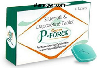
Super P-Force dosages: 160 mg
Super P-Force packs: 10 pills, 20 pills, 30 pills, 60 pills, 90 pills, 120 pills, 180 pills

Purchase 160 mg super p-force mastercard
These immature spines are slender processes wealthy in filamentous actin (F-actin) that seem to perform very like the filopodia that project from the expansion cone on the end of an extending axon (see Chapter 7) erectile dysfunction symptoms causes and treatments purchase 160mg super p-force mastercard. The morphology of dendritic spines is described based on the general shape of the neck and head regions latest erectile dysfunction drugs cheap 160mg super p-force visa. With Fragile X syndrome erectile dysfunction ayurvedic drugs in india buy super p-force 160mg online, backbone density is variable and the spines current are inclined to be longer and immature, often resembling filopodia. The size of the spine head seems to be associated with the synaptic strength, with larger heads being stronger. This is due partly to the elevated area obtainable for the postsynaptic neurotransmitter receptors. In many ways, spinogenesis appears to be linked to synaptogenesis, as a end result of the number of spines is correlated with the number of synapses that form. For example, dendritic spines type over a period of minutes, whereas the formation of mature synapses often takes place over a period of days or perhaps weeks. Moreover, in at least some mobile contexts, spinogenesis can happen in the absence of axonal contact. In both Weaver and Reelin mutant mice which are missing presynaptic cerebellar granule cells (see Chapter 5), the dendritic spines on postsynaptic Purkinje cells nonetheless develop and seem morphologically much like wild-type mice. Whether spinogenesis is directly linked to synapse formation or not, the significance of spinogenesis is noted by the massive variety of neurodevelopmental disorders related to decreases in dendritic spine quantity and altered morphology. For instance, decreased backbone density and alterations in spine morphology have been noted in schizophrenia, autism, Fragile X syndrome, Rett syndrome, and Down syndrome. Studies of those problems in mouse fashions or human post-mortem specimens have revealed a number of consistent adjustments associated with every disorder. The density of spines seems to be variable, no less than in those specimens noticed to date. In people with Rett syndrome, spine density is reduced and there are fewer spines exhibiting a mushroom-shaped morphology. As discussed later in the chapter, lack of synaptic connections is a traditional a half of postnatal development, but in those with Down syndrome the loss of dendritic spines is dramatically increased. The elevated motility is assumed to enhance the chance that a filopodium will initiate a synaptic connection. Eph/ephrin bidirectional signaling mediates presynaptic improvement Since 2001, a selection of research have revealed that ephrin ligands and their related Eph receptors affect pre- and postsynaptic growth. As noted in earlier chapters, ephrin ligands bind to Eph tyrosine kinase receptors to provoke signal transduction cascades within the receptor-bearing cell through forward signaling. In addition, the binding of the Eph receptors to the membrane-bound ligand also can induce signal transduction cascades within the ligand-bearing cell by way of the method of reverse signaling. In some mobile contexts, when Eph receptors in presynaptic neurons are activated by ephrin ligands on the postsynaptic neurons (forward signaling), clustering of presynaptic parts is observed. In other contexts, postsynaptic Eph receptors activate presynaptic ephrin ligands (reverse signaling) to induce clustering of presynaptic elements. Similarly, ahead and reverse signaling mediate improvement of postsynaptic components. Members of both the EphA/ephrin A and EphB/ ephrin B subclasses have been discovered to influence synaptogenesis. A number of experiments conducted in the laboratories of Matthew Dalva and Mark Henkemeyer are among those who have contributed to the current models of how ephrin signaling influences synapse formation. For instance, activation of EphB receptors by ephrin B ligands (forward signaling) has been shown to influence presynaptic growth of hippocampal and cortical neurons in numerous experimental preparations. An in vitro assay was developed in which ephrin B3 was expressed in nonneuronal cells. The presynaptic marker was only observed in the axons that contacted the ephrin B3-expressing nonneuronal cells. This suggested ephrin B3 mediated presynaptic differentiation of excitatory neurons by binding to endogenous EphB receptors on the hippocampal neurons. This discovering was in preserving with different research showing EphB activation results in presynaptic differentiation. Postsynaptic EphB receptors signaling through presynaptic ephrin B1 and ephrin B2 ligands (reverse signaling) additionally influence presynaptic development by regulating the clustering of synaptic vesicles and the maturation of active zone proteins. Evidence for ephrin-B-directed synaptic vesicle clustering was present in in vitro research of embryonic rat cortical neurons co-cultured with nonneuronal cells generated to categorical EphB receptors. These research famous that clustering of synaptic vesicles occurred on the sites of contact between the EphB-expressing nonneuronal cells and the cortical neurons. If the expression of both ephrin B1 and ephrin B2 have been reduced, an extra lower in synaptic vesicle accumulation was famous. Thus, only ephrin B1 and ephrin B2 appear to affect presynaptic vesicle clustering in cortical neurons. In vitro experiments have been carried out in which cortical neurons have been co-cultured with nonneuronal cells expressing EphB receptors. Syntenin-1 is one protein identified to work together with ephrin B throughout presynaptic growth. Syntenin-1 was first recognized as a melanoma differentiation gene and has since been discovered to be necessary not only for tumorigenesis, but additionally for receptor trafficking and synapse formation. One hypothesis about how this happens is that the ephrin Bs recruit syntenin-1 to the right presynaptic location so that syntenin-1 can then anchor synaptic vesicle proteins. Eph/ephrin signaling initiates a quantity of intracellular pathways to regulate the formation of postsynaptic backbone and shaft synapses A number of studies in cortical and hippocampal neurons have indicated that ephrin�Eph interactions additionally affect elements of postsynaptic development. For instance, reverse signaling initiated by presynaptic EphB receptors influences postsynaptic differentiation of ephrin-expressing postsynaptic neurons. In hippocampal neurons, EphB prompts postsynaptic ephrin B3 via reverse signaling. Thus, postsynaptic modifications that occur through ephrin B signaling appear to happen not only throughout synaptogenesis, but also throughout synapse maturation and refinement. Other signaling pathways have been recognized that regulate cytoskeletal dynamics in pre- and postsynaptic neurons. Proteins tagged with ubiquitin, a 76-amino acid peptide that attaches to other proteins, are transferred to one of many many proteasomes discovered within the nucleus or cytoplasm of the cell. This first turned apparent initially of the present century in studies of invertebrates. It is now recognized that ubiquitination mediates many cellular pathways that finally regulate synaptic size, stabilization, and elimination by rapidly degrading various pre- and postsynaptic proteins. The particular effect of ubiquitin on a target protein is dependent upon the length and configuration of the ubiquitin chains that connect to the targeted proteins. A ubiquitin chain is connected to a goal protein following a series of steps involving enzymes known as E1, E2, and E3 ubiquitin ligases. E1 ligases are the enzymes that activate ubiquitin then interact with E2 ubiquitin provider ligases.
Super p-force 160mg lowest price
One common mechanism for specifying neuronal versus nonneuronal cells is lateral inhibition impotence unani treatment in india purchase super p-force 160mg on-line, a process that relies on the level of Notch receptor exercise in a given cell erectile dysfunction 45 purchase super p-force 160mg line. Lateral inhibition designates future neurons in Drosophila neurogenic regions In the creating Drosophila nervous system erectile dysfunction drug has least side effects generic super p-force 160 mg visa, the areas of ectoderm that finally give rise to the neurons are called the neurogenic areas. Cells throughout the neurogenic area begin to express low levels of proneural genes, corresponding to atonal and members of the achaete-scute complicated (achaete, scute, lethal of scute, and asense). Thus, at the earliest phases the cells are equal, with each cell expressing low levels of proneural genes. In this instance, the center cell (blue) expresses a adequate degree of Delta to activate the Notch receptors in surrounding cells. What is clear is that when enough Delta expression is attained, the ligand can bind to Notch on an adjoining cell, initiating a signal transduction cascade that ultimately leads one cell within the pair to a neuronal fate and the opposite cell to a nonneuronal fate. The signaling pathway is initiated when the certain Notch receptor undergoes proteolysis. Thus, Delta binding to the Notch receptor initiates the pathway for inhibiting neural destiny in the Notch-activated cell. Conversely, further copies of those genes lead to additional neurons in the Drosophila nervous system. Lateral inhibition designates stripes of neural precursors within the vertebrate spinal wire Lateral inhibition additionally impacts the development of cells inside vertebrate neuroectoderm. In the Xenopus neural plate, for instance, the area of the future spinal twine contains three longitudinal stripes of neural precursors on each side of the midline. The stripes will finally give rise to the motor neurons (medial rows), intermediate zone neurons (center rows), or dorsal sensory interneurons (lateral rows) within the grownup spinal twine (see Chapter 4). However, before these stripe areas are established in the neural tube, proneural genes are expressed to set up which cells turn out to be neural precursors. Half segments of the neural plate illustrate how Notch signaling regulates the formation of neuronal stripes in Xenopus. Each section represents the stripes discovered on one side of the neural plate where three stripes of neural precursor cells (blue) emerge throughout late gastrulation. The blue row on the medial (M) facet will later form motor neurons, the row within the middle will type intermediate zone neurons, and the row on the lateral aspect (L) will kind dorsal interneurons (see additionally Chapter 4). Normally, Delta is restricted to the stripe regions in order that solely Notch receptors expressed on the cells of interstripes dn 6. When Delta is overexpressed, Notch is activated on cells of each stripe and interstripe regions, thus directing extra cells to a nonneuronal destiny. Thus, ngn1, a member of the atonal gene family, is important for establishing which cells have the potential to become neurons. The ngn1 gene induces downstream expression of NeuroD, another homolog within the atonal gene household, which is needed to regulate additional development of the neurons. A direct hyperlink between ngn1 and NeuroD expression was seen in research during which overexpression of ngn1 led to overexpression of NeuroD as nicely. Once ngn1 designates cells in the stripe areas because the neural precursors, lateral inhibition ensures that the additional development of neurons is restricted to the stripe regions. In the Xenopus spinal twine, it appears that the Delta ligand is expressed solely in cells inside the stripes, and this expression may be regulated by Xenopus achaete-scute homolog (Xash) genes, such as the Xash1 or Xash3. In distinction to the Delta ligand, the Notch receptor is expressed in cells of each the stripe and interstripe regions, though only the Notch receptors in the interstripe area will receive Delta signals. The neural precursors throughout the stripes subsequently obtain further signals to turn out to be specific neural types, corresponding to motor neurons and sensory interneurons. In a variety of areas of the Drosophila nervous system, the uneven distribution of Notch and Numb proteins additional restricts cell fate choices in precursor cells. Subsequently, the temporal expression of specific transcription components usually supplies further cues to affect the destiny options out there to the neuronal precursors. As launched in Chapter 5, the differential distribution of Numb and Notch can affect cell growth, with Numb inhibiting Notch receptor activation. Notch signaling can be important in sensory organs of the vertebrate nervous system. Examples are given later in this Chapter that describe the differentiation of sensory cells within the organ of Corti of the inside ear and within the retina of the eye. Thus, the same common signaling pathways are used to set up structurally various sensory areas in a number of species. As in different neuronal populations, the extent of Notch receptor activation influences cell fate. The absence of Numb leads to the production of socket and bristle cells (C) or only socket cells (D). These uneven divisions proceed for various lengths of time, depending on the Nb lineage. Similar to the vertebrate neurons, the apical proteins are wanted to direct the orientation of the mitotic spindles that decide the airplane of cell division as properly as direct basal proteins to the other pole. The apical proteins new neuroblast is prepared to divide once more as a result of the provision of adequate Notch signaling exercise. The apical proteins help orient the mitotic spindles to decide the aircraft of cell division. The third neuroblast (green) expresses Pdm, whereas the following (red) expresses Castor, and the ultimate Nb in this lineage (yellow) expresses Grainyhead. Rather, cell fate is decided by a mixture of transcription issue expression and cell location. If one transcription issue is absent, only the cell kind arising at that stage shall be eliminated. If a transcription issue is experimentally maintained, then these cell sorts will persist longer. Changes in transcription factor expression mediate the progressive growth of cerebellar granule cells Because the developmental occasions that result in the formation and migration of cerebellar granule cells are so nicely documented (see Chapter 5), a quantity of research have targeted on the indicators that regulate growth of this highly specialised group of cells. Manipulations of Notch activity in vivo revealed that if Notch activity is experimentally elevated, granule cells proliferate longer. Conversely, if Notch receptor exercise is inhibited, cells stop proliferating early and start to categorical Math1 (mouse Atonal homolog 1), a transcription issue characteristic of dedicated granule cells. Thus, a mixture of extrinsic alerts seems to regulate the expression of transcription factors and proteins that mediate the progressive development of cerebellar granule cells. Math1 expression is indicative of a committed granule cell fate and induces the expression of other transcription elements, including Zic1 and Zic2. Cerebellar granule cells must due to this fact integrate multiple alerts to progress from the granule cell precursor stage to a completely differentiated granule cell neuron. Temporal cues assist mediate the destiny of cerebral cortical neurons In the vertebrate cerebral cortex, the time of neurogenesis is linked to the migratory route and fate of newly generated neuronal precursors (see Chapter 5).

Generic 160 mg super p-force with visa
Early segmentation in the neural tube helps establish subsequent neural anatomical group As dn Box 3 impotence female buy super p-force 160mg amex. These three primary mind vesicles become the five secondary brain vesicles as development continues erectile dysfunction doctor near me generic 160 mg super p-force mastercard. The prosencephalon is split into the telencephalon and diencephalon erectile dysfunction first time order super p-force 160mg mastercard, whereas the rhombencephalon is split into the metencephalon and myelencephalon. Differences in spinal twine levels are more simply noticed along the dorsal-ventral axis (see Chapter 4). The expansions that kind along the A/P axis during the early phases of neural tube growth are often described as metameric segments, or neuromeres, a series of enlargements that provides regionally restricted areas for additional development. These are the prosencephalon (future forebrain), mesencephalon (future midbrain), and rhombencephalon (future hindbrain). In the forebrain, these expansions are called prosomeres and are numbered from 1-6 (p1-p6) starting on the junction of the forebrain (prosencephalon) and midbrain. In the hindbrain, the expansions kind rhombomeres which are numbered sequentially starting at the midbrain� hindbrain junction (r1-r6). The neuromeres are transient constructions which may be thought to present regionally restricted segments that produce distinctive indicators required for early cell improvement. The isthmic organizer is found at the boundary between the mesencephalon (midbrain) and metencephalon (hindbrain), where alerts are generated to affect development of these areas of the creating nervous system. The notochord extends from the mesencephalon to the spinal wire and provides signals that affect the event of cells in these extra posterior regions of the neural tube. In addition, the notochord extends along a lot of the neuroaxis and is a crucial source of alerts for patterning hindbrain and spinal twine regions. Specific molecules recognized in the varied signaling centers are described beneath. Younger organizer tissues induced anterior structures, whereas barely older tissues induced posterior regions. Together, these outcomes instructed that the organizer generated totally different alerts at totally different times in development to direct the formation of those areas alongside the A/P axis. Over the years, many scientists concluded that the anterior (forebrain-like) area of the neural tube is a "default" state for neural tissue, with further signals required to transform neural tissues into extra posterior areas. In the Nineteen Fifties, work by Pieter Nieuwkoop instructed a model by which the signal for neural induction, the "activator," was wanted to induce competent ectoderm to type anterior brain areas, however that progressively greater concentrations of other "reworking" alerts have been wanted to convert early neural tissue into hindbrain and spinal wire. Although identification of such molecules was elusive at the moment, the final mechanisms first proposed by this "activation�transformation" mannequin of Nieuwkoop have been largely supported by newer research. Additional indicators that restrict the activity of the reworking signals have additionally been discovered. Grafting studies by Otto Mangold and others demonstrated that when youthful (anterior) organizer tissue was grafted into a host embryo, the grafted organizer induced extra ectopic head buildings (A). It now seems that neural inducers first promote formation of primarily anterior-like tissue within the neural plate, then region-specific alerts emerge to refine every space along the A/P axis of the growing neural tube. The signals often overlap and work together by way of focus gradients established by morphogens-diffusible molecules that act on cells at a distance. Instead, indicators are produced at several areas and at numerous times so as to sample nearby areas while also proscribing the affect of molecules from adjoining territories. For example, indicators that specify midbrain structures have to be prevented from extending to more anterior or more posterior areas. Additionally, midbrain constructions must not be converted by signals originating within the forebrain or hindbrain regions. As shall be seen, the expression of regionally particular signals and the repression of adjacent indicators are equally important in patterning the A/P axis. Many of the genes and signal transduction pathways for A/P axis patterning are conserved throughout invertebrate and vertebrate species, though some differences in signaling mechanisms have been famous in the numerous animal models that may mirror, partially, the differences in mind anatomy. Further, different members of a given gene family might govern patterning in a specific A/P region within the completely different species. The examples that observe focus primarily on the gene families concerned in patterning the particular regions of the vertebrate neural axis, quite than species-specific relations. An inducer may be secreted such that those cells closest to the source of the inducer obtain the very best focus, while those further away obtain decrease concentrations. Inducers are thought to type primarily forebrain-like areas, while transforming indicators modify the results of the inducers to be able to affect growth of extra posterior-like regions. Various inhibitory signals restrict the unfold of inducers and transformers to assist maintain the correct boundaries of the morphogen indicators. Multiple indicators help sample the neural tube alongside the A/P axis and gradients of particular inducers, transformers, and inhibitors may function over long or quick distances to sample different segments. However, additional signaling molecules are necessary to delineate the telencephalon and diencephalon regions of the forebrain. The relative areas of signaling facilities that are necessary at later stages of improvement (the prechordal plate and notochord) are additionally proven. Forebrain segments are characterised by totally different patterns of gene expression the growing forebrain is often described as being segmented into divisions called prosomeres. Six totally different prosomeres have been recognized starting with prosomere 1 (p1) adjoining to the mesencephalon and progressing anteriorly to prosomere 6 (p6) within the telencephalon. These molecules play direct roles in forebrain patterning and are homologs of Drosophila genes that always have related patterning capabilities during fly development. For example, a quantity of experiments have demonstrated the need of Otx genes in forebrain development across species. However, if the Drosophila homolog, the Otd (orthodenticle) gene, replaces the missing Otx genes, the pinnacle buildings are rescued. Conversely, human Otx genes can rescue Drosophila Otd mutants, further demonstrating the extremely conserved nature of these genes and their capabilities in patterning forebrain and head constructions. Genes of the Pax family are also expressed widely in the growing neural tube and Pax6 is required for forebrain improvement. The results of Six3 and Pax6 are restricted to these more anterior regions partly by inhibitory alerts such as Irx3 and Wnt that originate within the midbrain. The prosomeres are the six neuromere segments located in the forebrain area anterior to the mesencephalon (p1-p6). Otx genes similar to Otx2 expressed in p1-p6 regulate development of the forebrain, as properly as the mesencephalon and different head buildings. Pax6 is expressed in p1-p6 and is required for regular forebrain, eye, and ear formation. Six3, which is localized to p3-p6, opposes expression of Irx3 in p1, p2, and more posterior regions. The interaction of neural inducers and Wnt in patterning forebrain versus midbrain areas has been proven in a quantity of experiments. For instance, when Wnt was overexpressed in the presence of a neural inducer (for example, chordin, follistatin, or noggin), genes and structures associated with anterior mind regions have been suppressed.


Buy cheap super p-force 160 mg on-line
It is now identified that the radial migration patterns described above are primarily utilized by the glutametergic erectile dysfunction natural treatment options cheap super p-force 160mg online, excitatory projection neurons (pyramidal neurons) of the cerebral cortex erectile dysfunction causes diabetes order 160 mg super p-force with mastercard. Such cues often direct cell migration by both attracting neurons to the correct location or repelling the neurons away from an incorrect pathway erectile dysfunction medication new zealand best 160mg super p-force. In addition, the interneurons are directed away from non-target regions by the repellent proteins of the semaphorin family described in Chapter 7. Thus, multiple engaging and repellent cues are used to guide tangentially migrating interneurons in order that they enter the proper cortical layer. Tangential migration remains less properly characterized than radial migration, but it has been observed within the forebrain, diencephalon, brainstem, and spinal wire. Many of the extracellular cues utilized by tangentially migrating neurons appear to be just like these used to guide newly fashioned axons to target cells (see Chapter 7). A coronal section via the embryonic telencephalon reveals the location of the lateral and medial ganglionic eminences. Cells from the lateral ganglionic eminence (red) migrate to areas related to the basal ganglia. Cells that migrate from the medial ganglionic eminence (green) travel alongside existing axons or cells to attain the neocortex. Tangential migration typically overlaps temporally with the radial migration of different neurons (purple) alongside radial glia (yellow). Some of the mechanisms and signaling molecules used in the cerebral cortex are also used within the cerebellar cortex. Cerebellar development also options a number of fascinating and noteworthy variations. Part of the rationale that the cerebellum has unique patterns of proliferation and migration is as a result of the group of the cerebellar cortex is quite different from that seen in the cerebral cortex. The cerebellar cortex is a extremely convoluted structure consisting of three layers. The granule cell layer is the innermost layer and contains the numerous, small granule neurons. In the mature cerebellum, the cell bodies of the Purkinje cells are discovered in the Purkinje cell layer, whereas their elabodn 5. The granule neurons found within the granule cell layer are notable for his or her small measurement and unbelievable number. Granule cells are essentially the most numerous neurons within the mammalian brain; in fact, they may constitute half of the entire variety of neurons within the mind of some species. Also dispersed within the adult cerebellar layers are various interneurons corresponding to Golgi, stellate, and basket cells. Cerebellar neurons arise from two zones of proliferation the cerebellum arises from the alar plate area in the caudal metencephalon. As described in Chapter four, the alar plate primarily offers rise to sensory structures, whereas the basal plate gives rise to motor constructions. In the caudal metencephalon, the alar plates broaden in a dorsomedial direction, start to cowl the thin roof plate, and prolong over the fourth ventricle. The higher, or rostral, rhombic lip area produces particular cerebellar cell populations mentioned under. The upper (rostral) region of the rhombic lips is the source of granule cells of the cerebellum. The lower (caudal) region of the rhombic lips offers rise to numerous nuclei of the pons. Panel D shows a cross section of the cerebellar primordium taken on the stage indicated by the dashed line in panel E. Initially, the Purkinje cell layer is comprised of several irregular rows of cells, nevertheless it later thins to the only layer seen within the adult cerebellum. A second group of cells (curved arrows) migrates from the rostral area of the rhombic lips, traveling over the surface of the cerebellar primordium until they lie beneath the pial membrane. A second group of neuronal precursors originates from the rostral portion of the rhombic lips. These cells stream tangentially over the surface of the cerebellar plate to type the external granule cell layer (sometimes referred to as the external germinal layer) that lies beneath the pial membrane. Jagged 1, a ligand for the Notch receptor, has additionally been shown to promote granule cell proliferation. As noted in the cerebral cortex, Notch receptor exercise is important for continued proliferation. The interaction between Bergmann glia and a granule cell are illustrated beginning with step three in panel A. Each cell then extends a third process into the molecular layer-the layer that ultimately consists of Purkinje cell dendrites and interneurons. From the Purkinje cell layer the Bergmann glia prolong processes to the pial floor. In the cerebellum, the place astrotactin was first found, this interplay occurs between the astrotactin-expressing granule cells and the Bergmann glia. Hatten and colleagues first identified this integral membrane protein within the late Eighties by producing antibodies to granule cell proteins. To examine the position of the newly recognized protein in granule cell growth, a cell tradition method was developed. Using this preparation, it was found that although granule cells might normally adhere to and migrate along a Bergmann glial cell in vitro, granule cell adhesion and migration had been blocked when anti-astrotactin antibodies had been added to the cultures. Further proof for the significance of astrotactin in cerebellar growth was seen in mice that lack astrotactin. The capacity of granule cells to continue migration, although at a slower fee, in the absence of astrotactin advised that different molecules may also mediate adhesion and migration of granule cells within the cerebellum. Instead, most Purkinje cells are clumped as aggregates in deeper layers of the cerebellum, nearer to the ventricular floor. Granule cells, which usually secrete Reelin protein, are additionally fewer in number and tons of fail to migrate past the present Purkinje cells (B). Nrg1 signaling seems to regulate Bergmann glial cell numbers as nicely as the migration of the granule cells alongside the radial processes. Several experiments have demonstrated that disrupting ErbB4 receptor perform or blocking Nrg1 expression leads to irregular Bergmann glia growth, together with a disruption in the length of the glial processes. These adjustments in Bergmann glial cell development therefore lead to altered granule cell migration. Mutant mice provide clues to the method of neuronal migration in the cerebellum As noted earlier in the chapter, spontaneously occurring mutations in mice can present perception to the mechanisms of cell migration and patterning. For example, reeler mice not only display altered layering in the cerebral cortex, as described above, but also show defects in Purkinje cell and granule cell migration within the cerebellar cortex.

Discount 160mg super p-force
One daughter carried two wild-type copies of the gene erectile dysfunction protocol list generic super p-force 160 mg with visa, and the opposite was homozygous for the mutation erectile dysfunction treatment in kenya cheap 160mg super p-force mastercard. As those two daughter cells underwent mitosis impotence blood pressure medication order super p-force 160 mg with mastercard, they created copies of themselves with their distinctive genetics. Since little cell migration happens during eye growth, these teams of cells stayed close to each other in patches known as clones. The resulting eyes were a mosaic of clones, and every cell in a clone shared a lineage, since all of them derived from a single cell. Therefore, rearranged chromosomes carried mutations known as markers, which resulted in cellular modifications that had been easy to see underneath a microscope. Eye cells with a wild-type w gene (w + cells) created pigment granules filled with simply seen pigments that give the eye its pink color. Cells with two mutant copies of the w gene (w � cells) made no pigment granules, so the eyes appeared white. When X-rays induced mitotic recombination in random components of the attention, each resulting mosaic eye was unique. Researchers looked a lots of of mosaic eyes and mapped which cell fates might come from the same clone. The researchers made diagrams showing the placement of w � cells in every ommatidium and seemed for patterns. What they found was that there was no relationship between cell fate and lineage within the fly eye. There have been no rules stating, for example, that if an R5 cell got here from a clone, the neighboring R4 cell had to come from the same clone. The fate of every cell was independent of whether or not or not it shared a lineage with another cell. Today we know that cell fate selections are decided by signaling between cells, but proof that environmental signaling occurred was a significant outcome on the time. Fly biologists also used mosaic evaluation to understand how these environmental alerts labored. Researchers had recognized mutations in two genes, named sevenless (sev) and bride of sevenless (boss), by which eyes had no R7 cells. To determine the signals encoded by these genes, scientists placed a marker mutation on the identical chromosome arm as the mutation they needed to follow. Then they could make clones that have been all mutant for sev or boss and which could be simply distinguished from the rest of the eye. This meant that sev was only required within the R7 cell and must be a signal-receiving gene. Filled circles characterize wild-type cells (w+) that carry both the white marker and the sev gene. By analyzing lots of of ommatidia, scientists established that the R7 cell must categorical the sev gene for an ommatidium to have an R7 cell (the middle cell in the backside row within each ommatidium on this example). The arrows point out the different cell patterns that have been noticed in these experiments. The purple arrow factors at an ommatidium in which R1�R5 and R8 are all wild kind, yet R7 is missing. The blue arrow points to an ommatidium by which all cells besides R7 are mutant, yet R7 is current. Collectively, these information indicated that sev is required solely in the presumptive R7 for R7 cell fate determination. The open dots point out cells that lack the wild-type gene boss, while the filled circles characterize people who carry the boss gene. In this panel, the blue arrow points to an ommatidium by which only R2 and R8 are wild kind, but R7 is current. The purple arrow factors to an ommatidium during which R2 is wild type, but R8 is mutant for boss. Together, these knowledge indicated that boss is particularly required in R8 for the R7 cell to form. The capacity to decipher so much info We now know that boss encodes a membrane-bound by way of manipulation of fly genetics demonstrates the signaling protein and that sev encodes its receptor on power of mosaic analyses. In reality, the R8 cell types first and is required to guarantee that the opposite cells to develop. Following R8, the R2 and R5 cells are the following to be determined and are formed on the same time. The expression levels of proneural genes similar to atonal1 and the activity of transcription elements Senseless and Rough contribute to the determination of R8. R8 is the first photoreceptor cell fate to be decided and is required for the formation of the other retinula cells. E(spl) inhibits atonal1 expression in those cells, thus preventing them from becoming R8 cells. Two of the three cells will produce the transcription components Rough and Senseless. Thus, the cell within the equivalence group that only expresses Senseless is ready to enhance the expression of atonal1 and turn out to be the R8 photoreceptor. Cell�cell contacts and gene expression patterns establish R1�R7 photoreceptor cell types the signaling pathways that regulate the willpower of the R7 photoreceptor had been among the many first to be found. In the Nineteen Seventies, Seymour Benzer and colleagues identified mutants that lacked the R7 photoreceptor cell. These mutants developed a cone cell in the ordinary location of the R7 photoreceptor. When levels of Senseless are greater than those of Rough, Senseless inhibits Rough. However, as growth continues, atonal1 and senseless expression become progressively extra restricted. In a subset of cells, Notch receptor activation leads to increased expression of Enhancer of cut up (E[spl]), which inhibits, in turn, the expression of atonal1 (tan cells), thus resulting in a lower in senseless expression as well. In cells missing excessive ranges of Rough, Senseless ranges are enough to promote expression of atonal1 and those cells undertake the R8 destiny (green cell in every equivalence group). The Sev receptor is expressed by the R7 photoreceptor cell and activated by the Bride of Sevenless (Boss) ligand expressed on R8. When Boss binds to Sev, the Ras sign transduction cascade is activated, which leads to the formation of R7. In the absence of the Sev receptor or if the Ras pathway is experimentally inactivated, a cone cell is produced as an alternative. However, the expression of other genes directs these cells to completely different photoreceptor fates. R2 and R5 (pink) require the expression of other transcription issue encoding genes, similar to rough (ro), to differentiate as non-R7 cells. The Sev receptor is activated by a membrane-bound ligand discovered on the surface of the centrally located R8 cell.
Buy 160mg super p-force
Bothwell M (1995) Functional interactions of neurotrophins and neurotrophin receptors erectile dysfunction organic order 160mg super p-force otc. Hamburger V (1939) Motor and sensory hyperplasia following limb bud transplantations in chick embryos impotence effect on relationship purchase 160mg super p-force with visa. Levi-Montalcini R erectile dysfunction videos quality 160 mg super p-force, Meyer H & Hamburger V (1954) In vitro experiments on the effects of mouse sarcomas 180 and 37 on the spinal and sympathetic ganglia of the chick embryo. Synaptic Formation and Reorganization Part I: the Neuromuscular Junction 9 T his chapter introduces two important elements of neurodevelopment: the method of synaptogenesis-the formation of latest synaptic contacts in the nervous system-and the method of synaptic reorganization-the subsequent strengthening or lack of a subset of those connections. The study of synapses, together with how they initially kind and later reorganize, has a protracted history-one often marked by vigorous debates and strong variations of opinion. As noted in previous chapters, neurobiologists within the late 1800s debated whether or not neurons communicated by way of a syncytial community or by way of connections between particular person cells. Evidence finally demonstrated that neurons communicated by way of small spaces-connections now termed synapses. In the primary half of the 20 th century, scientists engaged in another debate, this one concerning whether or not the communication at synapses occurred primarily through chemical or electrical alerts. These differing opinions have been typically referred to because the "war of the soups and the sparks," with the "soups" referring to chemical signals and the "sparks" to electrical indicators. As scientists examined the 2 hypotheses, it was finally determined that the majority of synapses utilize chemical alerts within the type of neurotransmitters to mediate neural communication. Chapters 9 and 10 due to this fact concentrate on the development of buildings related to chemical synapses. The institution of useful synapses is a dynamic process that takes place over an extended time frame. For instance, an extending, motile development cone must rework right into a nerve terminal capable of releasing a specific neurotransmitter, and the target cell should begin to produce the corresponding neurotransmitter receptors. All of these specialized proteins are additionally produced through the strategy of synaptogenesis. Perhaps surprisingly, after all of the effort to convert each synaptic companion into a highly differentiated cell, a subset of synaptic connections is lost in the course of the normal course of development. Thus, rather than induce differentiation in solely the subset of synaptic connections required for neural operate, the nervous system as a substitute over-produces extremely specialized synaptic contacts, then eliminates a portion of them. The stabilization of synapses occurs partly by maintaining synapses that kind useful partners. As the innervating neuron initiates action potentials within the goal cell, the cells eventually start to fire motion potentials in synchrony with one another. The phrase "cells that fireplace together, wire collectively" describes one frequent mechanism by which cells keep chosen synaptic connections. This article begins with an outline of the synaptic buildings present in synapses throughout the nervous system. As with other elements of nervous system development, lots of the molecules and signaling pathways recognized in synaptic improvement are conserved across vertebrate and invertebrate animal models. In most chemical synapses, an motion potential signals a presynaptic nerve terminal to release a neurotransmitter that diffuses across the synaptic space to bind to corresponding receptors on the surface of the postsynaptic associate cell, thereby initiating a response in that cell. Charles Sherrington first used the term "synapse" in 1887 to describe the small gap between speaking nerve cells. However, the chemical synapse was not visualized conclusively until the mid-1950s, when techniques for making ready tissues for electron microscopy grew to become sufficiently superior to present pictures of nerve cells. Stanley Bennett in 1955 had been the first to prove that neurons have been separated by a well-defined synaptic cleft. By the late twentieth century, additional advances in microscopy- significantly in the areas of time-lapse and fluorescence imaging-and new methods in cellular and molecular biology allowed scientists to identify tons of of intracellular synaptic specializations. The intricate elements of synaptic structures are at present studied at ranges of detail unimaginable even 25 years in the past. Reciprocal signaling by presynaptic and postsynaptic cells leads to the development of unique synaptic elements As an extending nerve fiber approaches a goal cell, the tip transforms from a motile progress cone to a presynaptic nerve terminal. Both pre- and postsynaptic partners produce various adhesion molecules to stabilize the newly fashioned synaptic connections. The exact alignment of pre- and postsynaptic components ensures neurotransmitters released by the synaptic vesicles attain the postsynaptic receptors as quickly as possible. The synaptic vesicles line up at the energetic zones- areas of proteins alongside the presynaptic membrane adjoining to the postsynaptic cell. Voltage-dependent Ca2+ channels open in response to an motion potential and initiate the release of neurotransmitter into the synaptic cleft. The neurotransmitter molecules bind to neurotransmitter receptors on the postsynaptic cell. Receptors are aligned instantly throughout from the lively zones so that neurotransmitter reaches the receptors rapidly. [newline]Also positioned in postsynaptic cells are ion channels that open in response to neurotransmitter binding and provoke the motion potential in that cell. Scaffolding proteins assist anchor the postsynaptic receptors and ion channels throughout from the presynaptic nerve terminal. Various adhesion molecules are positioned in pre- and postsynaptic cells that initiate or stabilize the synaptic contacts. Finally, glial cells encompass the pre- and postsynaptic contacts to provide alerts to maintain the synaptic elements and to remove mobile particles left by retracting nerve terminals. These primary synaptic parts are discovered at synapses throughout the nervous system. At most synapses in the central and peripheral nervous systems, the glial cells extend cellular processes that encompass pre- and postsynaptic cells to assist keep the required synaptic parts as nicely as take away mobile particles generated throughout synapse elimination. As scientists documented the formation of the varied synaptic components in the course of the early levels of synaptogenesis, a quantity of reviews noted that some postsynaptic specializations started to kind previous to the arrival of the presynaptic terminal. Such observations suggested that the postsynaptic cell develops first to provide alerts that direct the differentiation and connectivity of the presynaptic cell. However, other research reported that the presynaptic nerve terminal produces some synaptic elements previous to contact with the postsynaptic cell, suggesting that presynaptic differentiation was independent of postsynaptic indicators. In many circumstances, it was simply unclear whether or not pre- or postsynaptic parts were the first to come up. It now appears that both pre- and postsynaptic cells start to specific immature synaptic elements prior to contact, however solely differentiate organized, mature synaptic components in response to indicators obtained from the synaptic associate. Therefore, each pre- and postsynaptic cells provide reciprocal cues that induce maturation of synaptic parts to be positive that a proper synaptic connection is shaped, maintained, and reorganized, if necessary. Skeletal muscle tissue are attached to bones, usually through connective tissue called tendons, and are beneath voluntary control. The variety of muscle fibers that a single motor neuron innervates is identified as a motor unit, as a end result of stimulation of that neuron leads to contraction of all the muscle fibers it innervates. Each terminal department contacts a specialized area on the skeletal muscle referred to as a motor endplate, an oval-shaped region of a muscle cell that seems slightly elevated above the the rest of the cell surface on the time of innervation. Within the endplate area, the terminal branches finish as synaptic boutons (terminal finish bulbs), where they kind a synaptic reference to the muscle fiber. In grownup vertebrates, each motor neuron extends a myelinated axon that branches to set up contact with several skeletal muscle fibers.
Generic 160 mg super p-force overnight delivery
For the subunits to bind O2 impotence trials purchase super p-force 160mg mastercard, iron in the heme moieties must be in the ferrous state what age does erectile dysfunction usually start buy super p-force 160 mg mastercard. Methemoglobinemia has a quantity of causes together with oxidation of Fe2+ to Fe3+ by nitrites and sulfonamides erectile dysfunction diabetes uk order super p-force 160mg fast delivery. In fetal hemoglobin, the two chains are changed by chains, giving it the designation of twenty-two. The physiologic consequence of this modification is that HbF has the next affinity for O2 than hemoglobin A, facilitating O2 motion from the mom to the fetus. HbF is the normal variant current in the fetus and is gradually changed by hemoglobin A throughout the first year of life. Hemoglobin S is an irregular variant of hemoglobin that causes sickle cell disease. In hemoglobin S, the subunits are normal and the subunits are irregular, giving it the designation A2S2. In its deoxygenated kind, hemoglobin S varieties sickle-shaped rods in the pink blood cells, distorting the form of the purple blood cells. This deformation of the pink blood cells can lead to occlusion of small blood vessels, causing many of the symptoms of sickle cell disaster. Assume that for a standard hemoglobin concentration of 15 g/100 mL, the O2-binding capacity is 20. It is a given that at a traditional hemoglobin focus of 15 g/100 mL, O2-binding capacity is 20. Thus at a hemoglobin concentration of 10 g/100 mL, O2-binding capability is 10/15 of regular. If fewer than four molecules of O2 are sure to heme groups, then saturation is less than one hundred pc. For instance, if, on common, every hemoglobin molecule has three molecules of O2 sure, then saturation is 75%; if, on common, each hemoglobin has two molecules of O2 bound, then saturation is 50%; and if only one molecule of O2 is bound, saturation is 25%. Sigmoidal Shape (2) Next, calculate the precise quantity of O2 combined with hemoglobin by multiplying the O2-binding capability by the % saturation. O2 content of blood, as already described, is the sum of dissolved O2 (2%) and O2-hemoglobin (98%). A change within the value of P50 is used as an indicator for a change in affinity of hemoglobin for O2. An enhance in P50 displays a decrease in affinity, and a decrease in P50 reflects a rise in affinity. Due to constructive cooperativity, affinity is highest and O2 is most tightly certain (the flat portion of the curve). The high affinity makes sense because it could be very important have as much O2 as possible loaded into arterial blood within the lungs. Because oxyhemoglobin and deoxyhemoglobin have totally different absorbance characteristics, the machine calculates % saturation from absorbance at two totally different wavelengths. However, understanding % saturation, one can estimate PaO2 from the O2-hemoglobin dissociation curve. Such shifts replicate modifications within the affinity of hemoglobin for O2 and produce modifications in P50. Shifts can occur with no change in O2-binding capacity, during which case the curve moves proper or left, however the shape of the curve remains unchanged. Or, a right or left shift can occur in which the O2-binding capability of hemoglobin also modifications and, on this case, the shape of the curve changes. Together, these results lower the affinity of hemoglobin for O2, shift the O2-hemoglobin dissociation curve to the proper, and increase the P50, all of which facilitates unloading of O2 from hemoglobin within the tissues. The will increase in temperature also cause a proper shift of the O2-hemoglobin dissociation curve and a rise in P50, facilitating unloading of O2 within the tissues. Considering the example of exercising skeletal muscle, this impact also is logical. As heat is produced by the working muscle, the O2-hemoglobin dissociation curve shifts to the right, providing extra O2 to the tissue. This decrease in affinity causes the O2-hemoglobin dissociation curve to shift to the best and facilitates unloading of O2 in the tissues. Thus when the demand for O2 decreases, O2 is extra tightly bound to hemoglobin and fewer O2 is unloaded to the tissues. Decreases in temperature cause the alternative effect of increases in temperature- the curve shifts to the left. When tissue metabolism decreases, less warmth is produced and less O2 is unloaded within the tissues. This modification ends in increased affinity of hemoglobin for O2, a left shift of the O2-hemoglobin dissociation curve, and decreased P50. This elevated affinity is helpful to the fetus, whose PaO2 is low (approximately forty mm Hg). When the affinity is increased, unloading of O2 in the tissues is tougher. The implications for O2 transport are apparent: this impact alone would reduce O2 content material of blood and O2 delivery to tissues by 50%. Not solely is there reduced O2-binding capacity of hemoglobin, however the remaining heme websites bind O2 more tightly (Box 5. Proerythroblasts undergo further steps in improvement to kind mature erythrocytes (red blood cells). This distinguishing capacity is based on the fact that decreased renal blood circulate causes decreased glomerular filtration, which leads to decreased filtration and reabsorption of Na+. On a cold February morning in Boston, a 55-year-old man decides to warm his car within the storage. About 30 minutes later, his wife finds him tinkering at his workbench, confused and respiration quickly. This extremely excessive PaO2 does little to improve O2 delivery to the tissues as a end result of the solubility of O2 in blood is so low (0. This response is catalyzed by the enzyme carbonic anhydrase, which is present in most cells. If the H+ produced from these reactions remained free in resolution, it might acidify the purple blood cells and the venous blood. Therefore H+ have to be buffered so that the pH of the purple blood cells (and the blood) remains throughout the physiologic range. The H+ is buffered in the pink blood cells by deoxyhemoglobin and is carried in the venous blood in this type. Interestingly, deoxyhemoglobin is a better buffer for H+ than oxyhemoglobin: By the time blood reaches the venous end of the capillaries, hemoglobin is conveniently in its deoxygenated form. There is a helpful reciprocal relationship between the buffering of H+ by deoxyhemoglobin and the Bohr impact.

Cheap super p-force 160 mg on line
It begins when the membrane potential is -70 mV and continues till the membrane is absolutely repolarized again to -85 mV erectile dysfunction doctors in brooklyn buy generic super p-force 160 mg. The physiologic clarification for this elevated excitability is that the Na+ channels are recovered erectile dysfunction statistics 2014 cheap super p-force 160 mg otc. For comfort erectile dysfunction oil treatment order 160mg super p-force visa, the autonomic effects on coronary heart rate, conduction velocity, myocardial contractility, and vascular easy muscle are combined into one desk. Autonomic Effects on Heart Rate the consequences of the autonomic nervous system on heart price are referred to as chronotropic effects. The results of the sympathetic and parasympathetic nervous techniques on coronary heart price are summarized in Table 4. Briefly, sympathetic stimulation will increase heart fee and parasympathetic stimulation decreases coronary heart price. B, Sympathetic stimulation will increase the rate of part four depolarization and increases the frequency of motion potentials. C, Parasympathetic stimulation decreases the speed of phase four depolarization and hyperpolarizes the maximum diastolic potential to decrease the frequency of motion potentials. Once the membrane potential is depolarized to the threshold potential, an action potential is initiated. These 1 receptors are coupled to adenylyl cyclase via a Gs protein (see additionally Chapter 2). A 72-year-old woman with hypertension is being handled with propranolol, a -adrenergic blocking agent. The effects of the autonomic nervous system on conduction velocity are called dromotropic effects. Increases in conduction velocity are called optimistic dromotropic effects, and decreases in conduction velocity are known as adverse dromotropic effects. Recall, in considering the mechanism of those autonomic results, that conduction velocity correlates with the size of the inward present of the upstroke of the action potential and the rate of rise of the upstroke, dV/dT. The degree of coronary heart block may vary: In the 4-Cardiovascular Physiology � 143 milder forms, conduction of action potentials from atria to ventricles is just slowed; in more severe cases, action potentials will not be conducted to the ventricles at all. As a results of the sequence and the timing of the unfold of depolarization and repolarization in the myocardium, potential differences are established between different parts of the heart, which may be detected by electrodes positioned on the physique surface. The period of the P wave correlates with conduction time by way of the atria; for instance, if conduction velocity through the atria decreases, the P wave will unfold out. This truth could appear shocking as a result of the ventricles are a lot larger than the atria; nevertheless, the ventricles depolarize simply as quickly because the atria as a result of conduction velocity in the HisPurkinje system is far faster than within the atrial conducting system. It represents first ventricular depolarization to last ventricular repolarization. Not only will there be extra action potentials per time, but these motion potentials will have a shorter duration and shorter refractory periods. Because of the relationship between coronary heart price and refractory interval, will increase in heart price may be a consider producing arrhythmias (abnormal heart rhythms). As coronary heart rate will increase and refractory periods shorten, the myocardial cells are excitable earlier and more typically. The sarcomeres, which run from Z line to Z line, are composed of thick and thin filaments. The skinny filaments are composed of three proteins: actin, tropomyosin, and troponin. Actin is a globular protein with a myosin-binding web site, which, when polymerized, types two twisted strands. Tropomyosin runs alongside the groove of the twisted actin strands and capabilities to block the myosin-binding web site. Troponin is a globular protein composed of a fancy of three subunits; the troponin C subunit binds Ca2+. When Ca2+ is certain to troponin C, a conformational change occurs, which removes the tropomyosin inhibition of actin-myosin interplay. As in skeletal muscle, contraction occurs in accordance with the sliding filament mannequin, which states that when cross-bridges type between myosin and actin after which break, the thick and skinny filaments move past each other. The transverse (T) tubules invaginate cardiac muscle cells on the Z strains, are steady with the cell membranes, and performance to carry motion potentials to the cell interior. The T tubules type dyads with the sarcoplasmic reticulum, which is the location of storage and launch of Ca2+ for excitation-contraction coupling. Excitation-Contraction Coupling As in skeletal and clean muscle, excitation-contraction coupling in cardiac muscle translates the motion potential into the production of tension. The following steps are involved in excitation-contraction coupling in cardiac muscle. A longer cycle size signifies a slower heart fee, and a shorter cycle length signifies a faster heart price. Changes in coronary heart fee (and cycle length) change the length of the motion potential and, in consequence, change the durations of the refractory periods and excitability. For instance, if heart fee increases (and cycle length 4-Cardiovascular Physiology � one hundred forty five Cardiac motion potential measurement of the inward Ca2+ present through the plateau of the motion potential. Ca2+ release from the sarcoplasmic reticulum causes the intracellular Ca2+ concentration to improve even further. Ca2+ now binds to troponin C, tropomyosin is moved out of the way, and the interaction of actin and myosin can occur. Actin and myosin bind, cross-bridges type and then break, the thin and thick filaments move past each other, and rigidity is produced. A critically necessary idea is that the magnitude of the stress developed by myocardial cells is proportional to the intracellular Ca2+ focus. As a results of these transport processes, the intracellular Ca2+ focus falls to resting levels, Ca2+ dissociates from troponin C, actin-myosin interplay is blocked, and relaxation happens. Contractility Contractility, or inotropism, is the intrinsic capability of myocardial cells to develop drive at a given muscle cell length. Agents that produce an increase in contractility are stated to have constructive inotropic results. Positive inotropic agents enhance each the speed of tension growth and the peak rigidity. Agents that produce a decrease in contractility are said to have unfavorable inotropic effects. Negative inotropic brokers decrease each the speed of pressure improvement and the height rigidity. The cardiac action potential is initiated within the myocardial cell membrane, and the depolarization spreads to the inside of the cell via the T tubules. Entry of Ca2+ into the myocardial cell produces an increase in intracellular Ca2+ concentration. This process is called Ca2+-induced Ca2+ release, and the Ca2+ that enters through the plateau of the action potential known as the set off Ca2+.
References
- Stein, J., & Warfield, C. (1982). Phantom limb pain. Hospital Practices, 17, 166n167.
- Ismail R, Teh LK: The relevance of CYP2D6 genetic polymorphism on chronic metoprolol therapy in cardiovascular patients. J Clin Pharm Ther 2006;31:99- 109.
- Bartlett KH, Kidd SE, Kronstad JW. The emergence of Cryptococcus gattii in British Columbia and the Pacific Northwest. Curr Infect Dis Rep 2008;10 (1):58-65.
- King LR, Kozlowski JM, Schacht MJ: Ureteroceles in children. A simplified and successful approach to management, JAMA 249(11):1461n1465, 1983.

