Evista
Claire C. Caldwell, MD
- Department of Emergency Medicine, UMDNJ-Robert Wood Johnson
- Medical School, Camden, NJ, USA
Evista dosages: 60 mg
Evista packs: 30 pills, 60 pills, 90 pills, 120 pills, 180 pills, 270 pills, 360 pills
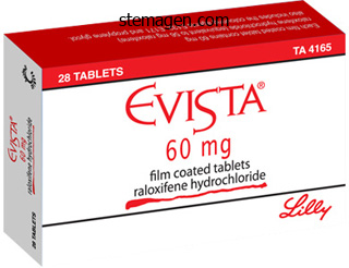
Order evista 60 mg mastercard
Mitochondrion Sarcolemma Nucleus Cross section of a myofiber Cross part of cardiocytes Cardiac muscle 7 women's health center in shelton ct order 60mg evista amex. Myocardial infarction Micrographs left and center from Damjanov I pregnancy quant levels cheap 60mg evista visa, Linder J: Pathology women's health center pembroke pines buy evista 60 mg otc. Intercalated disk Normal cardiac tissue consists of branching and anastomosing striated cardiocytes with a central nucleus and intracellular contractile myofilaments. Myocardial ischemia caused by occlusion of the coronary artery outcomes inside the first 24 hours within the necrosis of cardiocytes. Cardiocytes show an eosinophilic cytoplasm lacking the characteristic intracellular striations detected within the adjoining unaffected cardiocytes. The nuclei are pyknotic (Greek, pyknos, dense, thick; osis, condition) and irregularly shaped. Serum levels of those enzymes stay elevated days after the myocardial infarction. After 3 weeks (not shown), capillaries, fibroblasts, macrophages, and lymphocytes are observed within the necrotic area. If blood circulate is restored in lower than 20 minutes, an occasion generally known as reperfusion, cardiocyte cell viability is maintained. Timing is crucial for implementing early remedy to reestablish blood flow by using thrombolytic agents. A extra delicate marker is cardiocyte-specific troponin I not expressed in skeletal muscle. An enhance of troponin I within the serum of sufferers with acute coronary syndromes provides prognostic information on elevated threat of dying and permits remedy to decrease additional myocardial necrosis. Smooth muscle Smooth muscle may be found as sheets or bundles within the partitions of the gut, bile duct, ureters, urinary bladder, respiratory tract, uterus, and blood vessels. Smooth muscle differs from skeletal and cardiac muscle: easy muscle cells are spindle-shaped, 234 7. The perinuclear cytoplasm incorporates mitochondria, ribosomes, rough endoplasmic reticulum, a Golgi equipment, a latticework of thick myosin filaments, thin actin filaments, and intermediate filaments composed of desmin and vimentin. Actin and intermediate filaments insert into cytoplasmic and plasma membrane�associated constructions wealthy in -actinin, referred to as dense bodies. Invaginations of the plasma membrane, referred to as caveolae, act as a primitive T tubule system, transmitting depolarization alerts to the underdeveloped sarcoplasmic reticulum. Smooth muscle cell Basal lamina Cytoplasmic dense physique (equivalent to Z disks of striated muscle) Plasma membrane dense body Pinocytotic vesicle Actin-myosin bundle Basal lamina Caveola Caveolin-3 Gap junction Adjacent easy muscle cell Longitudinal section of easy muscle cells (muscularis of the stomach). Characteristics of clean muscle Smooth muscle is found in the walls of tubular organs, the walls of most blood vessels, the iris and ciliary physique (eye), and arrector pili muscle (hair follicles), amongst other websites. Caveolae, depressions of the plasma membrane, are everlasting constructions involved in fluid and electrolyte transport (pinocytosis). Caveolin-3, a protein encoded by a member of the caveolin gene household, is associated with lipid rafts. Complexes shaped by caveolin-3 certain to cholesterol in a lipid raft invaginate and kind caveolae. Depending on the section stage, a central nucleus is noticed in a number of the muscle cells. A basal lamina surrounds each muscle cell and serves to transmit forces produced by every cell. Mechanism of easy muscle contraction the association of the contractile proteins and the mechanism of contraction of clean muscle differ from these of skeletal and cardiac muscle: 1. Ca2+ ions that initiate contraction derive from outdoors the cell somewhat than from the sarcoplasmic reticulum. In clean muscle, actin filaments and associated myosin connect to cytoplasmic and plasma membrane Smooth muscle 7. Development of a caveola Fibroblast Smooth muscle cells Basal lamina Caveola Widely unfold tough endoplasmic reticulum Nucleus Mitochondria Lack of a basal lamina Nucleus Fibroblast surrounded by collagen fibrils Cytoplasmic dense body Polyribosomes Plasma membrane Plasma membrane dense physique Glycosphingolipid Cholesterol Invagination indicating the initial formation of a caveola Lipid raft Caveolin monomers assemble into homooligomers Cytoplasm A lipid raft is a area or domain of a membrane enriched in ldl cholesterol and sphingolipids. Lipid rafts are sites liable for cellular features, similar to vesicular trafficking and sign transduction. A lipid raft is a precursor of a caveola, a construction predominant in fibroblasts, adipocytes, endothelial cells, and muscle (striated and smooth). Caveola Src-like tyrosine kinase the detachment of a pinocytotic vesicle from the plasma membrane initiates vesicular trafficking. In addition, caveolae can concentrate signaling molecules, similar to Src-like tyrosine kinases, G protein, and nitric oxide. Dense our bodies are hooked up to the plasma membrane via desmin and vimentin intermediate filaments. When the actin-myosin advanced contracts, their attachment to the dense bodies causes cell shortening. The actin binding web site on the myosin head is uncovered and myosin can then bind to actin filaments to cause cell contraction. Smooth muscle can be stimulated to contract by nervous stimulation, hormonal stimulation, or stretch. For example, intravenous oxytocin stimulates uterine muscle contractions during labor. The Ca2+-calmodulin complicated prompts myosin light-chain kinase, which catalyzes phosphorylation of the myosin mild chain. When Ca2+ levels decrease, the myosin mild chain is enzymatically dephosphorylated, and the muscle relaxes. Concept mapping Muscle tissue Muscle Tissue Skeletal muscle Cardiac muscle Cardiac muscle cell/cardiocyte (central nucleus) Purkinje fibers Myofibril Intercalated disk Actin Smooth muscle Smooth muscle cell (central nucleus) General group Epimysium Perimysium Endomysium Microscopic organization Satellite cell Neuromuscular spindle Skeletal muscle cell (multinucleated, peripheral nuclei) Intrafusal fibers Myofibril Sarcomere Motor finish plate Myofilaments Z disks Actin Nebulin Titin Myosin Neuromuscular junction Intermediate Myosin filaments Sarcomere Transverse and longitudinal parts Z disks Myofilaments Diad (at the Z disk) Triad (at the A-I junction) Essential ideas Muscle Tissue Each skeletal muscle cell is surrounded by a plasma membrane (called sarcolemma). The sarcolemma projects lengthy processes, known as transverse tubules or T tubules, deep into the cytoplasm (called sarcoplasm). Each T tubule is flanked by sacs of the endoplasmic reticulum (called sarcoplasmic reticulum) forming a tripartite structure known as a triad, found on the junction of the A band and I band. The association of these two myofilaments generates a banding sample (or striation), attribute of skeletal and cardiac muscle tissue. Actin is related to the tropomyosinEssential ideas � There are three types of muscle: (1) Skeletal muscle. Skeletal muscle is surrounded by the epimysium, a layer of dense connective tissue. The perimysium, derived from the epimysium, surrounds bundles or fascicles of muscle cells, additionally referred to as muscle fibers. Each muscle fiber inside a fascicle is surrounded by the endomysium, a skinny layer of reticular fibers and extracellular matrix closely associated to a basal lamina enveloping every muscle cell. Skeletal muscle cells are multinucleated cells, ensuing from the fusion of myoblasts. The desmin-plectin complex forms a lattice with the alternative ends attached to costameres within the sarcolemma. This arrangement stabilizes the myofibrils within the sarcoplasm throughout muscle contraction. The size of the sarcomere decreases because actin and myosin slide past each other, represented by a reduction within the width of the I band and H band.
Syndromes
- Brain tumor
- Fluids
- Nicotinic acid
- Are there any other symptoms or problems?
- Feeling numbness or feeling tingling in your feet
- A healthy lifestyle, with good nutrition and enough rest and relaxation
- Mastoiditis (an infection of the bones around the skull)
Order evista 60mg on line
K+ Na+ Ca2+ Curare binding to the acetylcholine receptor prevents binding of acetylcholine and induces paralysis women's health magazine running tips trusted 60mg evista. Basal lamina Postsynaptic junctional fold Autoantibody binding to the acetylcholine receptor causes myasthenia gravis (fatigue with exercise) women's health center gainesville va order 60 mg evista with mastercard. Box 7-A Functional types of muscle fibers � A single action potential by way of a motor unit determines a twitch contraction womens health lynchburg va order evista 60mg without a prescription. Most skeletal muscle tissue include muscle fibers of the twitch kind capable of postural upkeep or transient bursts of intense activity. Type I muscle fibers are slowcontracting and fatigue-resistant (red fibers; wealthy in myoglobin and blood supply). The basal lamina incorporates acetylcholinesterase, which inactivates acetylcholine launched from the presynaptic buttons into acetate and choline. The basal lamina masking the Schwann cell becomes continuous with the basal lamina of the muscle fiber. Myasthenia gravis Myasthenia gravis is an autoimmune disease during which antibodies are produced against acetylcholine receptors. This blocks regular nerve-muscle interplay and results in progressive muscle weak point. Synaptic vesicle with acetylcholine Acetylcholine sure to the receptor Acetylcholinesterase related to the acetylcholine receptor Normal Myasthenia gravis Axon ending Mitochondria Muscle finish plate Muscle Autoantibody in opposition to the acetylcholine receptor prevents binding of acetylcholine Curare binds to the acetylcholine receptor and prevents binding of acetylcholine. Curare derivatives are utilized in surgical procedures in which muscle paralysis is important. Botulinum toxin, an exotoxin from the bacterium Clostridium botulinum, prevents the discharge of acetylcholine on the presynaptic finish. Muscle paralysis and dysfunction of the autonomous nervous system happen in cases of food poisoning mediated by botulinum toxin. Calcium controls muscle contraction In the absence of Ca2+, muscle is relaxed and the troponin-tropomyosin complicated blocks the myosin binding site on the actin filament. In the sarcomere, Ca2+ binds to troponin C and causes a change in configuration of the troponin-tropomyosin advanced. Creatine kinase is an enzyme found in soluble form in the sarcoplasm and in addition is a part of the Mline area of the H band. Muscle contraction Membrane depolarization 1 An motion potential passing alongside the sarcolemma reaches the T tubule system (triad in the skeletal muscle) answerable for transmitting the impulse deep throughout the muscle fiber. Internally, the net negative cost of the membrane modifications to a internet positive cost. This conformational change induces the ryanodine-sensitive Ca2+ channel present within the membrane of the sarcoplasmic reticulum to open and release Ca2+ saved in the terminal cisterna. Within the lumen of the sarcoplasmic reticulum, Ca2+ binds to the protein calsequestrin. Additional proteins embody syntrophins (, 1, 2 1 and a couple of subunits), dystrobrevin, and sarcospan. Dystrophin, syntrophins, and dystrobrevin are located within the sarcoplasm; dystroglycans, sarcoglycans, and sarcospan are transmembrane glycoproteins. The operate of dystrophin is to reinforce and stabilize the sarcolemma through the stress of muscle contraction by sustaining a mechanical hyperlink between the cytoskeleton and the extracellular matrix. Most patients die young (in their late teenagers or early twenties) because of an involvement of the diaphragm and other respiratory muscular tissues. Progressive muscle weak spot and wasting, sudden episodes of vomiting (caused by delayed gastric emptying), and belly pain are observed. Heterozygote female carriers could also be asymptomatic or have delicate muscle weak spot, muscle cramps, and elevated serum creatine kinase ranges. Muscular dystrophies A mutation in laminin-2 (which consists of, and chains), causes congenital muscle dystrophy. Dystroglycan- binds to the chain of laminin-2 (called merosin) and dystroglycan- binds to dystrophin. Dystroglycan advanced Costamere Basal lamina Sarcolemma Structural muscle proteins associated with mutations inflicting myopathies the Z disk is the insertion website of actin filaments of the sarcomere and performs a task in the transmission of pressure via the myofibril. Desmin filaments (intermediate filament protein) encircle the Z disks and are linked to them and to each other by plectin filaments. By this affiliation, desmin: (1) integrates mechanically the contractile motion of adjoining myofibrils and (2) hyperlinks the Z disk to the sarcolemma at costamere sites. The heat shock protein B-crystallin protects desmin filaments from stress-dependent damage. Note that desmin, plectin, and B-crystallin kind a network around the Z disks, thus defending the integrity of the myofibrils throughout mechanical stress. Mutations of desmin, plectin, and B-crystallin cause fragility of the myofibrils and their destruction after continuous stress. Sarcoglycan complicated Laminin-2 the elements of the sarcoglycan advanced are particular for cardiac and skeletal muscle. Defects in the components of the advanced cause autosomal recessive limb-girdle muscular dystrophies (known as sarcoglycanopathies). Sarcospan Dystrophin Actin Dystrophin reinforces and stabilizes the sarcolemma in the course of the stress of muscle contraction by maintaining a hyperlink between the cytoskeleton and the extracellular matrix. Cross part of a standard skeletal muscle fiber with the characteristic peripheral nucleus. Muscular dystrophies are a heterogeneous group of congenital muscle diseases characterised by extreme muscle weak point and atrophy and destruction of muscle fibers. Pathology: Satellite cells and muscle regeneration and will give rise to myogenic cells that can take part in muscle regeneration. The pluripotent nature of satellite cells and sidepopulation cells raises the potential of stem cell therapy of a variety of muscle accidents and degenerative diseases, including muscular dystrophy. Neuromuscular spindle and Golgi tendon organ Muscle development involves the chain-like alignment and fusion of dedicated muscle cell precursors, the myoblasts, to type multinucleated myotubes. Two important occasions occur during the dedication of the muscle cell precursor to myogenesis: 1. They are concerned in postnatal skeletal muscle maintenance, restore and regeneration They are hooked up to the surface of the myotubes. Satellite cells occupy a distinct segment, a selected web site the place they reside for an indefinite period of time, produce a cell progeny and self-renew. Satellite cells categorical 7 1 integrin, linking F-actin to the basal lamina, and M-cadherin, a calcium-dependent adhesion molecule attaching the satellite tv for pc cell to the sarcolemma of the subjacent muscle fiber. Satellite cells are mitotically quiescent in the grownup, but can reassume self-renewal and proliferation in response to stress or trauma. Quiescent satellite tv for pc cells express a receptor on their surface encoded by the proto-oncogene c-Met. In addition to satellite cells as progenitors of the myogenic cells in adult skeletal muscle, a inhabitants of stem cells in grownup skeletal muscle, known as sidepopulation cells, has the capability to differentiate into all main blood cell lineages in addition to myogenic satellite tv for pc cells.
Evista 60 mg for sale
Each spermatid has 23 single chromosomes lynn women's health center boca raton evista 60mg without prescription, the haploid quantity (1N) attribute of a gamete breast cancer 74 evista 60mg free shipping. The best proof indicates that presently menstrual endometrium order evista 60 mg with visa, germ cell mitosis ceases and no additional oocytes could be fashioned. If a main oocyte develops, it divides into two cells, a big egg (secondary oocyte) and a tiny first polar physique. Despite the dimensions distinction, the egg and polar body each contain 23 duplicated chromosomes (2N). If the secondary oocyte is chosen for ovulation, the second meiotic division takes place just earlier than the egg is launched from the ovary 4. Half the sister chromatids stay in the fertilized egg (zygote), while the other half are launched in a second polar body (1N). Gametogenesis in each women and men is under the control of hormones from the brain and from endocrine cells in the gonads. Some of these hormones are equivalent in men and women, but others are different. The endocrine pathways that regulate replica begin with secretion of peptide hormones by the hypothalamus and anterior pituitary. These trophic hormones management gonadal secretion of the steroid intercourse hormones, together with androgens, and the so-called female intercourse hormones estrogen and progesterone. Both sexes produce all three groups of hormones, but androgens predominate in males, and estrogens and progesterone are dominant in females. In men, most testosterone is secreted by the testes, however about 5% comes from the adrenal cortex. Males synthesize some estrogens, but the feminizing effects of these compounds are normally not apparent in males. Both testes and ovaries include the enzyme aromatase, which converts androgens to estrogens, the female sex hormones. In girls, the ovary produces estrogens (particularly estradiol and estrone) and progestins, significantly progesterone. Although primary control of gonadal function arises in the brain, the gonads also affect their very own function. Both ovary and testis secrete peptide hormones that feed again to act directly on the pituitary. Activins also promote spermatogenesis, oocyte maturation, and improvement of the embryonic nervous system. These gonadal peptides are produced in nongonadal tissues as properly, and their different capabilities are still being investigated. The suggestions pathways for trophic hormones comply with the overall patterns for negative suggestions [p. As steroid secretion will increase, unfavorable feedback usually inhibits gonadotropin release. However, in an unusual twist, higher concentrations of estrogen can exert both positive or unfavorable suggestions. Man-made chemical compounds with estrogenic properties include plastics, pesticides, industrial chemical compounds, and prescription drugs such as hormonal contraceptives. Others are anti-estrogens that block estrogen receptors or intervene with second messenger pathways or protein synthesis. Growing evidence suggests that some of these endocrine disruptors can adversely affect creating embryos and even have their effects handed right down to subsequent generations. The exterior genitalia include the penis and the scrotum, a saclike construction that incorporates the testes. The urethra serves as a typical passageway for sperm and urine, though not concurrently. The corpus spongiosum and two columns of tissue known as the corpora cavernosa represent the erectile tissue of the penis. The tip of the penis is enlarged right into a area known as the glans that at delivery is roofed by a layer of skin called the foreskin, or prepuce. In some cultures, the foreskin is eliminated surgically in a process referred to as circumcision. Opponents of circumcision declare that subjecting child boys to this surgical procedure is unnecessary. The scrotum is an external sac into which the testes migrate throughout fetal development. Environmental Factors Influence Reproduction Among the least-understood influences on reproductive hormones and gametogenesis are environmental results. In males, components that influence gametogenesis are difficult to monitor wanting requesting periodic sperm counts. Disruption of the traditional reproductive cycle in girls is simpler to study as a end result of physiological uterine bleeding within the menstrual cycle is definitely monitored. Factors that have an effect on reproductive operate in women include stress, dietary standing, and adjustments in the day�night cycle, such as people who happen with journey throughout time zones or with shift work. These may be naturally occurring compounds, such because the Male Reproduction 835 the failure of one or both testes to descend is named cryptorchidism crypto, hidden + orchis, testicle and occurs in 1�3% of new child males. Those that stay within the stomach by way of puberty become sterile and are unable to produce sperm. Because undescended testes are prone to turn into cancerous, authorities recommend that they be moved to the scrotum with testosterone remedy or, if needed, surgically. The bulbourethral glands and seminal vesicles empty their secretions into the urethra via ducts. The prostate gland is the best recognized of the three accessory glands due to its medical significance. Because the prostate gland completely encircles the urethra, its enlargement causes issue in urinating by narrowing the passageway. Fetal development of the prostate gland, like that of the exterior genitalia, is underneath the management of dihydrotestosterone. Nearly 19,000 men participated, with half of them receiving the drug and half receiving a placebo. The trial was stopped a year early after evaluation of the outcomes confirmed that the danger of growing prostate cancer fell by 25% within the men taking the drug. The growing spermatocytes stack in columns from the outer fringe of the tubule to the lumen. Between every column is a single Sertoli cell that extends from the outer fringe of the tubule to the lumen. The basolateral ends of the Sertoli cells rest on the basal lamina, making a basal compartment between the cells and the lamina.
Evista 60mg with visa
The two sides of the Y are identical womens health weekly discount 60mg evista amex, with one mild chain hooked up to one heavy chain pregnancy glucose test 60mg evista for sale. A hinge region between the arms and the stem permits versatile positioning of the arms as the antibody binds to the antigen menopause water retention cheap 60 mg evista overnight delivery. In anybody antibody molecule, the 2 mild chains are similar and the 2 heavy chains are similar. However, the chains range extensively among completely different antibodies, giving the antibody its specificity. Two lessons of immunoglobulins (IgM and IgA) are secreted as polymers: IgM is made up of five Y-shaped antibody molecules, and IgA has from one to four antibody molecules. Antibodies Work Outside Cells Most antibodies are discovered within the blood, where they make up about 20% of the plasma proteins in a healthy particular person. In most situations, the antibody binds first to the antigen, forming an antibody-antigen complex, also referred to as an immune advanced. This creates clumping of antigens, which facilitates their recognition and destruction by the immune system. One example where antibodies neutralize a bacterial toxin is infection by Corynebacterium diphtheria. In this illness, the bacterial toxin kills host cells, leaving ulcers that have a characteristic grayish membrane. Natural immunity to diptheria happens when the host produces antibodies that disable the toxin. When administered to a person, the vaccine triggers antibody manufacturing with out causing any signs of the illness. As a outcome, diphtheria has been virtually eradicated in nations with good immunization programs. In this manner, antibodies act as opsonins, tagging the immune complicated for destruction. The cellular signaling pathway for degranulation is similar to the discharge of vesicle contents in endocrine cells, neurons, and other cells: Receptor binding opens Ca2+ channels, and Ca2+ entry is the signal for exocytosis. Act as opsonins to tag antigens for phagocytosis by neutrophils and macrophages Hinge area allows motion of the arms. Trigger degranulation (b) Antigen Binding Antibodies have antigen-binding sites on the Fab areas. This nonspecific response of cytotoxic cells to antibody-antigen binding is called antibody-dependent cell-mediated cytotoxicity. The Fc receptors found on mast cells are particular for the Fc region of the IgE antibody family. When IgE antibodyantigen complexes bind to mast cell Fc receptors, the cells degranulate, releasing chemical compounds that mediate the inflammatory response. Complement proteins in flip trigger mast cell degranulation by binding to a unique set of Fc receptors. The Thymus Gland the thymus gland is a two-lobed organ located within the thorax simply above the center. During development within the thymus, these cells that might be selfreactive are eradicated. The main histocompat- T Lymphocytes Use Contact-Dependent Signaling Antibodies are usually effective only in opposition to extracellular pathogens because antibodies can bind only to soluble or uncovered antigens. Once a pathogen gets inside a number cell, it could not be "seen" by the humoral immune system. Defending the body against intracellular pathogens is the role of cytotoxic T lymphocytes, which carry out cell-mediated immunity. In this course of, cytotoxic T cells bind to cells that show international antigen 794 ibility complexes are a family of membrane protein complexes encoded by a particular set of genes. Although this destruction could appear to be an excessive response, it prevents the copy of intracellular invaders similar to viruses, some parasites, and a few bacteria. When granzymes enter the goal cell by way of perforin channels, they activate an enzyme cascade that induces the cell to commit suicide (apoptosis). Helper T cells also bind to B cells and promote their differentiation into plasma cells and reminiscence B cells. The details depend on the actual problem, but the primary sample is the same. The two pathways are interconnected, so cooperation and communication are important. There they often trigger an inflammatory response that represents the combined effects of many cells working to do away with the invader. If micro organism enter the lymph, infection-fighting takes place in lymph nodes as well. In addition to the nonspecific inflammatory response, lymphocytes interested in the realm produce antibodies keyed to the specific type of bacterium. Components of the bacterial cell wall are antigens that activate the complement system. Some products of the complement cascade are chemical indicators (chemotaxins) that entice leukocytes from the circulation to assist battle the infection. Cytokines secreted by mast cells act as further chemotaxins, attracting extra immune cells. Vasoactive chemical substances similar to histamine dilate blood vessels and enhance capillary permeability. The enhanced blood supply to the site creates the redness and heat of irritation. Plasma proteins that escape into the interstitial area pull water with them, resulting in tissue edema (swelling). The complement cascade ends with formation of membrane attack complex molecules that insert themselves into the bacterial wall of unencapsulated bacteria. The subsequent influx of ions and water lyses the bacteria, aided by the enzyme lysozyme. But if the bacteria are encapsulated, the capsule hides the bacteria from recognition by the macrophage receptors. Opsonins such as antibodies should coat the capsule before the bacteria can be identified and ingested by phagocytes. Molecules that act as opsonins embrace antibodies, complement, and acute-phase proteins. Some elements of the acquired immune response are called into play in bacterial infections. If antibodies towards the micro organism are already current, they improve the innate response by acting as opsonins and neutralizing bacterial toxins.
Order evista 60 mg on line
This temperature tracking is meant to determine whether or not or not she is ovulating womens health 7 minute workout order 60 mg evista with visa. Following ovulation menstruation age 9 generic 60 mg evista otc, physique temperature rises slightly and stays elevated by way of the remainder of the menstrual cycle menstrual odor cheap 60mg evista with visa. In the luteal phase, progesterone is dominant, though estrogen remains to be present. This level was chosen to begin the cycle because the bleeding of menstruation is an easily noticed physical signal. Just earlier than the beginning of every cycle, gonadotropin secretion from the anterior pituitary will increase. At the identical time, estrogen stimulates additional estrogen manufacturing by the granulosa cells. Clitoris Urethral opening Labium minus Labium majus Vagina Hymen (stretched) Anus (d) Structure of the uterus Endometrium is glandular epithelium whose construction varies with phases of the menstrual cycle. Outer connective tissue (e) Schematic cross part of an ovary, displaying all different levels of follicular growth. Oocyte Uterine cavity Uterine artery Ruptured follicle Dominant follicle Early tertiary follicle Secondary follicle Primary follicles Stroma Artery Vein Ovulated oocyte Corpus luteum Regressing corpus luteum 843 844 chaPter 26 Reproduction and Development fig. Primary oocyte 5 mm as much as 20�30 mm Meiosis resumes to kind secondary oocyte (2N) plus 2N polar body. Present Increase in cell numbers Increases in dimension None None Zona pellucida* Granulosa cells Antrum Precursor cells Appears Single layer. Present 3�6 layers Present Multiple layers None Converted to luteal cells Fills with migrating cells Disappears Converted to luteal cells None Luteal cells degenerate None Develops within granulosa layer and fills with fluid. Present Present Present Appears and begins to kind 2 layers Present Inner layer: secretory cells and small blood vessels Outer layer: connective tissue, easy muscle, giant blood vessels Blood vessels in the theca Basal lamina Theca Present Luteal cells degenerate Vascularization Appears Increases Increases Disappears *The zona pellucida is a glycoprotein coat that protects the ovum. Under the affect of estrogen from developing follicles, the endometrium begins to grow, or proliferate. This interval is characterised by a rise in cell number and by enhanced blood supply to deliver nutrients and oxygen to the thickening endometrium. As the follicular section ends, granulosa cells of the dominant follicle start to secrete inhibin and progesterone along with estrogen. Immediately before ovulation, the persistently high ranges of estrogen, aided by rising levels of progesterone, enhance pituitary Female Reproduction 845 fig. High levels of estrogen in the late follicular part put together the uterus for a attainable pregnancy. Just earlier than ovulation, the cervical glands produce copious amounts of skinny, stringy mucus to facilitate sperm entry. The prostaglandins might contribute to the follicle wall rupturing at its weakest point. Antral fluid spurts out together with the egg, which is surrounded by two to three layers of granulosa cells. The egg is swept into the Fallopian tube and carried away to be fertilized or die. This course of, generally identified as luteinization, entails biochemical and morphological modifications. The newly shaped luteal cells accumulate lipid droplets and glycogen granules of their cytoplasm and begin to secrete hormones. As the luteal phase progresses, the corpus luteum produces steadily rising amounts of progesterone, estrogen, and inhibin. Gonadotropin secretion, further suppressed by luteal inhibin production, stays shut down throughout many of the luteal phase. Under the affect of progesterone, the endometrium continues its preparation for being pregnant and turns into a secretory construction. Endometrial glands coil, and additional blood vessels grow into the connective tissue layer. These deposits will provide nourishment for a creating embryo while the placenta, the fetal-maternal connection, is developing. Thicker mucus creates a plug that blocks the cervical opening, stopping micro organism as well as sperm from entering the uterus. About two days after the corpus luteum ceases to operate, or 14 days after ovulation, the endometrium begins to slough its floor layer, and menstruation begins. Menstrual discharge from the uterus totals about forty mL of blood and 35 mL of serous fluid and mobile debris. There are normally few clots of blood in the menstrual flow because of the presence of plasmin [p. Menstruation continues for 3�7 days, well into the follicular section of the next ovulatory cycle. Hormones Influence Female Secondary Sex Characteristics Estrogens management the event of main intercourse characteristics in females, just as androgens control them in males. Estrogens additionally management the most outstanding feminine secondary intercourse traits: breast development and the feminine pattern of fats distribution (hips and upper thighs). Other feminine secondary intercourse traits, nonetheless, are governed by androgens produced in the adrenal cortex. Pubic and axillary (armpit) hair progress and libido (sex drive) are beneath the control of adrenal androgens. Late Luteal Phase and menstruation the corpus luteum has an intrinsic life span of roughly 12 days. The remnants of the corpus luteum become an inactive construction known as a corpus albicans albus, white Maintenance of a secretory endometrium depends on the presence of progesterone. When the corpus luteum degenerates and hormone manufacturing decreases, blood vessels in the surface concePt verify 16. What side effects would you are expecting in female athletes who take anabolic steroids to construct muscles On what day of the menstrual cycle will a woman with the next cycle lengths ovulate For aquatic animals that release gametes into the water, coordinated timing is every thing. Interaction between males and females of these species may be restricted to chemical communication by pheromones. In terrestrial vertebrates, inside fertilization requires interactive behaviors and specialized variations of the genitalia. For example, the feminine must have an inner receptacle for sperm (the vagina in humans), and the male should possess an organ (the penis in humans) that may place sperm in the receptacle. The human penis is flaccid (soft and limp) in its resting state, not able to penetrating the slender opening of the vagina.
Bittergurke (Bitter Melon). Evista.
- Diabetes, a skin condition called psoriasis, HIV/AIDS, stomach and intestinal disorders such as ulcers and constipation, kidney stones, liver disease, and skin abscesses and wounds.
- Dosing considerations for Bitter Melon.
- What is Bitter Melon?
- Are there safety concerns?
- Are there any interactions with medications?
- How does Bitter Melon work?
Source: http://www.rxlist.com/script/main/art.asp?articlekey=96773
Cheap evista 60 mg with visa
Abnormal plasma cells might accumulate in bones and bone marrow journal of women's health issues & care impact factor buy evista 60 mg with amex, causing bone destruction and affecting the manufacturing of normal blood cells women's health clinic douglasville ga trusted 60 mg evista. Some T and B cells turn into memory cells pregnancy upper back pain generic 60 mg evista overnight delivery, able to eliminate the same antigen if it recurs in the future. The secondary immune response (re-encounter with the same antigen that triggered their production) is more fast and of higher magnitude. Memory cells recirculate for many years and supply a surveillance system directed towards foreign antigens. B cells can present antigens, thus permitting direct interaction with T cells, which produce and secrete cytokines for plasma cell growth. Plasma cells are effector cells; they use antibodies to neutralize extracellular pathogens. In distinction, T cells are major effector cells for controlling or killing intracellular pathogens. IgG is essentially the most abundant immunogloblin and the only one to cross the placental barrier. It participates in opsonization, a mechanism that enhances phagocytosis of pathogens. However, the infected antigen-presenting cell lacks protectin and is vulnerable to the action of perforin. Perforin facilitates the supply of pro-apoptotic granzyme B proteases to the target cell. Inducing cell membrane harm by the release of pore-forming proteins (called perforins). These pores facilitate the unregulated entry of the pro-apoptotic protease granzyme, water, and salts. The cytolytic T cell protects itself by a membrane protein, protectin, that inactivates perforin, blocking its insertion into the cytolytic T cell membrane. When the cytolytic T cell receptor acknowledges an antigen on the floor of a target cell, Fas ligand is produced within the cytolytic T cell. Natural killer cells � Multiple myeloma is caused by the abnormal development of plasma cells in bone marrow and bone. An excessive grow of malignant plasma cells in bone and marrow causes bone fractures and prevents the manufacturing of regular blood cells in the marrow. Compression of the spinal cord by myeloma cells rising in vertebra can cause back ache, numbness, or paralysis. Renal failure may occur because of the buildup of immunoglobulins within the kidneys. The viral lipid envelope contains glycoproteins designated gp41 and gp120, encoded by the env viral sequence. General Pathology: Hypersensitivity reactions Hypersensitivity is a distinct immune response leading to dangerous host reactions rather safety in opposition to a pathogen. However, Infected cell the extent of antibody may be low, particularly during the early section of infection. Peptides Helper T cell Macrophage 2 Selected epitopes are exposed on the floor of macrophages and helper T cells bind to them. Corticosteroids are required to suppress inflammation decided by chronic reactions. Receptor aggregation induces three types of reactions: 1 Acute response (anaphylaxis, acute asthmatic response) inside seconds to minutes, triggered by mediators launched by mast cells and basophils. Type 2 hypersensitivity reactions are brought on by antibodies directed in opposition to plasma membrane-bound antigens leading to cytolysis. Examples are autoimmune hemolytic anemia and Rh incompatibility resulting in erythroblastosis fetalis (see Chapter 6, Blood and Hematopoiesis). Type 3 hypersensitivity is decided by the formation of soluble antigen-antibody complexes that activate the complement system. An instance is the Arthus response in response to intradermal injection of antigen (significant neutrophil infiltrate, erythema [redness of the skin] and edema). Type three hypersensitivity, and the resulting inflammatory injury caused by antigen-antibody complicated deposition on synovial membranes, are seen in rheumatoid arthritis (see Chapter 5, Osteogenesis), infectious arthritis and systemic lupus erythematosus. Type 4 hypersensitivity, also known as delayed hypersensitivity, includes antigen�T cell�macrophage interactions determining the formation of a granuloma. The Mantoux reaction in the tuberculin skin check is a basic delayed hypersensitivity response. This local response is manifested by erythema and edema in the injected pores and skin website within 48 hours. A continual granuloma represents an amplified tissue response that develops in response to a sustained immune response to released antigens quite than to the triggering pathogen itself. Helper T cells or cytotoxic T cells, macrophages and multinucleated big cells are the hallmark of chronic granulomas. We come back to Type four hypersensitivity and persistent granuloma when we address the method of chronic irritation. Complement offers a fast and environment friendly mechanism for eliminating pathogens to stop tissue harm or persistent an infection. Host tissues have cell surface�anchored regulatory proteins, which may inhibit complement activation and prevent unintended harm. The complement system consists of about 20 plasma proteins, synthesized mainly in the liver, that complement, or improve, a tissue response to pathogens. Binding of mannose-binding lectin to a bacterial carbohydrate moiety (lectin pathway). By spontaneous activation of C3, a proenzyme (inactive precursor) of the complement sequence (alternative pathway). The important molecule of the complement cascade is C1, a hexamer, known as C1q, with binding affinity to the Fc region of an immunoglobulin. When the globular domains of C1q bind to the Fc regions of immunoglobulins already sure to the surface of a pathogen, C1r is activated and converts C1s into a serine protease. The third step occurs when complement protein C2 is cleaved by C1s into C2a (discarded) and C2b. C2b binds to the already certain C4b, forming the advanced C4b-2b, additionally known as C3 convertase, on the floor of a pathogen. The fourth step takes place when complement protein C3 is cleaved by C3 convertase into C3a (discarded) and C3b. The C4b-2b-3b advanced, now designated C5 convertase, cleaves complement protein C5 into C5a (discarded) and C5b. The final steps consist within the binding of the opsonized pathogen to complement receptors on the floor of the phagocyte. Complement system C1q C1s C1 is the first part of the complement activation pathway. Nomenclature the letter "C" followed by a quantity designates the elements of the complement cascade. The merchandise of the cleavage of C1, C2, C3, C4, C5, and others are designated by lowercase letters: "a" is the small fragment; "b" is the larger fragment.
Cheap evista 60mg with visa
Chylothorax: Accumulation of chyle womens health visit cheap 60mg evista otc, a lipid-rich liquid transported from intestinal lacteals to systemic veins within the thorax via the thoracic duct pregnancy vs period order 60mg evista with amex. Obstruction or disruption of the thoracic duct by mediastinal tumors are the most typical reason for chylothorax womens health 2014 beauty awards evista 60mg sale. Pneumothorax: Accumulation of air within the pleural house indicates disruption of the visceral or parietal pleura after tracheobronchial rupture or focal pulmonary harmful processes. Visceral pleura Fibroelastic connective tissue with blood and lymphatic vessels and nerves Mesothelial cell lining of the visceral pleura Alveolus Pleura 13. Pleuresy Inflammation of the pleura, pleuresy, is usually secondary to an inflammatory illness in the lungs. The differential diagnosis of reactive mesothelial hyperplasia includes malignant mesothelioma. The linear association of the hyperplastic mesothelial lining, reflecting the mesothelial surface, differs with the invasion nature of malignant mesothelioma. The illustration reveals a fibrinous exudate masking a reactive mesothelial hyperplastic layer of the visceral pleura. The submesothelial space depicts intense vascularization and fibrosis, indicators of a persistent inflammatory strategy of the pleura. Fibrinous exudate Reactive mesothelial hyperplasia Fibrosis and vascularization of the submesothelial layer of the visceral pleura alveolar epithelium (bronchioalveolar carcinoma). Molecular screening of lung most cancers samples is extensively used for figuring out lung-cancer varieties and subtypes, estimating prognosis, and predicting the response to therapy. Squamous cell carcinoma, a tumor derived from the transformation of the respiratory epithelium into a squamous metaplastic epithelium. Mesothelioma the pleural mesothelioma (yellowish mass) has invaded the pericardium and enclosed the heart the pleura consists of two layers: 1. This connective tissue is continuous with the interlobular and interlobar septa of the lung. The visceral layer seals the lung floor, stopping leakage of air into the thoracic cavity. A very skinny liquid movie in between the visceral and parietal layers permits the graceful gliding of one layer against the other. The vascular provide to the parietal pleura derives from the systemic blood vessels. Branches of the phrenic and intercostal nerves are found in the parietal pleura; the visceral pleura receives branches of the vagus and sympathetic nerves supplying the bronchi. Pathology: Disorders of the pleura Lung Mesothelioma Heart Mesothelioma: glandulopapillary variant Macroscopy and microscopy from Damjanov I, Linder J: Pathology. Under regular conditions, the visceral pleura glides smoothly on the parietal pleura throughout respiration. However, during an inflammatory course of, characteristic friction sounds may be detected in the course of the physical examination. If fluid accumulates within the pleural cavity (hydrothorax), the lung collapses progressively and the mediastinum is displaced toward the other website. The presence of air within the pleural cavity (pneumothorax), attributable to a penetrating wound, rupture of the lung, or injections for therapeutic causes (to immobilize 434 thirteen. In the conventional lung, such a recoil is prevented by negative intrapleural strain and the shut association of the parietal and visceral layers of the pleura. Acute and continual inflammation of the pleura is secondary to a bacterial or viral inflammatory disease within the lungs. Mesothelioma is a tumor that originates in the mesothelial cell lining of the pleura, the peritoneum, and the pericardium. Organ imaging studies of the thorax can detect thickening of the pleura (asbestos plaques) and fluid containing tumoral cells. In basic, probably the most frequent explanation for neoplasm in the pleura are metastatic tumors from breast and lung causing pleural effusion containing cancerous cells detected by cytology. Essential concepts Respiratory System Paranasal sinuses (maxillary, frontal, ethmoidal, and sphenoidal sinuses) are lined by a thin pseudostratified columnar ciliated epithelium with few goblet cells. The mucosa of the olfactory area consists of pseudostratified ciliated columnar epithelium with goblet cells flanking the olfactory epithelium. The olfactory epithelium consists of three cell types: (1) Olfactory cells (bipolar neurons). The underlying lamina propria incorporates the superficial venous plexus, the glands of Bowman, and nerve bundles (called fila olfactoria). The olfactory cell has an apical area (the dendrite) characterised by a knob bearing nonmotile olfactory cilia. Olfactory cilia comprise odorant receptors that bind to odorant-binding proteins (produced by the gland of Bowman) carrying an inhaled odorant particle. On the alternative website of the ciliary dendritic area, olfactory cells kind small fascicles of unmyelinated axons surrounded by ensheathing glial cells. Axons penetrate the cribriform plate of the ethmoid bone and synapse with neurons within the olfactory bulb. The axons of the olfactory cells converge to a quantity of glomeruli and work together predominantly with dendrites of mitral cells. The olfactory bulb also accommodates interneurons known as granule cells and tufted cells. Axons from mitral cells and tufted cells kind the olfactory tract (olfactory nerve, or cranial nerve I), which carries olfactory information to the olfactory cortex. The inflow of Na+ throughout the plasma membrane generates an action potential conducted to the mind alongside the olfactory nerve. Olfactory cells have a life span of about 1 to 2 months and are changed throughout life by undifferentiated basal cells. Sensory endings of the trigeminal nerve, discovered within the olfactory epithelium, are liable for the harmful sensation brought on by irritants similar to ammonia. A nonkeratinizing stratified squamous epithelium covers the lingual surface of the epiglottis and the false and true vocal cords (also called folds). The relaxation is lined by a pseudostratified ciliated epithelium with goblet cells and seromucous glands within the lamina propria. The lamina propria of the true vocal cords has special characteristics of medical significance: (1) the superficial layer (under the stratified thirteen. The respiratory portion is lined by pseudostratified ciliated epithelium with goblet cells supported by a lamina propria consisting of connective tissue, seromucous glands, and a rich superficial venous plexus (called cavernous or erectile tissue). Incoming air is warmed by blood within the venous plexus and moistened by secretions of the seromucous glands and goblet cells. The superior, center, and inferior turbinate bones, or conchae, decide airflow disturbance to facilitate warming and moistening of air. Essential ideas 435 Concept mapping Respiratory System Respiratory system Air-conducting portion Nasal cavity Paranasal sinuses Olfactory gland of Bowman Olfactory cells Olfactory epithelium Supporting cells Larynx False vocal twine Stratified squamous epithelium Basal cells True vocal twine Vocal ligament and vocal muscle Space of Reinke C-shaped hyaline cartilage Trachea Respiratory epithelium Hyaline cartilage plates Bronchi Bronchioles Simple-to-cuboidal ciliated epithelium Cartilage plates absent Smooth muscle within the lamina propria Terminal bronchioles Respiratory portion Respiratory bronchioles Alveolar ducts Alveolar sac Alveoli Pulmonary lobule Pulmonary acinus Smooth muscle knobs Simple low cuboidal epithelium Ciliated Cartilage cuboidal and goblet epithelium cells absent Club cells squamous epithelium) consists of extracellular matrix and very few elastic fibers and fibroblasts. The mucus consists of: (1) A periciliary layer, involved with the apical area of the ciliated columnar cells. In addition to mucins, the mucus contains antimicrobial agents, immunomodulatory proteins, and protecting molecules.
Cheap 60mg evista visa
Tropoelastin menstrual tracker app cheap evista 60 mg with mastercard, containing desmosine formed in the extracellular area by oxidation of two lysines women's health center norman ok purchase evista 60 mg with amex. Packaging and secretion of tropoelastin pregnancy urine 60 mg evista sale, fibrillins, and fibulin Desmosine 3 Extracellular space 3 Co-assembly of fibulin 1, fibrillins, and tropoelastin produce elastic fibers (0. Fibulin 1 Assembly of single elastic fibers Fibrillins 1 and 2 Tropoelastin Bundle of several elastic fibers Fibroblast Single elastic fiber Single elastic fiber Fibrillins and fibulin 1 0. Cross-linking permits the stretching and recoil of tropoelastin, like rubber bands. Elastic fibers are produced throughout embryonic improvement and in adolescence however not a lot in adults. Although elastic fibers are resilient throughout human life, many tissues decrease elasticity with age, in particular the pores and skin, which develops wrinkles. Under the sunshine microscope, elastic fibers stain black or darkish blue with orcein, a pure dye obtained from lichens. Pathology: Marfan syndrome Marfan syndrome is an autosomal dominant disorder Elastic fibers 4. Heart-related problems might shorten the life span of individuals with Marfan syndrome. Etiology: an inherited defect in the gene encoding the protein fibrillin-1 is answerable for the Marfan syndrome. Fibrillin-1 is a component of tropoelastin, a microfibril predominant in the aorta, skin, ligaments, and the ciliary zonular fibers of the lens. An improve in proteoglycans between the elastic lamellae weakens the wall of the aorta. Tunica media (aorta) Chest deformity Long arm Arachnodactyly Elastic fibers Proteoglycans substitute the elastic lamellae during which the elastic tissue is weakened. Defects are predominantly noticed in three methods: the ocular, skeletal, and cardiovascular methods. Patients with Marfan syndrome show prolapse of the mitral valve and dilation of the ascending aorta. Dilation of the aorta and peripheral arteries may progress to dissecting aneurysm (Greek aneurysma, widening) and rupture. Medical remedy, corresponding to administration of -adrenergic blockers to reduce the pressure of systolic contraction to be able to diminish stress on the aorta, and restricted heavy exercise, enhance the survival fee of sufferers with Marfan syndrome. A mutation of the fibrillin 1 gene on chromosome 15 is responsible for Marfan syndrome. Fibrillin is present within the aorta, suspensory ligaments of the lens (see Chapter 9, Sensory Organs: Vision and Hearing), and the periosteum (see Bone). Monocytes flow into in blood and migrate into the connective tissue, the place they differentiate into macrophages. Macrophages migrate to the site of inflammation, attracted by certain mediators, particularly C5a (a member of the complement cascade; see Chapter 10, ImmuneLymphatic System). They contain abundant lysosomes required for the breakdown of phagocytic supplies. Active macrophages have numerous phagocytic vesicles (or phagosomes) for the transient storage of ingested materials. To present antigens to lymphocytes as part of inflammatory and immunologic responses (see Chapter 10, Immune-Lymphatic System). Phagosome 2 Lysosome 3 Antigen-presenting cell (macrophage) an activator of helper T cells, and tumor necrosis factor ligand, an inflammatory mediator (see Chapter three, Cell Signaling). Mature mast cells can launch ample proteases and proteoglycans stored in granules in addition to newly synthesized lipid-derived mediators (leukotrienes) after stimulation by chemokines and cytokines. Basophils depart the bone marrow with cytoplasmic granules; mast cells purchase them later when they rich their ultimate destination. These granules contain histamine, heparin, and chemotactic mediators to entice monocytes, neutrophils, and eosinophils circulating in blood to the site of mast cell activation. General Pathology: Mast hypersensitivity reactions cells and allergic an necessary function within the regulation of vascular permeability and bronchial easy muscle tone during allergic hypersensitivity reactions (for instance, in asthma, hay fever, and eczema). An IgE-sensitized mast cell releases Ca2+ from intracellular storage websites as properly as the content material of the cytoplasmic granules by a process often known as degranulation. The launch of histamine throughout asthma (Greek bronchial asthma, panting) causes dyspnea (Greek dyspnoia, difficulty with breathing) triggered by the histamine-induced spasmodic contraction of the smooth muscle surrounding the bronchioles and the hypersecretion of goblet cells and mucosal glands of bronchi. During hay fever, histamine increases vascular permeability resulting in edema (excessive accumulation of fluid in intercellular spaces). Mast cells within the connective tissue of skin launch leukotrienes that induce elevated vascular permeability associated with urticaria (Latin urtica, stinging nettle), a transient swelling within the dermis of the pores and skin. Plasma cells the secretion of specific vasoactive mediators plays Box 4-D Metachromasia: Highlights to remember � the granules of the mast cell have a staining property generally known as metachromasia (Greek meta, past; chroma, color). We focus on in Chapter 10, Immune-Lymphatic System, particulars of the origin of plasma cells. Immunoglobulins are glycoproteins, and therefore plasma cells have the three structural characteristics of cells lively in protein synthesis and secretion: 1. A prominent nucleolus At the sunshine microscopic stage, many of the cytoplasm of a plasma cell is basophilic because of the massive amount of ribosomes related to the endoplasmic reticulum. A clear space near the nucleus is slightly acidophilic and represents the Golgi apparatus. The nucleus has a attribute cartwheel configuration created by the actual distribution of heterochromatin. Proteoglycans contribute to the packaging and storage of histamine and proteases (mainly tryptase and chymase). Synthesize mediators derived from arachidonic acid via the cyclooxygenase, and lipoxygenase pathways. An example is the household of integrins with binding affinity for laminin and fibronectin. Noncollagenous glycoproteins have a widespread distribution in several connective tissues, although cartilage and bone comprise particular types of noncollagenous glycoproteins. We examine them later after we talk about the processes of chondrogenesis (formation of cartilage) and osteogenesis (bone formation). Within an acidic pH microenvironment, lysosomal hydrolytic enzymes turn into active and break down the antigen into small peptides. The core protein, in flip, is linked to a hyaluronan molecule by a linker protein. Proteoglycans have extremely high cost density and, subsequently, significant osmotic strain. These attributes enable a connective tissue mattress to resist compression because of the very high swelling capacity of these molecules. Proteoglycan combination Keratan sulfate (glycosaminoglycan) Chondroitin sulfate (glycosaminoglycan) Proteoglycans are extracellular protein complexes of glycosaminoglycans Proteoglycan aggregates are shaped by: 1. Several chains of glycosaminoglycans bound to the core protein type a proteoglycan.

Generic evista 60 mg amex
A pseudostratified epithelium traces a connective tissue rich in capillaries (the stria vascularis) women's health clinic pueblo co purchase evista 60mg without a prescription. Top view of the organ of Corti after eradicating the tectorial membrane Inner hair cell Inner hair cells (single row) Heads of inside pillar cells Border cells Inner phalangeal cells Border cells Spiral ganglion Tunnel Tectorial membrane Outer hair cells Hair bundle Phalangeal process Efferent terminal Afferent terminal Roof of the tunnel Tunnel Afferent terminal Outer phalangeal cells Inner pillar cell Outer pillar cell Outer phalangeal cells 306 9 pregnancy ultrasound at 6 weeks evista 60mg online. The tectorial membrane is an extracellular gel-like matrix contacting the stereocilia bundles of the outer hair cells women's health new zealand magazine discount 60 mg evista otc. As beforehand indicated, otogelin is important for the anchoring of the cupula and otolithic membrane to the sensory epithelium. Conversely, otogelin seems to be dispensable for the anchoring of the tectorial membrane to the spiral limbus. Functions of the organ of Corti Endolymph: High K+ content material the potential difference between endolymph and perilymph is +80 mV. The combined hinge movement of the tectorial membrane and the basilar membrane stimulates additionally the internal hair cells. Neurotransmitters launched at the basal area of each hair cell depolarize the afferent cochlear nerve fiber. Hinge point Perilymph: High Na+ content material Cochlear nerve fibers 2 A large potential difference between the endolymph and the inside of the hair cell (150 mV) enhances the response of the cell to the mechanical displacement of the stereocilia. Deafness and stability K+ is secreted by cells of the stria vascularis into the endolymph. A mutation within the gene encoding a protein of the K+ channel within the intermediate cells of the stria vascularis (Kcnj10 gene) determines a disruption in the production of endolymph and the degeneration of the organ of Corti. K+ channel proteins are present on the tip of the stereocilia of hair cells and regulate the move of K+ into hair cells to depolarize these cells. Gap junctions recycle K+ ions back to the endolymph of the cochlear duct following strimulation of the hair cells. Stereocilia Tectorial membrane Collagens Outer hair cell - and -tectorin Otogelin Inner hair cell A small variety of melanocytes in the stria vascularis, derived from the neural crest, are required for the strial function. Most importantly, stereocilia rigidity created by deflection opens transduction ion channels. Deflection of stereocilia toward the shortest stereocilia causes hyperpolarization. Processes of the bipolar sensory neurons of the spiral ganglion extend into the osseous spiral lamina, lose their myelin, pierce the basilar membrane, and synapse on the basal area of the internal and outer hair cells. Olivocochlear efferent fibers run along the basilar membrane to contact the inside and outer hair cells. Neurons of the auditory and vestibular ganglia fail to develop when the neurogenin 1 gene is deleted. The excessive concentration of K+ in the endolymph and the high focus of Na+ in the perilymph decide an electrical potential difference. Fluid movement in the scala tympani induces the movement of the basilar membrane causing the taller stereocilia to be displaced by the tectorial membrane. As a end result, ion channels on the stereocilia tip open driving K+ into the cell, which then becomes depolarized. Upon depolarization, an influx of Ca2+ to the basal region of the hair cells determines the release of neurotransmitters at the hair cell�cochlear nerve fiber synapse and technology of a stimulus. Changes in electrical potential between the perilymph and the hair cells happen in response to the magnitude of sound. Clinical significance: Deafness and steadiness Cytoskeletal elements in the apical domain of hair cells are relatively plentiful. Hair cells convert mechanical input, decided by the deflection of apical bundles of stereocilia embedded in the tectorial membrane and the otolithic membrane of the cupula, into an electromechanical input resulting in synaptic transmission. As previously indicated, the tectorial membrane, cupula and otolithic membrane comprise -tectorin, -tectorin and otogelin. A mutation within the gene for connexin 26, a component of gap junctions on the floor of supporting cells, is liable for deafness because the recycling of endolymph K+ from the intercellular spaces to the stria vascularis is disrupted. There are a quantity of mouse mutants with a lower in neural crest�derived melanocytes within the stria vas- cularis. Recall that melanocytes have a typical origin within the neural crest and are migratory cells. Essential ideas Sensory Organs: Vision and Hearing (1) the anterior chamber (between the corneal endothelium and the anterior surface of the iris). Aqueous humor (produced by the ciliary body) circulates from the posterior to the anterior chambers. Aqueous humor is drained from the trabecular meshwork into the canal of Schlemm located on the corneal-irideal angle. The ophthalmic artery (a department of the interior carotid artery) supplies vitamins to the attention and the orbit contents. Each optic vesicle, an outpocketing on the right and left sides of the diencephalon, turns into a two-layered optic cup. The outer layer becomes the pigmented epithelium; the inner neural layer turns into the retina. The surface of the ectoderm invaginates into the optical vesicle forming the lengthy run lens. The outer floor of the optic cup differentiates into the vascular choroid coat (which provides rise to the ciliary body, ciliary muscle, and ciliary processes), the sclera, and the cornea. The mesenchyme, extending into the invagination of the optic cup, types the vitreous element of the attention. The sclera is a thick layer of collagen and elastic fibers produced by fibroblasts. It consists of five layers: (1) A stratified corneal epithelium uncovered to the environment, (2) A supporting membrane or layer of Bowman. The ciliary physique, anterior to the ora serrata, consists of two parts: (1) the uveal portion (that consists of the supraciliaris portion of the choroid; the ciliary muscle, which controls the curvature of the lens by modifying the length of the suspensory ligaments; and fenestrated capillaries). The apical surfaces of those two layers face one another and secrete aqueous humor). It has an anterior floor with out epithelial lining (melanocytes and fibroblasts), and a posterior surface lined by a dual layer of pigmented cells. The stroma incorporates myoepithelial cells (dilator pupillae muscle) and easy muscle cells (sphincter pupillae). Filensin and crystallins (, and) are intermediate filament proteins discovered in the lens. Cataracts, an opacity of the lens, is attributable to a change in the solubility of those proteins. Accommodation entails the participation of the ciliary muscle, the ciliary physique, and the suspensory ligaments. When the ciliary muscle contracts, the tension of the ligaments is reduced (because the ciliary body strikes closer to the lens), and the lens acquires a spherical form (close vision). When the ciliary muscle relaxes, the strain of the ligaments increases (the ciliary body strikes away from the lens), and the lens becomes flat (distant vision). Hyperopia (or farsightedness) is when the eyeball is simply too shallow and the curvature of the lens is simply too flat; the picture of a distant object types behind the retina. Older individuals become farsighted as the lens loses elasticity, a condition generally known as presbyopia.
Evista 60 mg sale
Recurrent tumours menopause 18 year old discount evista 60mg with mastercard, when categorised after a diseasefree interval menopause chit chat order 60 mg evista amex, are identified by the prefix r womens health 092013 buy evista 60 mg with amex. They may be supplemented by the R classification, which offers with tumour standing after treatment. It displays the results of remedy, influences additional therapeutic procedures, and is a robust predictor of prognosis. The stage adopted is such as to ensure, as far as possible, that each group is type of homogeneous in respect of survival, and that the survival charges of these teams for each most cancers web site are distinctive. For pathological levels, if adequate tissue has been removed for pathological examination to consider the best T and N classes, M1 may be both medical (cM1) or pathological (pM1). However, if only a distant metastasis has had microscopic confirmation, the classification is pathological (pM1) and the stage is pathological. In this version the time period stage has been used as defining the anatomical extent of illness whereas prognostic group for classifications that incorporate different prognostic factors. Historically, age in differentiated thyroid cancer and grade in delicate tissue sarcoma are mixed with anatomical extent of illness to determine stage, and stage is retained quite than prognostic group in these two websites. Prognostic Factors Classification Prognostic factors could be categorised as these pertaining to: � Anatomic extent of disease: describes the extent of disease in the affected person on the time of prognosis. These could be: � predictive components � prognostic elements � companion diagnostic marker � Patient profile: this consists of phrases associated to the host of the cancer. These can be demographic factors, such as age and gender, or acquired, similar to immunodeficiency and efficiency status. This choice has stemmed from the shortage of an international standard staging system for many paediatric tumours. To enable stage information collection by population-based most cancers registries there needs to be settlement on cancer staging. Recognition of this led to a consensus meeting held in 2014 and resulted within the publication of suggestions on the staging of paediatric malignancies for the purposes of inhabitants surveillance. This has resulted within the International Histological Classification of Tumours, which accommodates, in an illustrated multivolume collection, definitions of tumour types and a proposed nomenclature. Paediatric cancer stage in populationbased cancer registries: the Toronto consensus principles and guidelines. The following are the procedures for assessing T, N, and M categories: T classes Physical examination, imaging, endoscopy, and/or surgical exploration N classes Physical examination, imaging, endoscopy, and/or surgical exploration M categories Physical examination, imaging, and/or surgical exploration Anatomical Subsites 1. Three of those nodes stations must be mediastinal, including the subcarinal nodes and three from N1 nodes/stations. This is based on the proof acquired earlier than treatment, supplemented or modified by the extra evidence acquired from surgery and from pathological examination. The pathological evaluation of the first tumour (pT) entails a resection of the primary tumour, or biopsy adequate to evaluate the best pT class. Removal of nodes adequate to validate the absence of regional lymph node metastasis is required for pN0. The pathological evaluation of distant metastasis (pM) entails microscopic examination. Pathologic staging is determined by the proven anatomic extent of disease, whether or not the primary lesion has been utterly removed. General Rule 3 states that scientific and pathological data could additionally be mixed when solely partial data is out there in both the pathological classification or the medical classification. Tumour 3 cm or much less in best dimension, surrounded by lung or visceral pleura, without bronchoscopic evidence of invasion more proximal than the lobar bronchus. The uncommon superficial spreading tumour of any size with its invasive part limited to the bronchial wall, which can lengthen proximal to the principle bronchus, can additionally be classified as T1a. T1mi Minimally invasive adenocarcinoma T1a Tumour 1 cm or less in biggest dimension T1b Tumour greater than 1 cm but no more than 2 cm in greatest dimension T1c Tumour greater than 2 cm however no more than three cm in biggest dimension T2 Tumour more than 3 cm but not more than 5 cm; or tumour with any of the following features. M1b Single extrathoracic metastasis in a single organ and involvement of a single distant (non-regional) node M1c Multiple extrathoracic metastases in one or several organs T Classification 1. Invasion of visceral pleura (T2) is outlined as "invasion beyond the elastic layer together with invasion to the visceral pleural surface". Tumour with direct invasion of an adjacent lobe, across the fissure or by direct extension at a point where the fissure is poor, should be categorized as T2a except other criteria assign a better T category. Vocal cord paralysis (resulting from involvement of the recurrent department of the vagus nerve), superior vena caval obstruction, or compression of the trachea or oesophagus could additionally be associated to direct extension of the first tumour or to lymph node involvement. If related to direct extension of the primary tumour a classification of T4 is recommended. If the first tumour is peripheral, vocal wire paralysis is normally related to the presence of N2 illness and ought to be categorized as such. The designation of "Pancoast" tumour pertains to the symptom complicated or syndrome brought on by a tumour arising within the superior sulcus of the lung that includes the inferior branches of the brachial plexus (C8 and/or T1) and, in some cases, the stellate ganglion. Some superior sulcus tumours are extra anteriorly situated, and trigger fewer neurological symptoms however encase the subclavian vessels. Direct extension to parietal pericardium is classified T3 and to visceral pericardium, T4. The unusual superficial spreading tumour of any dimension with its invasive component restricted to the bronchial wall, which can prolong proximal to the primary bronchus, is assessed as T1a. The classification of further tumour nodules in lung cancer relies upon upon their histological appearances. If limited to the lobe of the first tumour such tumours are classified as T3, when found in different ipsilateral lobes are designated as T4 and if discovered in the contralateral lung are designated M1a. Multiple tumours of similar histological appearance ought to only be thought of to be synchronous main tumours if within the opinion of the pathologist, primarily based on options similar to differences in morphology, immunohistochemistry and/ or molecular research, or, within the case of squamous cancers, are associated with carcinoma in situ, they characterize differing sub-types of the same histopathological cell kind. These circumstances are most commonly encountered when dealing with either bronchioloalveolar carcinomas or adenocarcinomas of mixed subtype with a bronchioloalveolar component. The highest T category and stage of illness ought to be assigned and the multiplicity or the number of tumours ought to be indicated in parenthesis. This distinction may require histopathological affirmation of cell kind from more than one tumour nodule, the place clinically appropriate. In the above classification lung differs from different websites within the software of General Rule 5 because the classification of further tumour nodules applies not solely to grossly recognizable tumours but in addition these that are microscopic or otherwise solely discovered on pathological examination, a not unusual discovering in lung most cancers. However, if such invasion is clearly limited to fats throughout the hilum, classification as T2a or T2b is suitable, relying upon dimension, unless different options dictate a higher T class. The regional lymph nodes are the intrathoracic, scalene, and supraclavicular nodes. In this nomenclature ipsilateral or contralateral node involvement in #1 would be classified as N3. Involvement of mediastinal nodes, if limited to the midline stations or ipsilateral stations (#2-9), can be categorised as N2. An exploratory evaluation suggested that nodal extent could be grouped into three categories with differing prognoses: i) involvement of a single N1 zone, designated as N1a, ii) involvement of more than one N1 zone, designated as N1b, or a single N2 zone, designated N2a, and iii) involvement of more than one N2 zone, designated as N2b. Nodal station #1 (Left/ Right) Description Low cervical, supraclavicular and sternal notch nodes Definition Upper border: decrease margin of cricoid cartilage Lower border: clavicles bilaterally and, in the midline, the upper border of the manubrium #L1 and #R1 limited by the midline of the trachea. The survival analyses performed on sufferers whose tumours were resected and had an adequate intraoperative nodal evaluation revealed four categories with different prognosis: i) involvement of a single N1 station, designated as N1a, ii) involvement of a couple of N1 station, designated as N1b, or involvement of one N2 station without N1 illness (skip metastasis), designated as N2a1, iii) involvement of one N2 station with N1 disease, designated as N2a2, and iv) involvement of multiple N2 station, designated N2b.
References
- Bosken CH, Wiggs BR, Pare PD, et al. Small airway dimensions in smokers with obstruction to airflow. Am Rev Respir Dis 1990;142:563-70.
- Yusuf S, Sleight P, Pogue J, et al. Effects of an angiotensinconverting-enzyme inhibitor, ramipril, on cardiovascular outcomes in high-risk patients: the Heart Outcomes Prevention Evaluation Study Investigators. N Engl J Med. 2000;342(3):145-153.
- Brunette D, McVaney K: Hypothermic cardiac arrest: an 11-year review of ED management and outcome. Am J Emerg Med 18:418, 2000.
- Bosch J, et al: Recombinant factor VIIa for upper gastrointestinal bleeding in patients with cirrhosis: a randomized, double-blind trial, Gastroenterology 127(4):1123-1130, 2004.
- Schiessel R, Novi G, Holzer B, et al. Technique and long-term results of intersphincteric resection for low rectal cancer. Dis Colon Rectum 2005;48:1858-67.
- Ball MA, Rebhun WC, Gaarder JE, Patten V. Evaluation of itraconazole-dimethyl sulfoxide ointment for treatment of keratomycosis in nine horses. J Am Vet Med Assoc. 1997;211(2):199-203.
- Papp E, Csermely P. Chemical chaperones: Mechanisms of action and potential use. Handb Exp Pharmacol. 2006;172:405-416.
- Evasovich, M.R., Clark, T.C., Horattas, M.C., Holda, S., Treen, L. Does pneumoperitoneum during laparoscopy increase bacterial translocation? Surg Endosc 1996;10: 1176-1179.

