

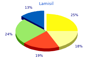
Lamisil
Andrew JP Lewington BSc MD FRCP
- Consultant renal physician
- St James’s University Hospital
- Honorary senior lecturer
- University of Leeds, Leeds, UK
Lamisil dosages: 250 mg
Lamisil packs: 30 pills, 60 pills, 90 pills, 120 pills, 180 pills, 270 pills

Buy lamisil 250mg otc
Their sparsely branched dendritic trees and the ramifications of their axons lie in a aircraft approximately perpendicular to the long axis of the folium-that is antifungal yard spray cheap 250mg lamisil, in the identical airplane as the Purkinje cell dendritic tree fungus parasite purchase lamisil 250mg online. Stellate cells are situated in the superficial molecular layer fungus clear discount 250 mg lamisil otc, and their axons synapse with the shafts of Purkinje cell dendrites. Both stellate and basket cells obtain excitatory synapses from parallel fibres passing through their dendritic timber. Their somata obtain synapses from Purkinje cell recurrent collaterals and from climbing and mossy fibres, in addition to from the parallel fibres. Basket cell axons increase in size away from their somata and run deep within the molecular layer simply above the Purkinje cells. Continuing for about 1 mm, every covers the territories of 10 to 12 Purkinje neurones. Branches from every basket cell axon additionally prolong within the path of the long axis of the folium to an extra three to six rows of Purkinje neurones, flanking the axon. It follows that as many as 72 Purkinje cell neurones might receive synapses from a single basket neurone. Most Golgi cell somata occupy the superficial zone of the granular layer, adjoining the Purkinje cell somata. In each planes they overlap the territories of several neighbouring Purkinje and Golgi cells. Some Golgi dendrites, nevertheless, divide within the granular layer and be part of cerebellar glomeruli, the place they receive excitatory synaptic contacts from mossy fibres. The axon of the Golgi cell arises from the bottom of the cell body or proximal dendrite and instantly divides right into a profuse arborization that extends by way of the complete thickness of the granular layer. The volume of the territory occupied by the axonal ramifications corresponds roughly to that of its dendritic tree within the molecular layer and it overlaps with the axonal arborizations of adjoining Golgi cells. The main synaptic input to Golgi cell dendrites is from parallel fibres within the molecular layer. Purkinje cell recurrent collaterals and mossy and climbing fibres also terminate on their proximal dendrites and, extra sparsely, on their somata. Each granule cell has a spherical nucleus, 5 to 8 �m in diameter, with a mere shell of cytoplasm containing a couple of small mitochondria, ribosomes and a diminutive Golgi complex. Granule cells give rise to three to five short dendrites that finish in claw-like terminals inside the synaptic glomeruli. The fine axons of granule cells enter the molecular layer and branch at a T-junction to form parallel fibres passing in opposite instructions over a distance of several millimetres. Terminals positioned alongside the parallel fibres give them a beaded look and are sites of synapses on the dendrites of Purkinje, stellate, basket and Golgi cells within the molecular layer. It has been estimated that 250,000 parallel fibres cross a single Purkinje dendritic tree, although every parallel fibre could not synapse with the dendritic tree it crosses. Two very different excitatory inputs serve the cerebellar cortex: climbing fibres and mossy fibres. Olivocerebellar fibres cross the white matter and enter the granular layer, where they department to form climbing fibres. There are about 10 occasions as many Purkinje cells as there are cells within the inferior olive, so each olivocerebellar fibre branches into roughly 10 climbing fibres. Individual climbing fibres pass alongside the soma of a Purkinje cell after which department to make quite a few synapses on the quick, stubby spines that protrude from the proximal segments of Purkinje cell dendrites. Mossy fibres take their origin from the spinal twine, trigeminal, dorsal column and reticular nuclei of the medulla and from the pontine tegmentum and basal pons. As every mossy fibre traverses the white matter, its branches diverge to enter several adjoining folia. Within every folium, these branches increase into grape-like synaptic terminals (mossy fibre rosettes) that occupy the centre of cerebellar glomeruli. Noradrenergic and serotoninergic fibres kind a rich plexus in all layers of the cerebellar cortex. The aminergic fibres are fantastic and varicose and kind intensive cortical plexuses; their launch of noradrenaline (norepinephrine) and serotonin is assumed to be non-synaptic, and their effects are paracrine, involving volumes of tissue. The serotoninergic afferents of the cerebellum take their origin from neurones within the medullary reticular formation, other than the raphe nuclei. The noradrenergic, coeruleocerebellar projection, when active, inhibits Purkinje cell firing not by direct motion however by -adrenergic receptor�mediated inhibition of adenylate cyclase in the Purkinje cells. Cerebellar afferents have been traced from dopaminergic cells within the ventral tegmental space, and dopamine D2 and D3 receptors are present within the molecular layer. A related plexus of thin, choline acetyltransferase�containing fibres is centred on the Purkinje cell layer. The connections of the cerebellum are organized in two perpendicular planes, corresponding to the planar organization of the cerebellar cortex. Efferent connections of the cortex are disposed in parasagittal sheets or bundles that connect longitudinal strips of Purkinje cells with specific cerebellar or vestibular nuclei. The climbing fibre afferents to a Purkinje cell zone from the inferior olive show an analogous zonal disposition. Cerebellar output is organized in modules, with a module consisting of a number of Purkinje cell zones, their cerebellar or vestibular goal nucleus and their olivocerebellar climbing fibre enter. Modular function is decided by the brain stem projections of the cerebellar or vestibular target nucleus. Mossy fibre afferent systems from precerebellar nuclei in the spinal twine and the mind stem terminate within the granular layer of certain lobules in transversely oriented terminal fields. The transverse lobular association of the mossy fibre afferents is enforced by the transverse orientation of the parallel fibres, that are axons of the granule cells and represent the second hyperlink in the mossy fibre�parallel fibre enter of the Purkinje cells. Parallel fibres cross and terminate on Purkinje cells belonging to a number of successive modules as they course via the molecular layer. Granule cell exercise generates easy spikes, which resemble the response of other neurones within the brain, whereas activation by a climbing fibre produces a prolonged depolarization on which several spike-like waves are superimposed. Whereas the Purkinje cell might fireplace simple spikes at a fee of hundreds per second, complicated spikes occur at very low frequencies, seldom greater than three or 4 per second. Like Purkinje cells, Golgi cells have a wealthy dendritic tree that extends through the molecular layer. Golgi cells regulate firing by presynaptic inhibition of the mossy fibre afferents, so they act as a governor, or fee limiter, of Purkinje cell exercise. Stellate and basket cells synapse instantly on Purkinje cells and are highly effective inhibitors of their exercise. The efferent pathways of those regions monitor the exercise within the corticospinal tract and in the subcortical motor methods descending from the vestibular nuclei and reticular formation. The inputs to the cerebellum and the outputs from it are organized in accordance with the same somatotopic patterns, however the orientation of these patterns is reversed.
Lamisil 250 mg low cost
Chronic Portal Venous Thrombosis Portal Vein Thrombolysis (Infusion Catheter Placement) (Left) Portal venography in a affected person with current liver transplantation reveals intensive filling defects within the principle portal vein fungus gnats hydroton discount 250 mg lamisil otc. An infusion catheter was advanced into the portal system through percutaneous transhepatic strategy antifungal review lamisil 250mg amex, traversing the thrombus antifungal amazon lamisil 250 mg low cost. After reviewing obtainable imaging, percutaneous transhepatic access of the portal system was obtained with a 22-g Chiba needle using ultrasound steerage. Step-By-Step: Portal Thrombolysis (Portal Venogram) Step-By-Step: Portal Thrombolysis (PostPharmacomechanical Thrombolysis) (Left) A 5-Fr sheath was advanced into the primary portal vein. Parenchymal tract embolization can be carried out by deploying contrast-soaked Gelfoam pledgets as the sheath is withdrawn. Step-By-Step: Portal Thrombolysis (Transhepatic Portal Vein Access) Step-By-Step: Portal Thrombolysis (Stenting and Thrombolysis) (Left) A guidewire and sheath have been advanced into the primary portal vein. A hepatic venogram shows the "spider net" sample of collaterals that kind across the occluded hepatic veins of sufferers with Budd-Chiari syndrome. Residual thrombus in the left portal vein was treated with systemic anticoagulation. Injected distinction provided a goal for the transjugular catheter, allowing transhepatic entry into a proper portal vein. These are typical vascular imaging findings which are seen in congestive hepatopathy. Lymphangiogram: Abdominal Lymphangiogram: Thoracic (Left) the thoracic duct is seen in the left hemithorax, turning laterally to be a part of with the left brachiocephalic vein. The lymphatic fluid will then enter the venous system on the junction of the thoracic duct and left brachiocephalic vein. A microcatheter has been advanced into the thoracic duct to the level of the mid chest. Sheybani A et al: Cerebral embolization of ethiodized oil following intranodal lymphangiography. The dye accumulates in the lymphatics just deep to the pores and skin, that are then accessed with a 30-g needle. Step-by-Step: Lymphography (Intranodal Access Setup) Step-by-Step: Lymphography (Fluoroscopic Monitoring at Pelvis) (Left) Rather than pedal access, nodal access can be achieved. The needle is then attached to an insufflator full of Ethiodol, which is injected at 2 mm Hg. Here, contrast fills the proper exterior iliac and common iliac lymphatic channels, plus inguinal and external iliac lymph nodes. Step-by-Step: Lymphography (Fluoroscopic Monitoring at Abdomen) Step-by-Step: Lymphography (Identification of Cisterna Chyli) (Left) Ethiodol distinction opacifies the proper paracaval lymphatics including paracaval lymph nodes. This sac-like dilatation is shaped by the confluence of the paralumbar (paraaortic/paracaval) lymphatics, that are joined by lymphatics that drain the intestinal tract. The thoracic duct continues from the cephalad end of the cisterna chyli and initially programs to the proper of midline earlier than ascending in the posterior mediastinum. The thoracic duct arises from the cisterna chyli and ascends to the base of the neck. Bilateral Groin Lymphangiogram Lymphangiogram in Upper Chest (Left) Postlymphography (A) frontal and (B) lateral pictures present that, within the chest, the thoracic duct courses posteriorly on the left. Its terminal end drains into the left subclavian vein at the internal jugular vein confluence. Contrast from the thoracic duct opacifies the venous system but also leaks into the pleural house. Thoracic Duct Embolization: Embolization With Coils and Glue Thoracic Duct Embolization: Needle Maceration of Cisterna Chyli (Left) Multiple embolization coils had been placed along the duct; the leak is not present. The decrease duct and needle entry into the cisterna chyli have been sealed with Trufill (n-butyl cyanoacrylate). This diverts chyle circulate into retroperitoneum, as proven by the extravasation, thereby permitting therapeutic of the leak within the thorax. Inadvertent arterial access superior to the inguinal ligament can result in intraabdominal bleeding. Access is inferior to the inferior epigastric artery, marking the inguinal ligament bordering common femoral & exterior iliac arteries. In addition to the superficial & deep femoral arteries, the deep circumflex iliac artery is seen. The 45-70ďż˝ angle of the percutaneous needle will result in arterial entry slightly superior to the hemostat. Step-by-Step: Femoral Artery Access (Superficial Anesthesia) Step-by-Step: Femoral Artery Access (Superficial Nick) (Left) Subcutaneous tissues are anesthetized with lidocaine. A small (1-2 cm) subcutaneous wheal is first delivered, helping information the situation of the longer term skin nick. After ready 5-10 seconds for the lidocaine to work, deep lidocaine (5-8 cc) is delivered medial & lateral to the deliberate access route. Step-by-Step: Femoral Artery Access (Blunt Dissection) Step-by-Step: Femoral Artery Access (Palpating Pulse) (Left) the shallow pores and skin nick prevents inadvertent chopping of the common femoral artery. The small incision can now be enlarged & deepened with blunt dissection of the gentle tissues, carried out with a hemostat. Instead, the femoral pulse was palpated above & beneath the skin nick previous to making an attempt access. If no blood is returned, proceed advancing the till contacting the femoral head (lidocaine can be delivered by way of the needle). As the needle enters the arterial lumen, a lack of resistance could also be felt & pulsatile brilliant red blood seen. Step-by-Step: Femoral Artery Access (Accessing Artery) Step-by-Step: Femoral Artery Access (Advancing zero. Fluoroscopy can be utilized to visualize the guidewire path & confirm arterial placement. After the needle is removed over the guidewire, handbook compression on the artery must be maintained to stop hematoma. Slight traction on the guidewire will straighten the wire & assist sheath development. Step-by-Step: Femoral Artery Access (Advancing Transitional Sheath) Step-by-Step: Femoral Artery Access (Advancing 0. The hemostatic valve minimizes blood loss, & the 3way stopcock allows for both distinction injection by way of the sheath & connection to a pressure bag for steady flushing of the sheath. Variants can be inadvertently punctured throughout tried entry of the femoral artery or vein. Spectral waveform suggests the proximal subclavian & axillary arteries are free of stenosis. More centrally (B), a catheter has been advanced over a J-tip guidewire into the subclavian artery.
Buy discount lamisil 250mg
An emergent celiac arteriogram revealed diffuse vasospasm fungus gnats tomato seedlings discount 250mg lamisil with visa, together with the hepatic artery and distinction extravasation from the hepatic artery toward the biliary stent fungus gnats and shore flies buy discount lamisil 250mg online. Arteriobiliary Fistula (Post Coil Embolization) Arteriobiliary Fistula (Superior Mesenteric Arteriogram) (Left) Despite the difficulty in accessing the severely spasmed hepatic artery fungus man buy 250 mg lamisil amex, the vessel was efficiently coiled. Autopsy confirmed diffuse ischemic bowel necrosis, doubtless a result of vasopressinmediated, diffuse vasoconstriction. Appropriate shape, length, and coiled diameter of a coil is operator and goal morphology dependent. Due to fears of bowel ischemia (possibly exaggerated), particles are used only in finish vessels, and provided that other embolization strategies are deemed unlikely to succeed. Embolic Agents: Covered Stents Embolic Agents: Gelfoam Slurry (Left) Covered, low-profile (self- or balloon-expandable) stents allow for efficient treatment of certain forms of hemorrhages, whereas preserving circulate within the injured vessel. Slurry is pushed backand-forth into a 2nd syringe by way of 3-way stopcock, which is narrowed until desired consistency is achieved. Embolic Agents: Gelfoam Slurry Duodenal Hemorrhage (Endoscopic View) (Left) Gelfoam slurry embolization is a fast and inexpensive methodology of embolization. Duodenal Hemorrhage (Post Coil Placement) (Left) A selective arteriogram after coil embolization of three tiny branches exhibits devascularization of the abnormal space of vascularity within the distal duodenum. Distal Duodenal Hemorrhage (Superior Mesenteric Angiogram) Distal Duodenal Hemorrhage (Superselective Angiogram) (Left) A superselective arteriogram with the catheter deciding on the defect, exhibits the duodenal hemorrhage to higher advantage. The distance between the defect and the duodenum could be very short and catheter stability is labile. Arterioportal Fistula (Post Coil Embolization) Arterioportal Fistula (Post Stent Placement) (Left) A selective superior mesenteric angiogram after coil embolization of the defect reveals decreased but persistent contrast extravasation. Provocative Angiography (Coil Embolization) Provocative Angiography (Postembolic Arteriogram) (Left) A selective completion angiogram of the proper colic artery via a microcatheter after coil embolization of 3 tiny aspect branches exhibits no additional hemorrhage and good antegrade move. Ileocolic Hemorrhage (Nuclear Scintigraphy) 386 Lower Gastrointestinal Hemorrhage Arterial Procedures Ileocolic Hemorrhage (Superior Mesenteric Arteriogram) Ileocolic Hemorrhage (Superselective Arteriogram) (Left) A selective superior mesenteric arteriogram by way of a sheath shows contrast extravasation from a branch of the ileocolic artery. Ileocolic Hemorrhage (Postembolic Arteriogram) Angiodysplasia of Cecum (Superior Mesenteric Arteriogram) (Left) Superior mesenteric arteriogram through a sheath shows decision of the hemorrhage after microcoil embolization. Selective superior mesenteric angiography shows a sausagelike vessel off of the ileocolic artery, with hemorrhage in the cecum indicative of angiodysplasia. Angiodysplasia of Cecum (Colonoscopic Appearance) Angiodysplasia of Cecum (Histologic Injected Specimen) (Left) Angiodysplasia is much more incessantly recognized by colonoscopy than by angiography. The endoscopic look is one of a discrete, small area of vascular ectasia with scalloped or frond-like edges and a visible draining vein. A selective inferior mesenteric angiogram exhibits focal extravasation within the mid descending colon. Diverticulitis (Superselective Arteriogram) Diverticulitis (Postembolization Arteriogram) (Left) A magnified view of angiogram was obtained post microcoil embolization by way of a superselective microcatheter. Endovascular or surgical remedy may be thought-about in acute hemorrhage/ischemia. Embolization of surgical anastomosis must be carried out with caution as the vascular changes associated to the anastomosis might increase the danger of postembolization ischemia. Complication of Embolization (Segmental Jejunal Infarction) Complication of Embolization (Segmental Jejunal Infarction) (Left) An intraoperative photograph following Gelfoam embolization reveals an infarcted phase of the jejunum, with adjoining normal bowel. Although problems can occur following embolization for lower gastrointestinal hemorrhage, the majority are clinically insignificant. By report, ischemic issues from transcatheter embolization require therapy in solely 0-6% of instances. Acute occlusive mesenteric ischemia from arterial embolus should be treated immediately. Unfortunately, the patent luminal diameter of the celiac artery is in all probability not significantly better. Emergency surgical revascularization, presumably together with bowel resection, have to be strongly considered in cases of impending (lactic acidosis or peritoneal signs) or actual bowel infarction. Diffuse, severe vasoconstriction is present, with nearly no bowel parenchymal opacification and marked bowel dilatation. Repeat angiography 36 hours later exhibits vital improvement within the vasoconstriction and the bowel opacification. Conservative administration with hydration and anticoagulation is usually the preliminary therapy for this scientific entity. Residual intimal hyperplasia remains within the stent, which must be closely monitored. Although this was a chronic underlying condition, the big territory concerned necessitated acute recanalization. The round density extrinsically narrowing the artery is the median arcuate ligament. The patient was asymptomatic and was thus systemically heparinized with no adverse sequelae. Noncovered stents sometimes observe via tortuous vessels simpler than would covered stents. Stent-assisted coiling is helpful for treating wide-necked aneurysms or pseudoaneurysms whereas preserving distal arterial perfusion. The celiac trunk is dilated and has a linear cleft suspicious for an arterial dissection. There is a subcapsular hematoma with central high attenuation, consistent with bleeding. Hepatic and splenic lacerations are managed conservatively but could require laparotomy or intervention. This exhibits instant filling of the splenic vein, indicating a traumatic arteriovenous fistula. There is diffuse arterial vasoconstriction as a outcome of the hypotension from active bleeding. It is necessary throughout embolization that the coils are positioned each proximal and distal to the lesion, as this prevents reperfusion in both an antegrade or retrograde style. In an actively bleeding hypotensive patient, coil embolization of the injured artery can rapidly improve hemodynamics. Atherosclerotic renal artery stenoses sometimes have an result on the proximal 1/3 of the renal artery, as on this example. Stents are reserved for complications of balloon angioplasty, similar to move limiting dissection or vessel rupture. Both stents project into aortic lumen by 1-2 mm, which is taken into account to be ideal positioning. Treatment of bilateral stenoses in one setting could also be performed if distinction volume is stored low. Renal Artery Stenosis (Post Treatment Angiogram) Ostial Atherosclerotic Stenosis (Treatment With Angioplasty) (Left) Graphic shows that balloon angioplasty of an ostial atherosclerotic stenosis is commonly ineffective, as the plaques will recoil upon deflation and elimination of the angioplasty balloon.
Buy lamisil 250 mg without a prescription
Placement of a balloon-retention G tube could require a peel-away 2-4 Fr sizes larger than the G tube and may not observe over a nonhydrophilic wire antifungal body cream generic lamisil 250mg visa. Gastrostomy Tube Placement (Position Confirmation) Gastrostomy Tube Placement (Final Tube Position) (Left) In this pigtail-type G tube candlesnuff fungus xylaria hypoxylon generic 250mg lamisil mastercard, the locking suture is pulled taut fungus gnat off uk generic lamisil 250mg fast delivery, forming the pigtail into a locked place, thereby securing the internal fixation mechanism of the G tube. Contrast injected via the tube outlines rugal folds within the decompressed abdomen. It is essential to ensure T-fasteners are deployed into the stomach lumen; not positioned inside the anterior stomach wall. The tract is dilated with the dilator advanced over the guidewire by the measured distance. Subsequently, a peel-away sheath is launched, by way of which the G tube is superior into the stomach. New gastric entry was obtained through the prevailing dermatotomy, toward the pylorus. Malpositioned Gastrojejunal Tube (New Access) Malpositioned Gastrojejunal Tube (Advancement to Jejunum) (Left) A hydrophilic wire and angled catheter have been advanced beyond the pylorus into jejunum. Gastrojejunal Tube (Normal Appearance) 732 Gastrostomy/Gastrojejunostomy Nonvascular Procedures Jejunostomy Tube (Over-The-Wire Exchange) Jejunostomy Tube (Retrograde Replacement) (Left) After exchanging a jejunostomy tube, inject distinction and observe the course of peristalsis to insure that the jejunostomy tube is downstream of the balloon. This balloon may be slightly overinflated in comparison with the diameter of the adjacent jejunum. Complication (Hemorrhage through Gastrostomy Tube) Complication (Hemorrhage via Gastrostomy Tube) (Left) the patient later presented with bleeding via the G tube. Of note, the G tube has been eliminated over a guidewire in order to eliminate any potential tamponade effect by the tube. This prognosis of achalasia is usually treated with surgical procedure or balloon dilatation, not stenting. Achalasia Stented Duodenal & Biliary Obstruction (Left) A patient with metastatic melanoma causing duodenal and biliary obstruction is proven after placement of duodenal and biliary stents. The mass was surgically resected and proved to be a large adenomatous polyp, precluding the need for either stent placement or balloon dilation. Review of the esophagram prior to stent placement is important, as this aids in the choice of the suitable stent type, diameter, and length. Esophageal Stricture Stenting (Initial Fluoroscopic Imaging) Esophageal Stricture Stenting (S/P Esophageal Stenting) (Left) Spot radiograph obtained throughout distinction esophagram shows that there has been placement of a coated Ultraflex esophageal stent. Covered stents placed for malignancy scale back the danger of tumor ingrowth when in comparability with noncovered stents. The stent is delivered by way of an over-thewire system, and the ends are flared to decrease migration risk. Stent placement can serve either as a palliative or a temporizing measure in malignant colonic obstruction. Biliary obstruction is a recognized complication of each noncovered and covered duodenal stents. Complication of Duodenal Stenting (Percutaneous Biliary Drainage) Complication of Duodenal Stenting (Percutaneous Biliary Stent Placement) (Left) Percutaneous biliary drainage was performed to decompress the obstruction. After gaining entry through a proper biliary radicle, a guidewire was superior via the interstices of the duodenal stent into the jejunum. Complication of Duodenal Stenting (Percutaneous Biliary Stent Placement) Complication of Duodenal Stenting (S/P Decompressive Biliary Stenting) (Left) After balloon dilatation, a noncovered Wallstent was deployed, extending from the intrahepatic biliary ductal bifurcation well into the duodenal stent. In cases of simultaneous duodenal and biliary obstruction, biliary and duodenal stents may be deployed facet by aspect at the outset. Variant Biliary Anatomy Abnormal Biliary Dilatation (Left) Spot radiograph demonstrates an internal/external biliary drain with the pigtail loop within the bowel. There is biliary ductal dilatation because of a standard duct stricture, via which the drain has been passed. Choledocholithiasis ďż˝ Biliary dilatation Diameter and size of balloon used Appearance pre/post dilatation ďż˝ Biliary stent Type, size, and diameter of stent(s) deployed Indicate if security catheter left indwelling ďż˝ Indicate if specimens despatched for microbiology, cytology Alternative Procedures/Therapies ďż˝ Radiologic Percutaneous cholecystostomy ďż˝ May be used to present access to distal common bile duct abnormality ďż˝ Surgical Open or laparoscopic surgical decompression ďż˝. An ultrasound probe with a sterile cover can be used to establish and align the target intrahepatic duct into sonographic view. Efflux of bile may be observed upon removal of the inner stylet to verify needle tip position. Site selection for the 2nd level of access is performed by inserting a hemostat over the potential site and evaluating beneath fluoroscopy to decide if an appropriate intercostal trajectory is available. The dermatotomy should be massive enough to permit the location of an 8-Fr drainage catheter. The 1st needle stays in place so that extra distinction can be injected if wanted. If the stricture had not been initially crossed with the 3-J guidewire, a combination of a hydrophilic guidewire and a Kumpe catheter might have been used to negotiate the stricture. Note that the first entry has still been preserved, which is helpful if the secondary entry is lost for any cause. Also note that there are embolization coils within the left lobe from remedy of prior hepatic harm. The drainage catheter is then advanced off of the cannula alongside the guidewire using fluoroscopic monitoring. The exterior hub of the catheter might be subsequently linked to a 3-way stopcock and drainage bag. A radiopaque ring demarcates essentially the most proximal sidehole, which is throughout the duct. Percutaneous transhepatic biliary drainage was subsequently carried out by way of a 2nd access. Cholangiography via Cholecystostomy 748 Transhepatic Biliary Interventions Nonvascular Procedures Dilation of Biliary Stricture (Imaging Prior to Crossing Lesion) Dilation of Biliary Stricture (Catheter Crossing Lesion) (Left) Spot radiograph from a 62-year-old man with a history of a prior Whipple process for pancreatic acinar cell carcinoma and new onset of jaundice exhibits contrast injection of a 5-Fr Kumpe catheter that had been introduced by way of a transhepatic method. Dilation of Biliary Stricture (Angioplasty Balloon Placement) Dilation of Biliary Stricture (Angioplasty Balloon Inflation) (Left) Balloon angioplasty catheter was introduced over an Amplatz guidewire by way of an entry sheath & superior to the extent of the stricture. Radiopaque markers demarcating the proximal & distal extent of the deflated balloon are positioned to encompass the stricture. Occasionally, the balloon may propel ahead into the bowel during inflation; countertraction on the catheter could also be necessary. Dilation of Biliary Stricture (Angioplasty Balloon Inflation) Dilation of Biliary Stricture (Appearance After Angioplasty) (Left) the balloon is inflated further and the "waist" is obliterated. The balloon is then deflated and the process is repeated for a complete of 3 dilatations. Algorithms differ amongst interventionalists; in a single regularly used algorithm, three treatment classes are employed, each 1-2 weeks apart. A 5-Fr Kumpe catheter has traversed a typical duct stricture, with the catheter tip in the bowel. The Wallstent is positioned to encompass the entire size of the stricture, with the ends of the stent extending at least 2-3 cm proximal and distal to the stricture margins.
Proven lamisil 250 mg
In addition fungus gnats bleach order lamisil 250 mg on-line, there could also be weak spot and wasting of the intrinsic muscles of the hand equipped by the ulnar nerve antifungal wood cheap 250mg lamisil mastercard, with clawing in excessive instances antifungal bacteria generic 250mg lamisil amex. Division of the ulnar nerve on the wrist paralyses all of the intrinsic muscles of the hand (except for the radial two lumbricals). The intrinsic muscle motion of flexing the metacarpophalangeal joints and extending the interphalangeal joints is subsequently lost. The unopposed action of the lengthy extensors and flexors of the fingers causes the hand to assume a clawed look with extension of the metacarpophalangeal joints and flexion of the interphalangeal joints. The clawing is much less intense in the index and center fingers due to their intact lumbricals, equipped by the median nerve. For a detailed account of the hand posture adopted in ulnar nerve lesions, see Smith 2002. There is sensory loss over the little finger and the ulnar half of the ring finger. In comparison with an ulnar nerve division on the elbow, the skin over the ulnar side of the dorsum of the hand is spared as a end result of the dorsal department of the ulnar nerve is given off approximately 5 cm proximal to the wrist joint. Branches of the superficial branch of the radial nerve reach the hand by curving across the wrist deep to the brachioradialis tendon. On the dorsum of the hand they normally communicate with the posterior and lateral cutaneous nerves of the forearm. Dorsal Digital Nerves Ulnar Nerve Division at the Wrist There are normally 4 or five small dorsal digital nerves. The first supplies the skin of the radial facet of the thumb and the adjoining thenar eminence and communicates with branches of the lateral cutaneous nerve of the forearm. The second provides the medial side of the thumb; the third, the lateral facet of the index finger; and the fourth, the adjoining sides of the index and center fingers. The pollicial digital nerves attain solely to the foundation of the nail; those within the index finger, midway alongside the middle phalanx; these to the middle and lateral parts of the ring finger might reach no farther than the proximal interphalangeal joints. The remaining distal dorsal areas of the skin in these digits are supplied by palmar digital branches of the median and ulnar nerves. The superficial terminal department of the radial nerve could provide the entire dorsum of the hand. The preganglionic sympathetic inflow to the upper limb is derived from neurones in the lateral horn of the higher thoracic spinal segments T2�6 (or 7). Fibres pass in white rami communicantes to the thoracic sympathetic chain and synapse in the stellate and second thoracic ganglia. Postganglionic fibres to the pores and skin are distributed through cutaneous branches of the brachial plexus. If the forearm is rotated into pronation, the wrist falls into flexion, and the fingers automatically lengthen. If the forearm is rotated into supination, the wrist extends and the fingers flex. This test, the wrist tenodesis check, is a helpful approach to study the limb for tendon harm. Wrist motion is managed principally by two wrist flexors (flexor carpi radialis and flexor carpi ulnaris) and three extensors (extensors carpi radialis longus and brevis, and extensor carpi ulnaris). It would be very limiting to have a pure hinge joint with collateral ligaments of fixed size. In this context, the wrist flexors and extensors could also be considered variable collateral ligaments that enable the joint to be set about numerous completely different axes. The basic mechanisms of hand closure are described earlier than special grips are thought-about. The involvement of the lengthy digital flexors on this movement is taken into account first, adopted by an evaluation of the position of the wrist. Flexor digitorum superficialis acts to flex principally the proximal interphalangeal joints, through its insertions into the middle phalanges. However, in every digit it also has an motion on the metacarpophalangeal joint, because the tendon passes anterior to that joint. The fact that each tendon arises from an individual muscle slip allows the clinician to test one finger at a time. The reader can confirm this by making an attempt to flex each digit individually whereas utilizing the other hand to hold the distal interphalangeal joints of the remaining fingers in extension. This take a look at is frequently used in scientific follow and is useful for the middle and ring fingers, the place flexion of one finger alone must be attributed to flexor digitorum superficialis. The index finger, however, has its own profundus musculotendinous unit and may therefore transfer independently underneath the action of this tendon. It also contributes, along with superficialis, to flexion on the proximal interphalangeal and metacarpophalangeal joints. These two long flexors (sometimes referred to as extrinsic flexors, as a outcome of the muscle bellies are exterior the hand) could be thought-about to act together to flex the finger. These small (intrinsic) muscle tissue have been described earlier in terms of their particular person actions. All three joints are then angulated to the same diploma, and the fingers form a normal arc of flexion. As the finger flexes, the lengthy extensor tendons (extensor digitorum, extensor indicis and extensor digiti minimi) aid the method by stress-free and allowing the extensor equipment to glide distally on the dorsa of the phalanges. Role of the Long Digital Flexors Making a Tight Fist It is feasible to observe and palpate the muscle groups which are lively in making a tight fist. The flexor compartment of the forearm is contracted tightly, and electromyographic evidence confirms that flexor digitorum profundus and flexor digitorum superficialis are energetic. The extensor compartment is tightly contracted, and the wrist extensors would certainly be anticipated to be active. Palpation of the long digital extensors on the again of the wrist exhibits that these are contracting as well. They due to this fact carry out the only task available to them and act together as an additional wrist extensor. In the thumb web, palpation confirms that the primary dorsal interosseous is contracting, as are all the opposite interossei and the thenar and hypothenar muscles. As the firm fist is swung ahead in anger, the brachioradialis stands out, and in the intervening time of impression, just about each muscle within the limb is in a state of contraction, aside from the lumbricals. The hand is opened from its relaxed balanced posture, such as when stretching out to reach an object. This motion is made up of extension of the distal interphalangeal, proximal interphalangeal and metacarpophalangeal joints. The laws of mechanics suggests that one motor could be required for every joint in a chain, along with some kind of controlling mechanism to make certain that the chain of joints moves collectively in a coordinated trend. In the hand, that is achieved via an extensor equipment that minimizes the number of motors required for motion by permitting the muscles to act on multiple joint and by linking completely different levels within the mechanism in order that the arc of motion is controlled. The tendons of extensor digitorum run distally over the metacarpal heads, forming the major component of the extensor equipment.
Purchase 250 mg lamisil
There is marked losing of the thenar eminence bilaterally (arrow) in a sophisticated case of carpal tunnel syndrome fungus habitat purchase lamisil 250 mg online. The major nerve regularly trifurcates to supply the pulp and skin of the terminal part of the digit antifungal jock itch cream lamisil 250 mg. Distal to the bottom of the proximal phalanx antifungal essential oil blend trusted lamisil 250mg, every proper digital nerve additionally offers off a dorsal branch to supply the pores and skin over the back of the center and distal phalanges. The correct palmar digital nerves to the thumb and lateral side of the index finger emerge with the lengthy flexor tendons from beneath the lateral edge of the palmar aponeurosis. They are arranged within the digits as described earlier, however in the thumb, small distal branches supply the pores and skin on the back of the distal phalanx only. Other Branches In addition to the branches of the median nerve already described, variable vasomotor branches provide the radial and ulnar arteries and their branches. Some of the intercarpal, carpometacarpal and intermetacarpal joints are thought to be equipped by the median nerve or its anterior interosseous department; the exact particulars are uncertain. Division leads to paralysis of the lumbricals to the index and center fingers and the thenar muscles (apart from adductor pollicis), in addition to loss of sensation to the thumb, index, middle fingers and radial half of the ring finger. The radial half of the hand turns into flattened as a result of wasting of the thenar muscular tissues and the adducted posture of the thumb. The first provides the medial aspect of the little finger; the second, the adjoining sides of the little and ring fingers; and the third, when present, supplies adjoining sides of the ring and center fingers. The final could also be replaced, wholly or partially, by a department of the radial nerve, which always communicates with it on the dorsum of the hand. In the little finger, the dorsal digital nerves extend solely to the bottom of the distal phalanx; within the ring finger, they lengthen solely to the base of the center phalanx. The most distal components of the little finger and of the ulnar facet of the ring finger are equipped by dorsal branches of the right palmar digital branches of the ulnar nerve. The most distal a half of the lateral aspect of the ring finger is provided by dorsal branches of the right palmar digital branch of the median nerve. The superficial terminal branch provides palmaris brevis and the medial palmar skin. It divides into two palmar digital nerves, which may be palpated in opposition to the hook of the hamate bone. One supplies the medial aspect of the little finger, and the opposite (a frequent palmar digital nerve) sends a twig to the median nerve and divides into two correct digital nerves to supply the adjoining sides of the little and ring fingers. The proper digital branches are distributed like these derived from the median nerve. The deep terminal department accompanies the deep department of the ulnar artery because it passes between abductor digiti minimi and flexor digiti minimi and then perforates the opponens digiti minimi to follow the deep palmar arch dorsal to the flexor tendons. As it crosses the hand, it provides the interossei and the third and fourth lumbricals. It ends by supplying adductor pollicis, first palmar interosseous and often flexor pollicis brevis. The medial a half of flexor digitorum profundus is provided by the ulnar nerve, as are the third and fourth lumbricals, which are linked with the Superficial Terminal Branch Median Nerve Division on the Wrist Deep Terminal Branch Ulnar Nerve Dorsal Branch the dorsal department arises approximately 5 cm proximal to the wrist. Similarly, the lateral part of flexor digitorum profundus and the primary and second lumbricals are provided by the median nerve. The deep terminal department is thought to give branches to some intercarpal, carpometacarpal and intermetacarpal joints; exact particulars are uncertain. Vasomotor branches, arising in the forearm and hand, provide the ulnar and palmar arteries. A mixed median and ulnar nerve palsy on the wrist ends in a full claw hand, with thenar and hypothenar flattening and thumb adduction and flexion. Causes of compression at this web site embrace ganglion, trauma and proximity of aberrant or accent muscle tissue. Symptoms embody pain within the hand or forearm and sensory adjustments within the palmar side of the little finger and ulnar half of the ring finger; nonetheless, sensation on the ulnar aspect of the dorsum of the hand is regular. Extensor digitorum has no insertion into the proximal phalanx and therefore exerts its extensor action on the metacarpophalangeal joint indirectly through more distal insertions. Acting at this insertion alone, extensor digitorum can lengthen both metacarpophalangeal and proximal interphalangeal joints collectively. The interossei are additionally energetic in hand opening as a result of they have a tendency to enhance extension of the proximal interphalangeal joint. At one extreme, with no interosseous contribution, the lengthy extensor exerts all its motion on the metacarpophalangeal joint; this results in full extension or even hyperextension, whereas the proximal interphalangeal joint remains flexed (the As the fingers wind up to make a fist, the wrist tends to lengthen, significantly when drive is utilized. On its own, digital flexion would require the long tendons to move proximally of their sheaths, and the flexor muscular tissues in the forearm would shorten. Dorsiflexion of the wrist tends to produce a lengthening of the same muscle tissue, which in regular use is nearly sufficient to steadiness the shortening because of finger flexion; the online effect is a really slight shortening (approximately 1 cm) of the long flexors within the forearm. The wrist can due to this fact be seen as a mechanism for maximizing pressure, as a end result of it allows the fingers to flex while maintaining the resting size of the extrinsic muscular tissues near the peak of the force�length curve. It is, in fact, possible to wind up the fingers with the wrist held in a neutral place, however the grip is somewhat weaker. Flexion of the fingers on gripping tends to lead to a distal excursion of the lengthy extensors. The net impact is a very small proximal excursion of the lengthy extensor tendons on gripping, mirroring the impact on the flexor surface. Thus, within the proximal part of the extensor equipment, the hand possesses a variable mechanism that permits different amounts of relative metacarpophalangeal or proximal interphalangeal joint motion. In contrast, the more distal a part of the extensor equipment acts as an automatic or mounted mechanism, whereby the 2 interphalangeal joints, proximal and distal, move together. The lateral slips of the extensor equipment come up from extensor digitorum and pass distally on both side of the central slip and thus over the proximal interphalangeal joint. A helpful analogy is to contemplate this association as consisting of two pulleys of different sizes on one axle. The central slip can be thought to be a twine that passes over the larger wheel, and every lateral slip as a cord that passes over the smaller wheel. There is an extra mechanism by which the lateral slips move laterally during flexion of the proximal interphalangeal joint. The effect of this lateral movement is to additional reduce the distance between the lateral slips and the joint axis, thereby lowering the amount of tour on the proximal interphalangeal joint much more and permitting extra excursion on the distal joint. When the hand flexes, this mechanical linkage system permits each interphalangeal joints to flex together in a coordinated method. The extensor growth also receives contributions from the interossei and lumbricals, which approach the digits from the webs and join the corresponding enlargement in the proximal section of the digit. Apart from the parts of the extensor growth involved with joint perform, the entire structure requires further anchorage.
Purchase lamisil 250 mg without a prescription
The tonsil is exposed by incision of the anterior faucial pillar anti fungal diet yogurt generic 250mg lamisil, shelled out alongside the connective-tissue plane between the parenchyma and pharyngeal muscle fungi definition biology online effective lamisil 250mg, and indifferent at its inferior pole anti fungal acne treatment purchase lamisil 250 mg on-line. Heavy postoperative bleeding could happen on the day of the tonsillectomy, through the first week after the operation, and even later in rare instances. Chronic pharyngitis the typical appearance of a granulating irritation involving the posterior wall of the pharynx (hypertrophic form). This leads to frequent arousals from sleep and gasping for air, stopping a normal sleep pattern. Factors that slim the pharyngeal airway or lead to decreased muscle tone (Table 5. Witnesses moreover report irregular loud night breathing with durations of apnea followed by "gasping" and loud loud night breathing. Diagnosis: Mirror examination might demonstrate an elongated uvula, a narrow velopharyngeal passage, and a cumbersome soft palate with a small oropharyngeal lumen. It is also frequent to find a hyperplastic tongue base and hyperplasia of the palatine tonsils. Systemic arterial hypertension Erythropoiesis Central arousal Loss of deep sleep, fragmented sleep Return to sleep Resumption of respiratory Probst-Grevers-Iro, Basic Otorhinolaryngology� 2006 Thieme All rights reserved. Polycythemia Daytime fatigue, drowsiness, intellectual deterioration 5 Pharynx and Esophagus 121 Table 5. Treatment: General treatment measures include weight discount, abstinence from alcohol and nicotine, and avoiding big meals, especially at evening. It is also important to establish a daily sleep�wake cycle and avoid the usage of sedatives. One nonsurgical remedy possibility is the Esmarch splint, an occlusive splint that advances the decrease jaw. By transferring the tongue base and adjoining pharynx ahead, this system widens the airway within the unstable portion of the oropharynx. Surgical remedy choices are tailored to the specific pathology inflicting the apnea. Surgical treatment requires very careful affected person selection, because many sufferers will derive little or no benefit from the operation. The result of the M�ller maneuver could be helpful in choosing patients for a surgical procedure on the soft palate. Some sufferers will also require adjuvant intranasal surgical procedure (septoplasty, septorhinoplasty, turbinate reduction). Nasal obstruction Decreased muscle tone � Dysgnathia � Acromegaly � Turbinate hyperplasia � Septal deviation � Nasal polyps � Deformities of the external nose � Tumors of the nose � Alcohol � Nicotine � Drugs (sedatives, hypnotics, muscle relaxants) Other � Sleep deprivation � Shift work � Hypothyroidism � Sex (males predominate) � Genetic predisposition: familial occurrence � Sleeping in a supine position airway must also be examined for possible septal deviation, turbinate hyperplasia, or other abnormalities. When a wholesome patient performs this maneuver, it causes little or no decrease within the cross-sectional space of the pharyngeal lumen. Objective measuring strategies: Screening: Reports given by the patient and witnesses could be objectified by recording O2 saturation, respiratory sounds, and coronary heart fee on an outpatient basis during sleep. Besides dysphagia and odynophagia, the signs may include blood-tinged saliva and a fetid breath odor. The resulting tissue defect could also be closed primarily with native pedicled flaps or by utilizing microvascular free tissue transfers, relying on the scale and placement of the defect. Postoperatively, radiation should often be delivered to the tumor site and lymphatic pathways. Alternatives for the treatment of superior tumors (T3, T4) are primary radiotherapy or combined radiation and chemotherapy. Tumors Benign Tumors and Precancerous Lesions the incidence, incidence, clinical options, and treatment of those lesions are covered in 4. Malignant Tumors the overwhelming majority of malignant tumors of the oropharynx are squamous cell carcinomas. Etiology: In most sufferers, persistent nicotine and alcohol abuse have a major etiologic role in the growth of oropharyngeal cancers. Congenital malformations corresponding to brachyesophagus and tracheoesophageal fistulas are managed by a thoracic or pediatric surgeon, whereas esophageal tumors are treated by a basic surgeon or stomach surgeon. Injuries and Foreign Bodies Caustic Ingestion Etiopathogenesis: Caustic ingestion in children is type of all the time unintended, caused by consuming an alkaline or acidic liquid that has not been properly stored. While acids cause a coagulation necrosis with the denaturation of proteins, alkalis cause a colliquative necrosis with liquefaction of the necrotic tissue. There is a long-term threat of most cancers developing in esophageal strictures attributable to caustic ingestion. Symptoms: Acute cases present with extreme pain within the mouth and pharynx and possibly within the retrosternal and epigastric areas. Patients with an esophageal perforation may also present with subcutaneous emphysema within the neck or a pneumomediastinum. Symptoms such as excessive fever and retrosternal or interscapular ache in these instances are accompanied by typical shock signs with an elevated pulse price, a fall in blood strain, cold sweats, and pallor. In the lengthy term, patients may develop an esophageal stricture with progressive dysphagia. Diagnosis: Acute evaluation begins with a mirror examination of the oral cavity, oropharynx, hypopharynx, and larynx. The mucosa initially appears erythematous and edematous and later could present epithelial defects and a whitish fibrin coating. It is also important to get hold of radiographs of the chest and abdomen to exclude a perforation of the esophagus or stomach. Follow-up: Caustic accidents to the esophagus require long-term follow-up with imaging studies and periodic esophagoscopy. It is essential to stabilize the airway, substitute fluids, correct electrolyte imbalances, relieve pain, and provide sedation. Treatment also wants to embody excessive doses of corticosteroids as well as antibiotics to prevent superinfection. Conservative therapy of esophageal strictures An esophageal stricture is handled with a dilator passed over a guide wire. In instances with extreme caustic damage, early dilation should be started just one week after the harm. The dilator ought to by no means be launched blindly however ought to always be handed over a guide wire. It could then be essential to carry out a gastric pull-up or interpose a free segment of jejunum with microvascular anastomosis, depending on the size of the esophageal section that has been eliminated. Whenever an ingested international body is suspected, rigid esophagoscopy must be carried out without delay to retrieve the international object. This may require a transcervical incision or thoracotomy, depending on the location of the overseas physique. Rupture of the Esophagus Synonym: Boerhaave syndrome Boerhaave syndrome refers to a spontaneous rupture within the left posterolateral portion of the terminal esophagus simply above the esophageal hiatus brought on by forceful vomiting or retching.
Cheap 250mg lamisil amex
Musculature of the pharynx and esophagus Various muscular tissues contribute to the wall construction of the pharynx and esophagus fungus in mulch order 250mg lamisil amex. Constrictor pharyngis superior Constrictor pharyngis medius Constrictor pharyngis inferior Killian triangle Killian-Jamieson region Laimer triangle Esophageal musculature Uvula Stylopharyngeus muscle Palatopharyngeus muscle Cricopharyngeus muscle pharyngis inferior (cricopharyngeal part) run instantly horizontally fungal rash lamisil 250mg generic, creating anatomical weak spots within the pharyngeal wall (Laimer and Killian triangles anti yeast antifungal diet cheap 250mg lamisil fast delivery, 5. These weak spots are sites of predilection for the development of pulsion (Zenker) diverticula in the hypopharynx. Three additional pairs of exterior muscular tissues are distributed to the pharyngeal wall and assist in controlling vertical movements of the pharynx: the stylopharyngeus, the salpingopharyngeus, and the palatopharyngeus. Neurovascular Supply the pharynx receives its blood provide from the territory of the exterior carotid artery (branches of the facial artery, maxillary artery, ascending pharyngeal artery, lingual artery, and superior thyroid artery). The lymphatic drainage of the upper parts of the pharynx is thru the retropharyngeal lymph nodes, while the lower parts drain to the parapharyngeal or deep cervical nodes. The plexus itself is located on the outer aspect of the constrictor pharyngis medius muscle. The retropharyngeal space incorporates smaller arterial and venous vessels and, most notably, the retropharyngeal lymph nodes that drain the nasopharynx. Esophagus the esophagus begins on the higher esophageal sphincter, located on the level of the C6 and C7 vertebrae (inferior border of the cricoid cartilage). The three physiologic constrictions of the esophagus are clinically necessary due to the tendency for ingested international bodies to become lodged at these levels. The parapharyngeal house is divided anatomically into two components, the retropharyngeal space and the lateral pharyngeal house. The latter in turn is subdivided by the common connective-tissue sheath of the muscles arising from the stylohyoid course of (stylopharyngeal aponeurosis) right into a prestyloid and a retrostyloid half. It contains the lateral and medial pterygoid muscles, lingual nerve, optic ganglion, and maxillary artery. Anatomy of the esophagus a b zero Thyroid cartilage Cricoid cartilage Cricopharyngeus muscle Trachea Aorta Sternum Esophagus Heart Diaphragm Epiglottis Cricoid cartilage Upper esophageal constriction Trachea Midesophageal constriction Aorta Piriform recess Thyroid cartilage Cricopharyngeus muscle a the esophagus extends from C6/C7 to the gastric cardia at the degree of the T10 vertebra. The thoracic esophagus is supplied by the aorta and intercostal arteries, and the abdominal esophagus by the left gastric artery and left inferior phrenic artery. Thoracic and belly drainage is to the azygos and hemiazygos veins and esophageal veins. Lymphatic drainage is to the lymph nodes of the posterior mediastinum and pulmonary hilum. Nerve supply: the upper, cervical a half of the esophagus is supplied with branches from the recurrent nerve and the decrease part with unnamed branches from the vagus nerve. Below the tracheal bifurcation is the esophageal plexus, shaped by the 2 vagus nerves. Secondary peristalsis is additionally triggered in the esophagus by the strain of the bolus against the esophageal wall. Through the coordinated action of those mechanisms, the bolus is transported into the stomach within 7�10 seconds. This tissue is structurally similar to lymph nodes but lacks afferent lymphatic vessels. The tonsils are named for their location, consisting of a pharyngeal tonsil, the paired palatine tonsils, and the unpaired lingual tonsil at the base of the tongue. Additionally, smaller condensations of lymphoepithelial tissue are discovered in the pharyngeal recess. While the pharyngeal tonsil is covered primarily by multiple rows of ciliated epithelium, the palatine and lingual tonsils are coated by stratified, nonkeratinized squamous epithelium. Physiology of Swallowing Normal swallowing requires a coordinated interplay of varied anatomic constructions in the oral cavity, pharynx, larynx, and esophagus. From a practical standpoint, the voluntarily initiated oral section of swallowing is distinguished from an "involuntary" pharyngeal phase and esophageal part, which are managed by way of reflex mechanisms. This is accomplished mainly by urgent the meals in opposition to the onerous palate with the tongue. The pharyngeal phase begins when the bolus comes into contact with receptors within the throat (especially on the tongue base), eliciting an involuntary swallowing reflex. The intensive nerve provide highlights the complexity of swallowing as properly as the potential vulnerability of this process. While the involuntary swallowing reflex is triggered in the course of the pharyngeal section, the velum is elevated to close off the nasopharynx. This is accompa- Structure of the Palatine Tonsil the palatine tonsil has special immunologic significance among the many tissues of the tonsillar ring owing to its distinctive morphology. Its surface is invaginated by crypts-fold-like tissue indentations which might be lined by porous epithelium and substantially increase the floor space of the tonsil. This association facilitates contact between impressed or ingested antigens and the subepithelial lymphatic tissue. Physiology of swallowing a Oral phase b Pharyngeal part Bolus c Esophageal phase Bolus Bolus the circled numbers check with the textual content (p. Within the lymphatic tissue, main follicles are fashioned during embryonic improvement and differentiate into secondary follicles after start. The secondary follicles primarily include B lymphocytes at various levels of differentiation, along with scattered T lymphocytes. Besides the lymph follicles, there are additionally extrafollicular areas with B and T lymphocytes that enter the lymphatic tissue through the postcapillary venules. Functional Importance of the Tonsils within the Immune System the palatine tonsil specifically is taken into account to be an "immune organ" that plays a big role within the defense towards higher respiratory infections. Accordingly, this tissue has the flexibility to mount specific immune reactions in response to numerous antigens. The exercise of this lymphatic organ is especially pronounced throughout childhood, when immunologic challenges from the environment induce hyperplasia of the palatine tonsils. While the tonsils turn into less important immunologically with ageing, the tonsillar tissue continues to carry out immune capabilities even at a sophisticated age, although this could not alter the choice to remove the tonsils if a sound indication for tonsillectomy exists (see Chronic Tonsillitis, p. While the tonsils are "learning" their immune perform throughout childhood, extreme tonsillar hyperplasia ("kissing tonsils") might develop, leading to functionally vital narrowing of the faucial isthmus, with consuming difficulties and obstructed respiration. Especially Probst-Grevers-Iro, Basic Otorhinolaryngology� 2006 Thieme All rights reserved. Tonsillar hyperplasia Mantle zone Germinal zone b the dimensions and exercise of the germinal facilities are maximal at roughly 6�8 years of age, reflecting the immunologic exercise of the palatine tonsil. Tonsillar hyperplasia develops in lots of children during this "lively immunization phase. Consequently, there ought to be little hesitation in recommending tonsillectomy, even in young children. The secondary follicles in the lymphatic tissue, which are important for tonsillar immune perform, function a light-colored germinal zone and a dark mantle zone with mature lymphocytes. This development is due largely to the arrival of computed tomography and magnetic resonance imaging, while conventional radiographs have turn into largely obsolete in the investigation of diseases of the pharynx.
References
- Cappell MS, Friedel D: Acute nonvariceal upper gastrointestinal bleeding: Endoscopic diagnosis and therapy. Med Clin North Am 92:511, 2008.
- Papatsoris AG, Buchholz N: A novel thermo-expandable ureteral metal stent for the minimally invasive management of ureteral strictures, J Endourol 24:487, 2010.
- Jadersten M, Malcovati L, Dybedal I, et al. Erythropoietin and granulocyte-colony stimulating factor treatment associated with improved survival in myelodysplastic syndrome. J Clin Oncol. 2008;26:3607-3613.
- Kugel RD: The Kugel repair for groin hernias. Surg Clin N Am 83:1119, 2003.
- Quasha AL, Loeber N, Feeley TW, et al: Postoperative respiratory care: A controlled trial of early and late extubation following coronary-artery bypass grafting, Anesthesiology 52:135, 1980.
- Weber R, Ruppik M, Rickenbach M, et al. Decreasing mortality and changing patterns of causes of death in the Swiss HIV cohort study. HIV Med 2013;14:195-207.